ABSTRACT
Objective: The aim of the present study was to compare the long-term outcomes of the laparotomy (LT) and laparoscopic surgery and to evaluate the results according to low, intermediate, and high-risk groups of endometrial cancer (EC).
Methods: We identified 801 patients with EC and these patients were classified as group 1, who underwent LT (n=515); and group 2, who underwent laparoscopy (LS) (n=286).
Patient's demographics, clinical characteristics such as stage, grade, histopathologic type, lymphovascular space invasion, myometrial invasion, lymph node involvement, and risk groups, peri- and post-operative outcomes, and survival outcomes were compared between the groups according to risk classification. Survival outcomes were assessed using Kaplan- Meier method.
Results: The demographic characteristics of both groups were similar except age. Shorter hospital stay and fewer complications were observed in group 2. The overall survival (OS) were similar in the low, low-intermediate, high-intermediate and high-risk groups (p=0.269, 0.476, 0.078, and 0.085; respectively) for LS compared to LT. The covariate analysis revealed that the death and recurrence risks were approximately twice higher in the LT group than in the LS group (odds ratio [OR]=1.9; 95% confidence interval [CI]=1.2–3.1 for OS; OR=2.0;
95% CI=1.2–3.3 for disease-free survival).
Conclusion: The results of our study support the well-known positive aspects of LS as well as safe and effective use in cases of intermediate and high-risk EC.
Keywords: Endometrial Cancer; Laparoscopy; Laparotomy; Prognostic Factors;
Risk Assessment
INTRODUCTION
Minimally invasive surgery (MIS) has inevitably entered into our practice. Laparoscopic surgery in endometrial cancer (EC) treatment has become increasingly common for 15 years.
Surgery is a curative treatment for the majority of patients with EC. Laparoscopic surgery in early-stage EC is an effective and safe approach resulting in similar oncologic outcomes compared to laparotomy (LT) [1-3]. Many laparoscopic studies in this regard were performed with a relatively small number of patients, short median follow-up and in patients with
Original Article
Received: Jul 19, 2018 Revised: Oct 5, 2018 Accepted: Nov 9, 2018 Correspondence to Umran Kucukgoz Gulec
Department of Obstetrics and Gynecology, Cukurova University Faculty of Medicine, Balcalı Mahallesi, Çukurova Üniversitesi Rektörlüğü, Saricam/Adana 01330, Turkey.
Email: [email protected] Copyright © 2019. Asian Society of Gynecologic Oncology, Korean Society of Gynecologic Oncology
This is an Open Access article distributed under the terms of the Creative Commons Attribution Non-Commercial License (https://
creativecommons.org/licenses/by-nc/4.0/) which permits unrestricted non-commercial use, distribution, and reproduction in any medium, provided the original work is properly cited.
ORCID iDs Mehmet Ali Vardar
https://orcid.org/0000-0003-0616-6733 Umran Kucukgoz Gulec
https://orcid.org/0000-0003-3094-1381 Ahmet Baris Guzel
https://orcid.org/0000-0002-9498-7592 Derya Gumurdulu
https://orcid.org/0000-0002-7129-3424 Ghanim Khatib
https://orcid.org/0000-0002-0163-1141 Gulsah Seydaoglu
https://orcid.org/0000-0002-0899-894X
Mehmet Ali Vardar ,1 Umran Kucukgoz Gulec ,1 Ahmet Baris Guzel ,1 Derya Gumurdulu ,2 Ghanim Khatib ,1 Gulsah Seydaoglu 3
1Department of Obstetrics and Gynecology, Cukurova University Faculty of Medicine, Adana, Turkey
2Department of Pathology, Cukurova University Faculty of Medicine, Adana, Turkey
3Department of Biostatistics, Cukurova University Faculty of Medicine, Adana, Turkey
Laparoscopic surgery for low,
intermediate and high-risk
endometrial cancer
Presentation
This study was presented as poster and e-poster at 20th International Meeting of the European Society of Gynecological Oncology (ESGO 2017), November 4–7, Vienna.
Conflict of Interest
No potential conflict of interest relevant to this article was reported.
Author Contributions
Conceptualization: V.M.A., G.U.K., G.A.B., S.G.; Data curation: G.U.K., S.G.; Formal analysis: S.G.; Investigation: V.M.A., G.U.K., G.A.B., G.D., K.G., S.G.; Methodology: G.U.K., S.G.; Resources: V.M.A., G.U.K., G.A.B., G.D.;
Supervision: V.M.A., G.U.K., G.A.B., S.G.;
Validation: S.G.; Visualization: G.U.K.; Writing - original draft: V.M.A., G.U.K.; Writing - review &
editing: V.M.A., G.U.K., G.A.B., G.D., K.G., S.G.
early stage EC. However, EC is a disease with several histopathologic subtypes and different tumor biology and thus having different clinical outcomes. New risk groups were defined by the European Society for Medical Oncology (ESMO)-European Society of Gynaecological Oncology (ESGO)-European SocieTy for Radiotherapy & Oncology (ESTRO) consensus in 2016 as a guide for adjuvant therapy use [4]. Lymphovascular invasion is also assessed in this novel risk classification because of its prognostic importance indicated in the recent studies [5,6]. In that classification, a total of 4 groups are formed by dividing the intermediate group into intermediate and high-intermediate. There are limited data regarding the feasibility and safety of laparoscopic management of EC with intermediate and high-risk factors such as advanced stage and type 2 EC.
In this study, we aimed to evaluate long-term oncologic outcomes in patients with low, intermediate, and high-risk EC treated by laparoscopic surgery and to compare the surgical and long-term survival data in LT cases at the same time. To our knowledge, there is no study evaluating laparoscopic surgery considering the risk classification of EC in the literature.
MATERIALS AND METHODS
The medical records of the patients with EC, who underwent surgery and were followed in the department of gynecologic oncology, were reviewed between the January 2005 and 2016. We identified 801 patients with EC and these patients were classified as group 1, who underwent LT (n=515), and group 2, who underwent laparoscopy (LS) (n=286). Informed consent was obtained from all patients before surgery. All patients diagnosed with EC based on endometrial biopsy were examined through transvaginal ultrasonography and chest X-ray in accordance with our clinical approach. The presence of a distant metastasis was excluded via computed tomography in the high-risk group. The main exclusion criteria to perform a laparoscopic surgery were severe cardiopulmonary disease, a contraindication for prolonged Trendelenburg position, and large uterus that could not be removed vaginally intact. Age, high body mass index (BMI), and prior pelvic surgery were not the exclusion criteria for LS. All EC patients were evaluated with an intraoperative frozen section according to histopathologic type, grade, and depth of myometrial invasion. An algorithm was
followed for EC surgery in accordance with the Mayo clinic surgical algorithm for EC [7,8].
Totally hysterectomy and bilateral salpingo-oophorectomy were performed on the patients with endometrioid adenocarcinoma, stage 1a, International Federation of Gynecology and Obstetrics (FIGO) grade 1 and 2 tumors (low-risk factors), tumor diameter of <2 cm by LT or LS whereas pelvic lymphadenectomy (PLND) was performed on the patients with tumor diameter >2 cm. Pelvic-paraaortic lymphadenectomy (PPALND)±omentectomy (for serous or mixed type carcinomas) was performed in cases with grade 3, more than 50% myometrial invasion, type 2 histopathology, and higher stages than stage 1a. The operations were performed by the same gynecologic oncology surgical team (V.M.A., G.A.B., G.U.K.).
The demographic parameters were age, BMI, nulliparity, and the history of infertility.
Surgical and treatment parameters were the type of surgery, conversion rate, the duration of surgery (from skin incision to skin closure, except the frozen section time), estimated blood loss, decrease in hemoglobin (Hb) level, postoperative hospitalization time (day), intraoperative complications (bleeding was defined as a blood loss exceeding 1,000 mL or requiring blood transfusion), postoperative complications, and adjuvant therapy modalities. Clinical and pathological variables were stage, grade, histopathologic type,
myometrial invasion (as <50% and ≥50%), lymph nodes involvement, lymphovascular space invasion (LVSI), the number of lymph nodes yielded, comorbidities, and survival outcomes.
Overall survival (OS) and disease-free survival (DFS) were assessed. The surgical stage was determined considering histopathologic results according to FIGO 2009 guidelines. All patients received antibiotic prophylaxis preoperatively and thromboprophylaxis during the hospitalization.
Risk classification was done based on ESMO-ESGO-ESTRO consensus report [4]. Patients with grade 1 or 2 endometrioid, myometrial invasion less than 50%, stage 1, and LVSI negative were classified as low risk; patients with stage 2, 3, 4 endometrioid tumors, grade 3 endometrioid and myometrial invasion more than 50%, regardless of LVSI and type 2 tumors were classified as high risk; patients with stage 1 endometrioid, grade 1–2, ≥50%
myometrial invasion, and LVSI negative were classified as intermediate risk; and patients with stage 1 endometrioid, grade 3, <50% myometrial invasion regardless of LSVI or stage 1 endometrioid, grade 1–2, LVSI positive, regardless of myometrial invasion were classified as high-intermediate risk group (model 1). High-intermediate and high-risk group were combined and formed as a new risk group (model 2). Thus, the groups with a high risk of recurrence and death were evaluated together. The requirement of adjuvant therapy was considered for the risk group stratification. Therefore, adjuvant radiotherapy (RT) or chemotherapy (CT) were suggested to the intermediate and high-risk patients. The adjuvant therapy order was individualized according to the characteristics of the patients and pathology. RT was administered in the form of brachytherapy and/or external pelvic RT. CT that consisted of a platin-based regimen for 6 cycles was given to appropriate patients.
Survival analysis was completed on 763 patients with known outcomes. OS was defined as the time (months) between the date of surgery/diagnosis and the date of death or last follow-up.
DFS was defined as the time (months) from surgery to the first evidence of recurrent disease or death. Patients known to be alive or lost in the follow-up at the time of analysis were censored at their last follow-up.
The variables were presented as mean±standard deviation, median or number (%). The variables were tested to determine whether or not they were normally distributed by using visual (histograms, probability plots) and analytical methods (Shapiro-Wilk's test).
Independent t-tests and χ2 test were used for comparisons between the groups. The effect of clinical variables, histopathologic subtypes and risk groups on OS and DFS of the patients were analyzed using the Kaplan-Meier method. Differences in the survival curves were calculated using the log-rank test. The Cox proportional hazard model was used to assess the significance of multiple variables. Data were analyzed using the SPSS software version 20.0 (IBM Corp., Armonk, NY, USA).
RESULTS
The laparoscopic group consisted of 286 patients and the LT group consisted of 515 patients.
The demographic data and the comparison of the variables are presented in Table 1. The mean age was 58.1±10.7 years and 56.0±11.1 years in the LT group and in the LS group, respectively (p=0.011). BMI was similar in both of the groups (p=0.234). There was morbid obesity (BMI>40kg/m2) in 25% of the LS group and 30% of the LT group. Approximately 15%
of patients had an infertility history for both groups.
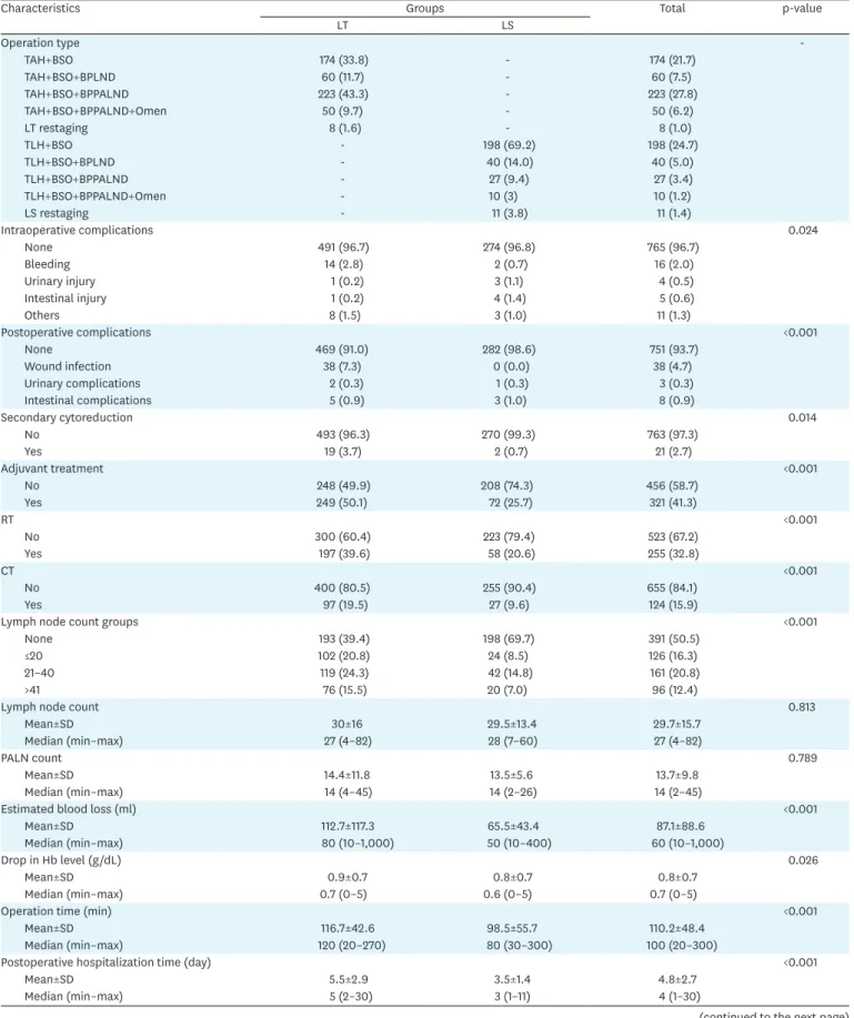
The clinical and pathological characteristics and risk classification of the groups are summarized in Table 2. Comorbidity was similar with high percent in both groups (45% of patients). Type 2 EC rate was 17% in the LT group and 14.8% in the LS group (p=0.326). The details of treatment and surgical procedures and operative outcomes are shown in Table 3. In eleven patients, the surgery technique was changed to LT from LS due to surgical obligations or complications (3.7%). Laparoscopic PLND was performed for 88 patients and PPALND was performed for 48 patients. Laparoscopic lymphadenectomies had a longer operation time than LT (119 vs. 141 minutes for hysterectomy + PLND and 142 vs. 187 minutes for hysterectomy + PPALND). Estimated blood loss and decrease in Hb levels were significantly lower in the LS group than those in the LT group (p≤0.001 and 0.026, respectively).
Postoperative hospitalization time was lower in LS than LT (p<0.001). Moreover, not only postoperative complication rate but also intraoperative complication rate was lower in LS than LT (p<0.001 and 0.024, respectively). There were a total of 5 major vessel injuries, 4 of them in laparoscopic surgery (one deep epigastric artery, 2 common iliac vein and one vena cava inferior). The main postoperative complication was wound infection in the LT group (7.5%). Secondary cytoreduction was performed in 19 patients in the LT group (3.7%) and in 2 patients in the LS group (p=0.014). There was no difference between the groups according to lymph nodes yield (p=0.813). Adjuvant therapy was given to 50% of patients in the LT group and 25% of patients in the LS group (p<0.001). The median follow-up time was 46 months in the LT group and 32 months in the LS group. There was a tumor recurrence in 34%
of the LT group and 15% of the LS group. Five-year survival rate was 98% for the LS group and 93% for the LT group.
The mean overall and DFS based on the risk groups and prognostic factors such as stage, grade, myometrial invasion, LVSI, and histopathological type are demonstrated in Table 4. There was no difference between the groups in terms of DFS and OS in the risk groups of model 1, whereas there were significant differences regarding OS and DFS in high-risk groups on the Table 1. Demographics and descriptive variables between the study groups
Variables Groups Total p-value
LT LS
Age groups
<44 61 (11.8) 35 (12.2) 96 (12.0) 0.004
45–54 122 (23.7) 94 (32.9) 216 (27.0) -
55–64 178 (34.6) 101 (35.3) 279 (34.8) -
>65 154 (29.9) 56 (19.6) 210 (26.2) -
Age
Mean±SD 58.1±7.4 56.0±11.1 57.4±10.9 0.011
Median (min–max) 58 (28–91) 56 (27–86) 58 (27–91) -
BMI groups
<30 42 (22.7) 61 (29.6) 103 (26.3) 0.234
30–34 37 (20.0) 48 (23.3) 85 (21.7) -
35–39 51 (27.6) 45 (21.8) 96 (24.6) -
>40 55 (29.7) 52 (25.2) 107 (27.4) -
BMI
Mean±SD 35.8±7.4 35.0±7.4 35.4±7.4 0.330
Median (min–max) 35 (19–60) 34 (17–57) 35 (17–60) -
Nulliparity
Yes 76 (17.0) 44 (16.4) 120 (16.7) 0.918
Infertility
None 431 (85.9) 233 (85.3) 664 (85.7) 0.847
Present 71 (14.1) 40 (14.7) 111 (14.3) -
Data are shown as number (%) not otherwise specified.
BMI, body mass index; LS, laparoscopy; LT, laparotomy; SD, standard deviation.
model 2. The patients with high risk in the LS group had improved OS and DFS outcomes compared to the LT group (p=0.007 and 0.002, respectively). The patients with advanced stage disease in the laparoscopy group had an advantage for DFS (p=0.048). OS was similar for low Table 2. Comparison of the clinical and pathological features between the study groups
Characteristics Treatment Total p-value
LT LS
Stage -
1a 240 (46.7) 210 (73.4) 450 (56.3)
1b 99 (19.3) 45 (15.7) 144 (18.0)
2 66 (12.8) 7 (2.4) 73 (9.1)
3a 65 (12.6) 19 (6.6) 84 (10.5)
3b 4 (0.8) 1 (0.3) 5 (0.6)
3c 38 (7.4) 4 (1.4) 42 (5.3)
4 3 (0.4) 0 (0.0) 2 (0.3)
Stage groups <0.001
1a 240 (46.6) 210 (73.4) 450 (56.3)
1b 99 (19.3) 45 (15.4) 144 (17.9)
2 66 (12.8) 7 (2.8) 73 (9.1)
3a+3b+3c+4 110 (21.4) 24 (8.4) 134 (16.8)
Grade <0.001
1 181 (43.3) 159 (66) 340 (51.6)
2 170 (40.6) 67 (28) 237 (36)
3 67 (16.0) 15 (6) 82 (12.4)
Myometrial invasion <0.001
None 73 (14.5) 85 (29.7) 158 (20.1)
<50% 216 (43.0) 135 (47.2) 351 (44.5)
>50% 213 (42.4) 66 (23.1) 279 (35.4)
Lympovascular space invasion <0.001
No 274 (55.6) 215 (76.5) 489 (63.2)
Yes 219 (44.4) 66 (23.5) 285 (36.8)
Histology 0.002
Endometrioid 418 (82.4) 241 (85.2) 659 (83.4)
Serous 26 (5.1) 4 (1.4) 30 (3.8)
Clear 9 (1.8) 1 (0.4) 10 (1.3)
Mixed 28 (5.5) 29 (10.2) 57 (7.2)
Carcinosarcoma 26 (5.1) 8 (2.8) 34 (4.3)
Histologic type 0.326
Type 1 418 (82.4) 241 (85.2) 659 (83.4)
Type 2 89 (17.6) 42 (14.8) 131 (16.6)
Risk group model 1 <0.001
Low 221 (42.9) 185 (64.7) 406 (50.6)
Intermediate 113 (21.9) 38 (13.3) 151 (18.9)
Intermediate-high 37 (7.2) 35 (12.2) 72 (9.0)
High 144 (28.0) 28 (9.8) 172 (21.5)
Risk group model 2 <0.001
Low 221 (42.9) 185 (64.7) 406 (50.6)
Intermediate 113 (21.9) 38 (13.3) 151 (18.9)
High 181 (35.2) 63 (22.0) 246 (30.5)
Comorbidities 0.484
No 261 (51.7) 152 (54.3) 413 (52.6)
Yes 244 (48.3) 128 (45.7) 372 (47.4)
Recurrence <0.001
No 340 (66.0) 242 (84.6) 582 (72.7)
Yes 175 (34.0) 44 (15.4) 219 (27.3)
Status <0.001
Alive 371 (72.0) 265 (92.7) 636 (79.4)
Exitus 106 (20.6) 21 (7.3) 127 (15.9)
Unknown 38 (7.4) 0 (0.0) 38 (4.7)
Data are shown as number (%).
LS, laparoscopy; LT, laparotomy.
Table 3. Features of treatment and operative variables according to the groups
Characteristics Groups Total p-value
LT LS
Operation type -
TAH+BSO 174 (33.8) - 174 (21.7)
TAH+BSO+BPLND 60 (11.7) - 60 (7.5)
TAH+BSO+BPPALND 223 (43.3) - 223 (27.8)
TAH+BSO+BPPALND+Omen 50 (9.7) - 50 (6.2)
LT restaging 8 (1.6) - 8 (1.0)
TLH+BSO - 198 (69.2) 198 (24.7)
TLH+BSO+BPLND - 40 (14.0) 40 (5.0)
TLH+BSO+BPPALND - 27 (9.4) 27 (3.4)
TLH+BSO+BPPALND+Omen - 10 (3) 10 (1.2)
LS restaging - 11 (3.8) 11 (1.4)
Intraoperative complications 0.024
None 491 (96.7) 274 (96.8) 765 (96.7)
Bleeding 14 (2.8) 2 (0.7) 16 (2.0)
Urinary injury 1 (0.2) 3 (1.1) 4 (0.5)
Intestinal injury 1 (0.2) 4 (1.4) 5 (0.6)
Others 8 (1.5) 3 (1.0) 11 (1.3)
Postoperative complications <0.001
None 469 (91.0) 282 (98.6) 751 (93.7)
Wound infection 38 (7.3) 0 (0.0) 38 (4.7)
Urinary complications 2 (0.3) 1 (0.3) 3 (0.3)
Intestinal complications 5 (0.9) 3 (1.0) 8 (0.9)
Secondary cytoreduction 0.014
No 493 (96.3) 270 (99.3) 763 (97.3)
Yes 19 (3.7) 2 (0.7) 21 (2.7)
Adjuvant treatment <0.001
No 248 (49.9) 208 (74.3) 456 (58.7)
Yes 249 (50.1) 72 (25.7) 321 (41.3)
RT <0.001
No 300 (60.4) 223 (79.4) 523 (67.2)
Yes 197 (39.6) 58 (20.6) 255 (32.8)
CT <0.001
No 400 (80.5) 255 (90.4) 655 (84.1)
Yes 97 (19.5) 27 (9.6) 124 (15.9)
Lymph node count groups <0.001
None 193 (39.4) 198 (69.7) 391 (50.5)
≤20 102 (20.8) 24 (8.5) 126 (16.3)
21–40 119 (24.3) 42 (14.8) 161 (20.8)
>41 76 (15.5) 20 (7.0) 96 (12.4)
Lymph node count 0.813
Mean±SD 30±16 29.5±13.4 29.7±15.7
Median (min–max) 27 (4–82) 28 (7–60) 27 (4–82)
PALN count 0.789
Mean±SD 14.4±11.8 13.5±5.6 13.7±9.8
Median (min–max) 14 (4–45) 14 (2–26) 14 (2–45)
Estimated blood loss (ml) <0.001
Mean±SD 112.7±117.3 65.5±43.4 87.1±88.6
Median (min–max) 80 (10–1,000) 50 (10–400) 60 (10–1,000)
Drop in Hb level (g/dL) 0.026
Mean±SD 0.9±0.7 0.8±0.7 0.8±0.7
Median (min–max) 0.7 (0–5) 0.6 (0–5) 0.7 (0–5)
Operation time (min) <0.001
Mean±SD 116.7±42.6 98.5±55.7 110.2±48.4
Median (min–max) 120 (20–270) 80 (30–300) 100 (20–300)
Postoperative hospitalization time (day) <0.001
Mean±SD 5.5±2.9 3.5±1.4 4.8±2.7
Median (min–max) 5 (2–30) 3 (1–11) 4 (1–30)
(continued to the next page)
and intermediate risk between the groups, but it was higher in the laparoscopy group than in the laparotomy group for high-risk patients (Fig. 1).
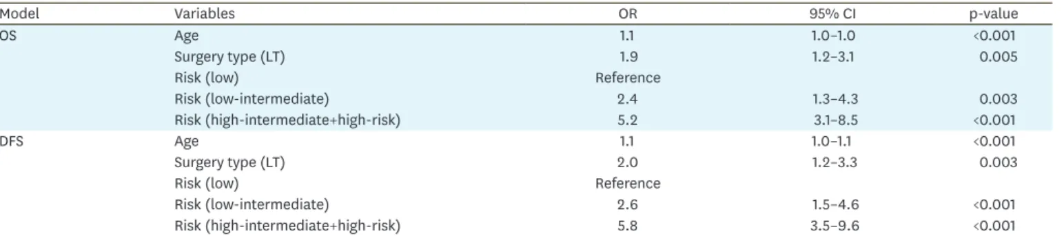
The results of the regression analysis standardized by age and risk groups revealed that LS or LT as a surgical option was found to be a prognostic risk factor (Table 5). There was an increased risk for death and recurrence in the LT group compared to the LS group (odds Table 4. The results of the survival analysis based on the risk groups and prognostic factors between and within the study groups
Variables LT LS Total p for OS p for DFS
Total/dead,
No. (%) OS/DFS
(mean) Total/dead,
No. (%) OS/DFS
(mean) Total/dead,
No. (%) OS/DFS
(mean) Risk group model 1
Low 201/17 132.2/132.2 185/5 127.5/127.5 386/22 133.1/133.2 0.289 0.269
Intermediate 107/22 115.9/104.2 38/5 108.5/108.0 145/27 117.5/105.9 0.493 0.476
High-intermediate 35/11 79.0/76.8 35/4 85.2/85.2 70/15 87.0/86.6 0.119 0.078
High 142/56 67.1/58.7 28/7 72.6/64.3 170/63 69.1/61.1 0.079 0.085
p-value <0.001 <0.001 <0.001
Risk group model 2
Low 201/17 132.7/132.2 185/5 127.5/127.5 386/22 133.0/133.2 0.289 0.269
Intermediate 107/22 115.2/104.2 38/5 108.0/108.4 145/27 105.9/105.9 0.493 0.476
High 177/67 70.1/65.3 63/11 79.1/74.9 249/78 77.9/71.4 0.007 0.002
p-value <0.001 <0.001 <0.001
Stage
1a 225/23 129.4/129.4 210/6 126.8/126.8 435/29 131.3/131.4 0.087 0.074
1b 92/17 118.4/105.9 44/7 104.0/105.4 136/24 118.0/105.9 0.927 0.981
2 58/17 81.7/77.2 8/1 89.0/78.6 66/18 84.0/79.5 0.313 0.318
3a+3b+3c+4 109/48 60.0/55.2 24/7 76.6/70.3 133/55 63.6/58.9 0.048 0.054
p-value <0.001 <0.001 <0.001
Grade
1 176/17 130.1/129.9 179/8 124.1/124.8 335/25 130.5/130.5 0.326 0.283
2 176/38 113.5/112.2 77/9 103.6/104.0 253/47 115.8/115.1 0.220 0.166
3 70/31 66.8/61.6 16/4 70.6/67.9 86/35 71.1/65.9 0.142 0.151
p-value <0.001 <0.001 <0.001
Myometrial invasion
None 68/5 131.4/131.5 85/3 126.0/126.0 153/8 132.9/133.0 0.804 0.787
<50% 199/29 123.6/123.6 135/5 120.4/120.4 334/34 127.5/127.6 0.022 0.019
>50% 205/67 93.7/82.9 66/13 96.2/97.3 271/80 97.4/86.9 0.047 0.034
p-value <0.001 <0.001 <0.001
LVSI
No 252/33 125.0/124.8 215/9 126.5/126.5 467/42 128.4/128.4 0.020 0.014
Yes 212/60 86.1/84.0 66/12 85.3/86.6 278/72 87.8/86.0 0.153 0.125
p-value <0.001 <0.001 <0.001
Histopathological type
Type 1 389/62 120.9/120.1 241/14 122.9/123.4 630/76 123.8/123.4 0.006 0.002
Type 2 89/38 61.6/59.0 42/6 72.2/78.8 131/44 70.2/70.6 0.003 <0.001
p-value <0.001 <0.001 <0.001
Total 485/106 111.2/110.4 286/21 118.7/120.3 771/127 116.3/116.1 0.001 <0.001
DFS, disease-free survival; OS, overall survival; LS, laparoscopy; LT, laparotomy; LVSI, lymphovascular space invasion.
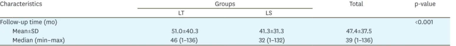
Characteristics Groups Total p-value
LT LS
Follow-up time (mo) <0.001
Mean±SD 51.0±40.3 41.3±31.3 47.4±37.5
Median (min–max) 46 (1–136) 32 (1–132) 39 (1–136)
Data are shown as number (%) not otherwise specified.
BPLND, bilateral pelvic lymphadenectomy; BPPALND, bilateral pelvic-paraaortic lymphadenectomy; BSO, bilateral salpingo-oophorectomy; CT, chemotherapy;
Hb, hemoglobin; LS, laparoscopy; LT, laparotomy; Omen, omentectomy; PALN, paraaortic lymph node; RT, radiotherapy; SD, standard deviation; TAH, total abdominal hysterectomy; TLH, total laparoscopic hysterectomy.
Table 3. (Continued) Features of treatment and operative variables according to the groups
ratio [OR]=1.9; 95% confidence interval [CI]=1.2–3.1 for OS; OR=2; 95% CI=1.2–3.3 for DFS) considering the results of covariate analysis. Age, stage, histopathological type, and operation type were determined as independent prognostic factors in multivariate analysis for both of DFS and OS (Table 6).
Time (mo) Risk_group=Low
p=0.289 p=0.493
140 100
40 20 0.2
0
OS
0.4 0.6 0.8 1.0
120 80
60 LS
LT Treatment
Time (mo) Risk_group=Intermediate
140 100
40 20 0.2
0
OS
0.4 0.6 0.8 1.0
120 80
60 LS
LT Treatment
p=0.007
Time (mo) Risk_group=High
100 40
20 0.2
0
OS
0.4 0.6 0.8 1.0
120 80
60 LS
LT Treatment
Fig. 1. The survival curves between the study groups based on the risk groups. High-risk group refers to the sum of the high-intermediate risk group and the high-risk group.
Table 5. Results of the co-variate analysis
Model Variables OR 95% CI p-value
OS Age 1.1 1.0–1.0 <0.001
Surgery type (LT) 1.9 1.2–3.1 0.005
Risk (low) Reference
Risk (low-intermediate) 2.4 1.3–4.3 0.003
Risk (high-intermediate+high-risk) 5.2 3.1–8.5 <0.001
DFS Age 1.1 1.0–1.1 <0.001
Surgery type (LT) 2.0 1.2–3.3 0.003
Risk (low) Reference
Risk (low-intermediate) 2.6 1.5–4.6 <0.001
Risk (high-intermediate+high-risk) 5.8 3.5–9.6 <0.001
CI, confidence interval; DFS, disease-free survival; LT, laparotomy; OR, odds ratio; OS, overall survival.
DISCUSSION
The current retrospective cohort study conducted in a single tertiary reference center in the south of Turkey aimed to evaluate the efficiency and reliability of the laparoscopic approach in different risk groups of ECs. In this study, we showed that laparoscopic surgery was as effective and safe as abdominal approach for the treatment of EC. By comparing the operative results between the LS and LT groups that had similar demographic characteristics, a significant superiority was found in the LS cohort in terms of short-term results such as complications, blood loss, and the length of hospital stay in accordance with the literature [2,9-11]. The benefits of the laparoscopic surgery may be particularly marked in women with obesity and comorbidity. In our laparoscopic cohort, there was a higher rate of obese women (twenty-five percent of the LS group had morbid obesity) and approximately half of the patients had comorbidity. Laparoscopic surgery can substantially reduce the rate of postoperative complications compared to LT surgery in such cases [12]. The most common postoperative complication was wound site infection in our cohort.
Most of studies on the laparoscopic management of the EC were performed on the low-risk EC cohort such as stage 1. A meta-analysis including 8 randomized controlled trials showed that LS was associated with similar OS and DFS for early stage endometrioid adenocarcinoma [13]. Efficacy and safety of LS in terms of survival outcomes in early stage EC is at least controversial. The results of LAP2 survival outcomes [1] and meta-analysis of randomized controlled trials [3,9] highlight the importance of LS in the management of early stage EC.
Similarly, we demonstrated that there was no significant difference between the groups in terms of OS and DFS in the low-risk patients. Furthermore, LS has similar survival outcomes to those of LT not only in early stage but also in low-risk patients.
For intermediate risk group, there is no study performed on intermediate group alone in the literature. This group mainly consists of stage 1b patients. In the LAP2 study [1], stage 1b accounted for 12% of LT group and also LS group (n=204). Our laparoscopic cohort consisted of patients with stage 1b at similar rate (15%; n=45). There was no difference between the groups in terms of DFS and OS in both intermediate and high-intermediate groups in our study. In the last guide [4], MIS is recommended in the low and intermediate risk EC (level of evidence 1, the strength of recommendation: A). Even in this groups of patients, long-term survival analysis should be further supported by randomized controlled studies.
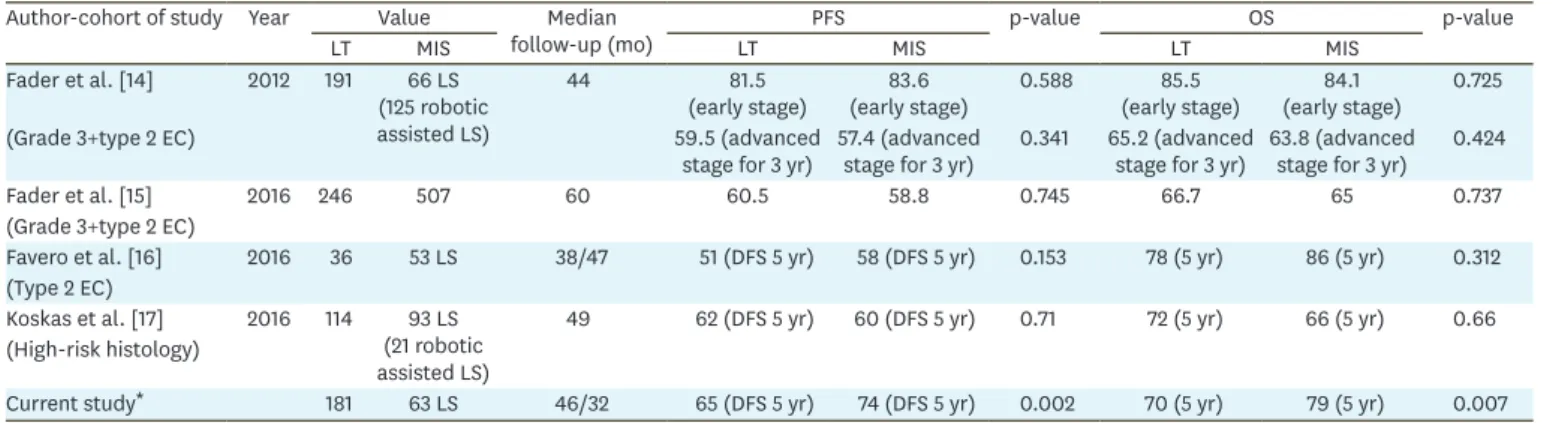
Table 6. Studies of MIS management of high-risk histopathological type EC Author-cohort of study Year Value Median
follow-up (mo) PFS p-value OS p-value
LT MIS LT MIS LT MIS
Fader et al. [14] 2012 191 66 LS (125 robotic assisted LS)
44 81.5
(early stage) 83.6
(early stage) 0.588 85.5
(early stage) 84.1
(early stage) 0.725
(Grade 3+type 2 EC) 59.5 (advanced
stage for 3 yr) 57.4 (advanced
stage for 3 yr) 0.341 65.2 (advanced
stage for 3 yr) 63.8 (advanced stage for 3 yr) 0.424
Fader et al. [15] 2016 246 507 60 60.5 58.8 0.745 66.7 65 0.737
(Grade 3+type 2 EC)
Favero et al. [16] 2016 36 53 LS 38/47 51 (DFS 5 yr) 58 (DFS 5 yr) 0.153 78 (5 yr) 86 (5 yr) 0.312
(Type 2 EC)
Koskas et al. [17] 2016 114 93 LS (21 robotic assisted LS)
49 62 (DFS 5 yr) 60 (DFS 5 yr) 0.71 72 (5 yr) 66 (5 yr) 0.66
(High-risk histology)
Current study* 181 63 LS 46/32 65 (DFS 5 yr) 74 (DFS 5 yr) 0.002 70 (5 yr) 79 (5 yr) 0.007
DFS, disease-free survival; EC, endometrial cancer; LS, laparoscopy; LT, laparotomy; MIS, minimally invasive surgery; OS: overall survival; PFS, progression free interval.
*High-intermediate and high-risk group.
There is a lack of data on the laparoscopic management of high-risk patients. According to our literature review, 4 recent studies have investigated laparoscopic management of patients with grade 3 and type 2 EC [14-17]. In 2012, a multicenter retrospective study, which is the first on laparoscopic management of high grade and type 2 endometrial carcinomas (n=191), concluded that high-risk histopathologic types were not a contraindication for MIS [14].
The post-hoc analysis of the LAP2 including early stage patients with grade 3 endometrioid and type 2 (n=507) showed that patients with high-grade uterine cancers managed by LS had similar rates of recurrence and survival to those managed by LT [15]. In this study, type 2 cancer recurred 2 times more than grade 3 endometrioid cancer. Favero et al. [16]
studied the feasibility, operative outcomes and oncologic safety of laparoscopic surgery in patients with type 2 EC (n=53). They concluded that LS was associated with better oncologic outcomes than LT with no statistical significance [16]. Indeed, with increasing number of patients, a statistical significance could be achieved. And the last study on high-risk EC by Koskas et al. [17] compared 93 laparoscopic and 21 robotic surgeries with 114 laparotomic surgery. In accordance with other studies, DFS and OS were not different between the surgical groups [17]. Survival outcomes of the retrospective studies of MIS management of high-risk histopathological type EC were shown in the Table 6. These 4 trials studied the patients with high-risk histopathology. We evaluated not only patients with type 2 cancer and grade 3 endometrioid adenocarcinoma but also patients with the high-risk which was also the subject of other risk factors such as advanced stage, myometrial invasion, and LVSI.
Despite the similar type 2 proportions between the groups (18% vs. 15%) in our cohort, higher superiority of LS in survival analyses occurred for type 2 cancer. Type 2 and grade 3 endometrioid cancer constituted less than 20 percent of all ECs and even in the early stage, the prospects for distant metastasis have increased [18]. The patients with type 2 EC are usually older than those with type 1 cancer and the probability of systemic disease is increased. Adjuvant therapy is frequently recommended in this high-risk group with a high likelihood of recurrence. Therefore, it is important that surgical morbidity is less than in LS and adjuvant therapy can be started immediately. Although there was no difference in survival analysis between the groups on the risk groups. As a result of our study, LS seems to be superior to LT in terms of survival outcomes in multivariate analysis. However, that result should be interpreted carefully because of the retrospective structure and bias in group selection. It is clear that we need more effective and reliable data on the laparoscopic management of high-risk patients.
Our study includes all negative aspects of retrospective study design. Another limitation of our study is that we cannot give the initiation time of adjuvant treatment. However, the main strength of our study was the survival analysis according to risk group classifications which were based on the most important factors (stage, grade, myometrial invasion, LVSI, and histopathology) affecting the prognosis. In this way, we attempted to minimize the effect of non-randomized and biased patient's selection on the survival analysis.
Additionally, we had a relatively large cohort of the groups for a single center and longer follow-up period.
The results of our study suggest that LS can be used effectively and safely in patients with intermediate and high-risk EC. Long-term survival analysis should be supported by randomized controlled studies to demonstrate that LS may be an acceptable alternative to LT for EC patients with high-risk.
REFERENCES
1. Walker JL, Piedmonte MR, Spirtos NM, Eisenkop SM, Schlaerth JB, Mannel RS, et al. Recurrence and survival after random assignment to laparoscopy versus laparotomy for comprehensive surgical staging of uterine cancer: Gynecologic Oncology Group LAP2 Study. J Clin Oncol 2012;30:695-700.
PUBMED | CROSSREF
2. Palomba S, Falbo A, Mocciaro R, Russo T, Zullo F. Laparoscopic treatment for endometrial cancer: a meta-analysis of randomized controlled trials (RCTs). Gynecol Oncol 2009;112:415-21.
PUBMED | CROSSREF
3. Palomba S, Falbo A, Russo T, Zullo F. Updating of a recent meta-analysis of randomized controlled trials to assess the safety and the efficacy of the laparoscopic surgery for treating early stage endometrial cancer. Gynecol Oncol 2009;114:135-6.
PUBMED | CROSSREF
4. Colombo N, Creutzberg C, Amant F, Bosse T, González-Martín A, Ledermann J, et al. ESMO-ESGO- ESTRO consensus conference on endometrial cancer: diagnosis, treatment and follow-up. Int J Gynecol Cancer 2016;26:2-30.
PUBMED | CROSSREF
5. Weinberg LE, Kunos CA, Zanotti KM. Lymphovascular space invasion (LVSI) is an isolated poor prognostic factor for recurrence and survival among women with intermediate- to high-risk early-stage endometrioid endometrial cancer. Int J Gynecol Cancer 2013;23:1438-45.
PUBMED | CROSSREF
6. Jorge S, Hou JY, Tergas AI, Burke WM, Huang Y, Hu JC, et al. Magnitude of risk for nodal metastasis associated with lymphvascular space invasion for endometrial cancer. Gynecol Oncol 2016;140:387-93.
PUBMED | CROSSREF
7. Mariani A, Webb MJ, Keeney GL, Haddock MG, Calori G, Podratz KC. Low-risk corpus cancer: is lymphadenectomy or radiotherapy necessary? Am J Obstet Gynecol 2000;182:1506-19.
PUBMED | CROSSREF
8. Mariani A, El-Nashar SA, Dowdy SC. Lymphadenectomy in endometrial cancer: which is the right question? Int J Gynecol Cancer 2010;20:S52-4.
PUBMED | CROSSREF
9. Zullo F, Falbo A, Palomba S. Safety of laparoscopy vs laparotomy in the surgical staging of endometrial cancer: a systematic review and metaanalysis of randomized controlled trials. Am J Obstet Gynecol 2012;207:94-100.
PUBMED | CROSSREF
10. Walker JL, Piedmonte MR, Spirtos NM, Eisenkop SM, Schlaerth JB, Mannel RS, et al. Laparoscopy compared with laparotomy for comprehensive surgical staging of uterine cancer: Gynecologic Oncology Group Study LAP2. J Clin Oncol 2009;27:5331-6.
PUBMED | CROSSREF
11. Zhang H, Cui J, Jia L, Hong S, Kong B, Li D. Comparison of laparoscopy and laparotomy for endometrial cancer. Int J Gynaecol Obstet 2012;116:185-91.
PUBMED | CROSSREF
12. Gunderson CC, Java J, Moore KN, Walker JL. The impact of obesity on surgical staging, complications, and survival with uterine cancer: a Gynecologic Oncology Group LAP2 ancillary data study. Gynecol Oncol 2014;133:23-7.
PUBMED | CROSSREF
13. Galaal K, Bryant A, Fisher AD, Al-Khaduri M, Kew F, Lopes AD. Laparoscopy versus laparotomy for the management of early stage endometrial cancer. Cochrane Database Syst Rev 2012:CD006655.
PUBMED
14. Fader AN, Seamon LG, Escobar PF, Frasure HE, Havrilesky LA, Zanotti KM, et al. Minimally invasive surgery versus laparotomy in women with high grade endometrial cancer: a multi-site study performed at high volume cancer centers. Gynecol Oncol 2012;126:180-5.
PUBMED | CROSSREF
15. Fader AN, Java J, Tenney M, Ricci S, Gunderson CC, Temkin SM, et al. Impact of histology and surgical approach on survival among women with early-stage, high-grade uterine cancer: an NRG Oncology/
Gynecologic Oncology Group ancillary analysis. Gynecol Oncol 2016;143:460-5.
PUBMED | CROSSREF
16. Favero G, Anton C, Le X, Silva E Silva A, Dogan NU, Pfiffer T, et al. Oncologic safety of laparoscopy in the surgical treatment of type II endometrial cancer. Int J Gynecol Cancer 2016;26:1673-8.
PUBMED | CROSSREF
17. Koskas M, Jozwiak M, Fournier M, Vergote I, Trum H, Lok C, et al. Long-term oncological safety of minimally invasive surgery in high-risk endometrial cancer. Eur J Cancer 2016;65:185-91.
PUBMED | CROSSREF
18. Creasman WT, Kohler MF, Odicino F, Maisonneuve P, Boyle P. Prognosis of papillary serous, clear cell, and grade 3 stage I carcinoma of the endometrium. Gynecol Oncol 2004;95:593-6.
PUBMED | CROSSREF