Veterinary Science Diversity of swine Bordetella bronchiseptica isolates evaluated by RAPD analysis and PFGE
Eun-Kyung Shin
1, Yeon-Soo Seo
2, Jeong Hee Han
2, Tae-Wook Hahn
2,*
1
South Branch, Gangwon Veterinary Service, Wonju 200-170, Korea
2
School of Veterinary Medicine, Kangwon National University, Chuncheon 200-701, Korea
The degree of genetic diversity in 45 Bordetella ( B. )
bronchiseptica strains comprised of a vaccine strain (N = 1), reference strains (N = 3) and field isolates (N = 41) was evaluated using random amplified polymorphic DNA (RAPD) fingerprinting and pulsed-field gel electrophoresis (PFGE). Three candidate primers were selected for RAPD analysis after screening 20 random decamer oligonucleotides for their discriminatory abilities. The OPA-07, OPA-08 and OPA-18 primers yielded 10, 10, and 6 distinct fingerprint patterns, respectively. The most common identical RAPD pattern was produced by OPA- 07 which was shared by 32 isolates (71.1%), the pattern produced by OPA-08 was shared by 26 isolates (57.8%), and the pattern produced by OPA-18 was shared by 40 isolates (88.9%). The RAPD patterns of the vaccine strain and the 3 reference strains did not match any of the patterns produced by the field isolates when primers OPA-07 and OPA-08 were used. PFGE using the restriction endonuclease Xba I produced a total of 15 patterns consisting of 4 PFGE types (A, B, B1 and C, differing by ≥ 4 bands) and 11 A subtypes (differing by ≤ 3 bands). Most of the field isolates exhibited identical type A and B patterns, suggesting that they were related. The vaccine strain and the three reference strains showed different PFGE patterns as compared to the identical type A strains.
Key words: Bordetella bronchiseptica, genetic diversity, PFGE, RAPD
Introduction
Bordetella are Gram-negative bacteria that cause respiratory tract infections in humans and animals. Species in the genus
Bordetella are close phenotypically, possess common antigens and share a high degree of DNA similarity [3].
Bordetella ( B .) bronchiseptica infects many domestic and wild animal species. In pigs, for example, B. bronchiseptica
is known to play a role in development of atrophic rhinitis (AR) and porcine respiratory disease complex [2]. AR is an infectious disease of pigs characterized by purulent nasal discharge, shortening or twisting of the snout, atrophy of the turbinate bones and reduced growth rate [13]. Many aspects of the biology of B. bronchiseptica have been studied, including colony morphology [20], hemolysin production [4], hemagglutination [5], and plasmid content [11], however reports regarding genetic typing of B. bronchiseptica are scarce. Serotyping has demonstrated that B. bronchiseptica
isolates from pigs differ from those of other animal species, however, these results were based on only a few B.
bronchiseptica isolates from each animal species tested [4,5,11,19,20]. Phenotypic typing based on expression of cellular characteristics may vary according to culture or experimental conditions, and is being gradually replaced by bacterial genomic analysis [6].
A number of molecular methods, including restriction enzyme analysis (REA) [24], Random amplified polymorphic DNA (RAPD) fingerprinting [15], ribotyping [12,21,27] and macro-restriction analysis by pulsed-field gel electrophoresis (PFGE) [6] have been used to study differences in epidemiology between different strains of B. bronchiseptica , and the results obtained using these methods suggest that considerable genomic diversity exists between strains. REA performed on 195 B. bronchiseptica isolates from 12 different host species worldwide showed 48 distinct fingerprint patterns after Hinf I digestion and 39 fingerprint profiles after Alu I digestion [24]. Ribotyping of B.
bronchiseptica isolates obtained from several different animal species revealed that the isolates fell into distinct groups [21]. PFGE provides a highly reproducible restriction profile of large bacterial DNA fragments and therefore a means for discriminating between B. bronchiseptica isolates in epidemiologic studies [6,16]. Binns et al . [6] identified 17 PFGE types with numerous subtypes within a collection of 164 isolates, predominantly from cats. Keil and Fenwick [15] combined RAPD analysis and ribotyping to evaluate
*Corresponding author
Tel: +82-33-250-8671; Fax: +82-33-244-2367
E-mail: [email protected]
genetic diversity among 26 canine B. bronchiseptica
isolates. Although many molecular methods have been used to study B. bronchiseptica isolates from different hosts, there are few reports on the typing of B. bronchiseptica isolates from swine [6,21,24]. To our knowledge, this study is the first to provide genotyping data obtained by both RAPD and PFGE analyses of a large number of Korean swine B.
bronchiseptica field isolates and is also the first study to combine these methods to classify B. bronchiseptica isolates from swine. The purpose of this study was to evaluate the genetic diversity of B. bronchiseptica field isolates using RAPD and PFGE in comparison to a vaccine strain and 3 standard strains of B. bronchiseptica .
Materials and Methods
Bacterial strains
Forty-five B. bronchiseptica strains comprised of 1 vaccine strain, 3 reference strains and 41 field isolates were evaluated. The field isolates were obtained from 6-month- old slaughtered pigs from the provinces of Gangwon and Gyeonggi Province in Korea between October 2001 and October 2002. All isolates were identified as B. bronchiseptica
using Smith-Baskerville medium [26] and standard methods [8,14]. Three reference strains (ATCC 19395, 10580, and 4617) and the B. bronchiseptica vaccine P4 strain (HAP- VAC; Choongang Vaccine Laboratory, Korea) were minimally passed and stored in a Microbank (KOMED, Korea) at
− 70
oC until used.
DNA preparation for RAPD
Bacterial isolates were inoculated into 2 ml fresh brain heart infusion broth (Difco, USA) and incubated at 37
oC for 24 h. Genomic DNA from each strain was obtained using a DNeasy Tissue Kit (QIAGEN, Germany) according to the manufacturer’s instructions. A set of 20 commercially available primers (Oligo 10-mer kit A; QIAGEN, Germany) was screened to identify suitable primers for RAPD analysis of swine B. bronchiseptica isolates. Primers OPA-07 (5'- GAAACGGGTG-3'), OPA-08 (5'-GTGACGTAGG-3') and OPA-18 (5'-AGGTGACCGT-3') resulted in informative fingerprints and were used to evaluate the remaining strains.
RAPD PCR consisted of 50 ng of total B. bronchiseptica DNA, 5 mM MgCl
2(Promega, USA), 12 pmole primer, 2.5 units of GoTaq DNA polymerase (Promega, USA), and 500 mM dNTPs (Takara, Japan) in 25 mM Tris-HCl (pH9.0)-25 mM NaCl in a volume of 25 µ l was subjected to the following conditions: 2 min of initial denaturation at 95
oC followed by 45 cycles of 1 min of denaturation at 94
oC, 1 min of annealing at 33
oC, 2 min of extension at 72
oC. Reactions were performed using a UNO-II thermalcycler (Biometra, Germany). Following PCR, 10 µ l of the reaction mixture
was analyzed by gel electrophoresis in a 2.0% agarose gel containing 500 ng/ml ethidium bromide. A 100-bp DNA ladder (Jeil Biotechservice, Korea) was used to determine molecular size. The agarose gels were photographed under UV light using a Biocapt (Vilber Lourmat, France) and the DNA bands were analyzed using Bio-Profil Bio 2D software (Vilber Lourmat, France), which was also used to construct dendrograms of the isolates.
Preparation of DNA for PFGE
The methods used to conduct PFGE essentially followed the ‘pulse Net’ system protocol described by the Centers for Disease Control [7,9]. Briefly, B. bronchiseptica were cultured in Luria Bertani agar (Difco, USA) plates and incubated at 37
oC overnight. Colonies were then harvested and suspended in TE suspension buffer (100 mM Tris-HCl and 100 mM EDTA, pH 7.5). The turbidity of the bacterial cell suspension was set to 20% transmittance using a colorimeter (BioMeieux, France). Proteinase K and 1.2%
Seakem Gold agarose (FMC Bioproducts, USA) were then mixed with the cell suspension and dispensed into disposable plug molds (Bio-Rad, USA). ES buffer (0.5 M EDTA, pH 9.0, 1% sodium-lauroyl-sarcosine) and proteinase K were added to the plugs, which were then incubated in a 55
oC water bath for 1 h. After proteolysis, the plugs were washed once for 15 min in sterile distilled water then 4 times for 30 min in TE buffer (10 mM Tris-HCl and 1 mM EDTA, pH 7.5) preheated to 50
oC. Washed plugs were stored in TE buffer (10 mM Tris-HCl and 1 mM EDTA, pH 7.5) at 4
oC until ready for restriction enzyme digestion. The stored plugs were cut into 2-1 mm wide slices with a razor blade and the 2 halves transferred to a tube containing the restriction enzyme Xba I (30 U; Promega, USA) and digested at 37
oC for 3 h. After incubation, the enzyme mix was aspirated from the tube and replaced with 500 µ l of TE washing buffer.
PFGE DNA obtained from bacteria was electrophoresed using a contour homogeneous field electrophoresis system (DR II;
Bio-Rad, USA). Digested plugs were electrophoresed in 1%
Seakem gold agarose gel (FMC Bioproducts, USA) with 0.5X TBE buffer. The electrophoresis conditions were as follows: initial switch time, 2.2 s; final switch time, 55.0 s;
run time, 15.5 h; gradient, 6.0 V/cm; buffer temperature, 14
oC. A standard lambda DNA ladder (Bio-Rad, USA) was used as a size marker. After electrophoresis, the gel was stained with ethidium bromide staining solution for 30 min, then destained in water for 20 min. The stained gel was viewed on an UV transilluminator and photographed with Polaroid film and scanned using a Bio-Pn 05 System (Vilber Lourmat, France).
Data analysis of PFGE
PFGE DNA patterns were compared using Tenover’s
criteria [28]. The most frequently repeated PFGE pattern
was designated type “A”. Because all type A patterns were identical it was used as the standard to differentiate the other banding patterns. Subtypes A1 to A11 differed from the
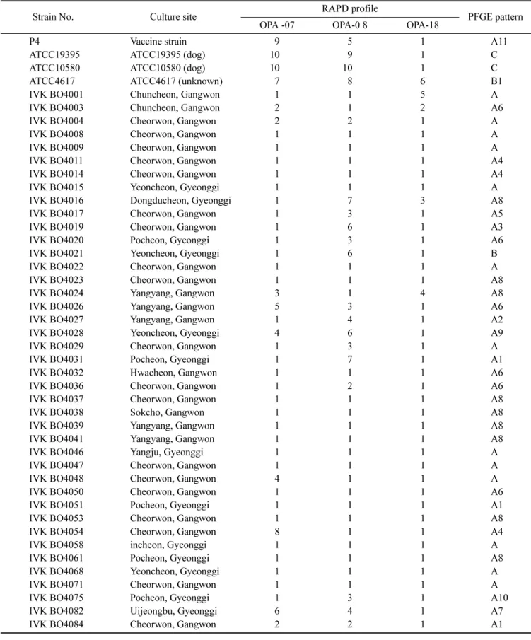
Table 1. Summary of the properties of B. bronchiseptica : culture site, RAPD and PFGE type of the tested strains
Strain No. Culture site OPA -07 RAPD profile OPA-0 8 OPA-18 PFGE pattern
P4 Vaccine strain 9 5 1 A11
ATCC19395 ATCC19395 (dog) 100 9 1 C
ATCC10580 ATCC10580 (dog) 100 100 1 C
ATCC4617 ATCC4617 (unknown) 7 8 6 B1
IVK BO4001 Chuncheon, Gangwon 1 1 5 A
IVK BO4003 Chuncheon, Gangwon 2 1 2 A6
IVK BO4004 Cheorwon, Gangwon 2 2 1 A
IVK BO4008 Cheorwon, Gangwon 1 1 1 A
IVK BO4009 Cheorwon, Gangwon 1 1 1 A
IVK BO4011 Cheorwon, Gangwon 1 1 1 A4
IVK BO4014 Cheorwon, Gangwon 1 1 1 A4
IVK BO4015 Yeoncheon, Gyeonggi 1 1 1 A
IVK BO4016 Dongducheon, Gyeonggi 1 7 3 A8
IVK BO4017 Cheorwon, Gangwon 1 3 1 A5
IVK BO4019 Cheorwon, Gangwon 1 6 1 A3
IVK BO4020 Pocheon, Gyeonggi 1 3 1 A6
IVK BO4021 Yeoncheon, Gyeonggi 1 6 1 B
IVK BO4022 Cheorwon, Gangwon 1 1 1 A
IVK BO4023 Cheorwon, Gangwon 1 1 1 A8
IVK BO4024 Yangyang, Gangwon 3 1 4 A8
IVK BO4026 Yangyang, Gangwon 5 3 1 A6
IVK BO4027 Yangyang, Gangwon 1 4 1 A2
IVK BO4028 Yeoncheon, Gyeonggi 4 6 1 A9
IVK BO4029 Cheorwon, Gangwon 1 3 1 A
IVK BO4031 Pocheon, Gyeonggi 1 7 1 A1
IVK BO4032 Hwacheon, Gangwon 1 1 1 A6
IVK BO4036 Cheorwon, Gangwon 1 2 1 A6
IVK BO4037 Cheorwon, Gangwon 1 1 1 A8
IVK BO4038 Sokcho, Gangwon 1 1 1 A8
IVK BO4039 Yangyang, Gangwon 1 1 1 A8
IVK BO4041 Yangyang, Gangwon 1 1 1 A8
IVK BO4046 Yangju, Gyeonggi 1 1 1 A
IVK BO4047 Cheorwon, Gangwon 1 1 1 A
IVK BO4048 Cheorwon, Gangwon 4 1 1 A
IVK BO4050 Cheorwon, Gangwon 1 1 1 A6
IVK BO4051 Pocheon, Gyeonggi 1 1 1 A1
IVK BO4053 Cheorwon, Gangwon 1 1 1 A8
IVK BO4054 Cheorwon, Gangwon 8 1 1 A4
IVK BO4058 incheon, Gyeonggi 1 1 1 A
IVK BO4061 Pocheon, Gyeonggi 1 1 1 A8
IVK BO4068 Yeoncheon, Gyeonggi 1 1 1 A
IVK BO4071 Cheorwon, Gangwon 1 1 1 A
IVK BO4075 Pocheon, Gyeonggi 1 3 1 A10
IVK BO4082 Uijeongbu, Gyeonggi 6 4 1 A7
IVK BO4084 Cheorwon, Gangwon 2 2 1 A1
major type A pattern by less than three bands; the major type B pattern differed by 5-6 bands; and the type C pattern differed by 7 bands. After manual inspection, a dendrogram was constructed using Bio-Profil Bio 2D (Vilber Lourmat, France) software to depict the relatedness of each type.
Results
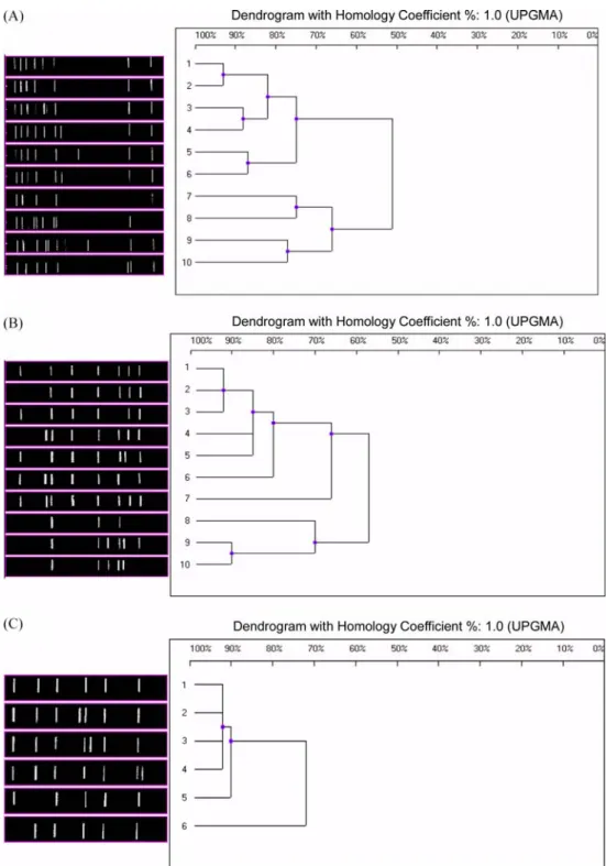
RAPD The 3 different primers, OPA-07, OPA-08 and OPA-18 produced different numbers of patterns in the RAPD analyses of 45 B. bronchiseptica strains (Table 1). OPA-07, OPA-08, and OPA-18 yielded 10, 10, and 6 patterns, respectively (Table 2).
The 10 distinct DNA patterns produced by OPA-07 fingerprinting were designated as OPA-07(1) through OPA- 07(10) (Fig. 1A). Fingerprint OPA-07(1) was the most common RAPD pattern, shared by 32 of the 45 isolates (71.1%) although the vaccine strain and the 3 reference strains did not produce this pattern. The P4 vaccine strain was defined by fingerprint OPA-07(9), the ATCC 19395 and 10580 strains by OPA-07(10), and ATCC 4617 by OPA- 07(7).
The 10 distinct DNA patterns produced by OPA-08 fingerprinting were designated as OPA-08(1) through OPA- 08(10) (Fig. 1B). Fingerprint OPA-08(1) was the most common RAPD pattern, shared by 26 isolates (57.8%). The P4 vaccine strain was defined by fingerprint OPA-08(5), the ATCC 19395 and 10580 strains by OPA-08(9) and OPA- 08(10), respectively, and ATCC 4617 by OPA-08(8).
The 6 distinct RAPD patterns produced by OPA-18 fingerprinting were designated as OPA-18(1) through OPA- 18(6) (Fig. 1C). Fingerprint OPA-18(1) was the most common RAPD pattern, shared by 40 of the 45 isolates (88.9%), including the vaccine strain and 2 of the reference strains, ATCC 19395 and ATCC 10580. The third reference
strain, ATCC 4617, was defined by fingerprint OPA-18(6).
These results indicate there is considerable heterogeneity among B. bronchiseptica strains based on RAPD analysis.
Although common fingerprints exist, most notably OPA- 07(1), OPA-08(1), and OPA-18(1), overall classification of the isolates is dependant upon the primer used. Moreover, the field isolates appear to be genetically distinct from the vaccine and reference strains.
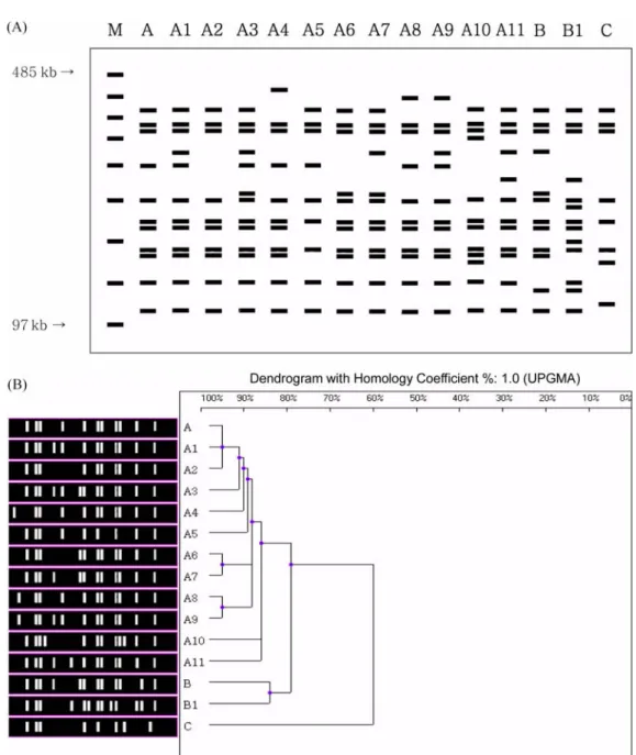
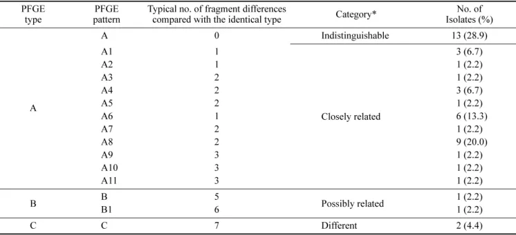
PFGE The same 45 B. bronchiseptica strains were also tested by PFGE (Table 1). PFGE of Xba I-digested genomic DNA produced patterns of well-resolved bands ranging in size from 100 to 390 kb (Fig. 2A). The majority of isolates produced between 8 and 13 bands, yielding a diverse array of DNA profiles. A total of 15 distinct PFGE patterns were observed, including 4 major types (differing by > 4 bands;
A, B, B1 and C) and 11 A subtypes (differing by ≤ 3 bands;
A1 to A11) (Fig. 2A). The most common PFGE pattern, which includes 13 isolates (28.9%), was named ‘identical type A’ (Table 3). Subtypes A1 to A11 were closely related to identical type A and differed from identical type A by only 1 to 3 bands. Types B and B1 differed from identical type A by 5 and 6 bands, respectively, however, they may still be classified as being related to type A strains based on Tenover’s criteria [28]. Type C and identical type A differed by seven bands and can therefore be considered different isolates.
The P4 vaccine strain used in Korea has a type A11 PFGE pattern (it differs by 3 bands from identical type A), both ATCC 19395 and ATCC 10580 have type C patterns (they differ by 7 bands) and ATCC 4617 strain has a type B1 pattern (it differs by 6 bands).
Dendrogram analysis of B. bronchiseptica DNA digested with Xba I showed the relationship of each strain to one another in comparison to the PFGE profile (Fig. 2B). A PFGE profile relatedness diagram was constructed using the unweighted pair group method of average linkage (UPGMA).
Three major relatedness clusters can be recognized. Subtypes A1 to A11 are closely related to identical type A (86%
homology), and these subtypes include most of the field isolates. Types B and B1 are less closely related to identical type A (79% homology). Type C is different from identical type A (60% homology).
Discussion
The molecular epidemiology of B. bronchiseptica was investigated using a variety of techniques, including electromorphotyping [18] and ribotyping [21], which have shown a lack of genetic diversity among field isolates.
Genomic analysis by RAPD and PFGE has been successful for many bacteria, including various Bordetella species [15,17,31,32].
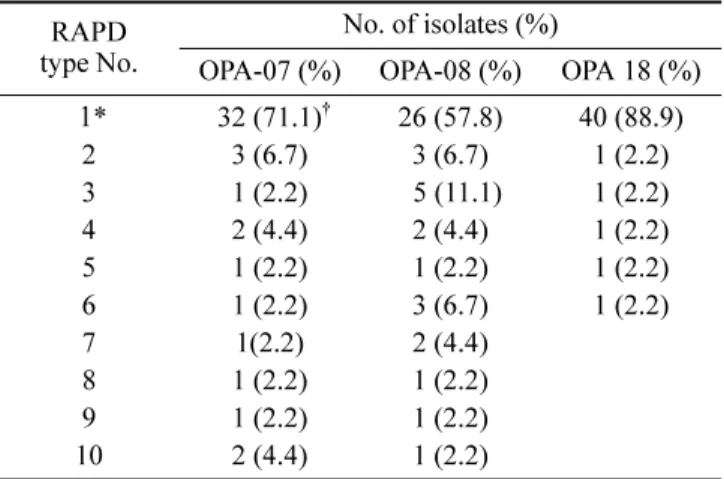
Table 2. RAPD patterns of B. bronchiseptica type No. RAPD No. of isolates (%)
OPA-07 (%) OPA-08 (%) OPA 18 (%) 1*
032 (71.1)
†26 (57.8) 40 (88.9)
2 3 (6.7) 3 (6.7) 1 (2.2)
3 1 (2.2) 05 (11.1) 1 (2.2)
4 2 (4.4) 2 (4.4) 1 (2.2)
5 1 (2.2) 1 (2.2) 1 (2.2)
6 1 (2.2) 3 (6.7) 1 (2.2)
7 1(2.2) 2 (4.4)
8 1 (2.2) 1 (2.2)
9 1 (2.2) 1 (2.2)
10 2 (4.4) 1 (2.2)
*The most common identical RAPD pattern.
†