Elevated Serum C-Reactive Protein as a Prognostic Marker in Small Cell Lung Cancer
Soojung Hong,
1,2Young Ae Kang,
1Byoung Chul Cho,
1,2and Dae Joon Kim
3Departments of 1Internal Medicine, 3Thoracic and Cardiovascular Surgery, and 2Yonsei Cancer Center, Yonsei University College of Medicine, Seoul, Korea.
Received: November 3, 2010 Revised: January 26, 2011 Accepted: February 22, 2011
Corresponding author: Dr. Dae Joon Kim, Department of Thoracic and Cardiovascular Surgery, Yonsei University College of Medicine, 50 Yonsei-ro, Seodaemun-gu,
Seoul 120-752, Korea.
Tel: 82-2-2228-8126, Fax: 82-2-393-3652 E-mail: [email protected]
∙ The authors have no financial conflicts of interest.
© Copyright:
Yonsei University College of Medicine 2012 This is an Open Access article distributed under the terms of the Creative Commons Attribution Non- Commercial License (http://creativecommons.org/
licenses/by-nc/3.0) which permits unrestricted non- commercial use, distribution, and reproduction in any medium, provided the original work is properly cited.
Purpose: Elevated C-reactive protein (CRP) is associated with poor prognosis in several tumor types. The purpose of this study was to investigate serum CRP as a prognostic marker in small cell lung cancer (SCLC). Materials and Methods: The pretreatment serum CRP level was measured in 157 newly diagnosed SCLC pa- tients, and correlation between serum CRP level and other clinical parameters was analyzed. Multivariate analyses were performed to find prognostic markers using Cox’s proportional hazards model. Results: The initial CRP concentration was with- in the normal range in 72 (45.9%) patients and elevated in 85 (54.1%) patients.
There was a significant correlation between serum CRP level and the extent of dis- ease (p<0.001), weight loss (p=0.029) and chest radiation (p=0.001). Median overall survival (OS) in the normal CRP group was significantly longer than with the high CRP group (22.5 months vs. 11.2 months, p<0.001). Extent of disease (p<0.001), age (p=0.025), and performance status (p<0.001) were additional prognostic factors on univariate analysis. On multivariate analysis, elevated serum CRP level was an independent prognostic factor for poor survival (HR=1.8; p=0.014), regardless of the extent of disease (HR=3.7; p<0.001) and performance status (HR=2.2; p<0.001).
Conclusion: High level of CRP was an independent poor prognostic serum marker in addition to previously well-known prognosticators in patients with SCLC.
Key Words: C-reactive protein, prognosis, small cell lung cancer
INTRODUCTION
Small cell lung cancer (SCLC), which accounts for 20-25% of all lung cancers, is highly sensitive to radiotherapy and chemotherapy. Traditionally, well-known prognostic factors of SCLC include extent of disease, performance status, and weight loss. Several laboratory factors, such as neuron-specific enolase (NSE), cy- tokeratin-19 fragments (CYFRA 21-1), carcinoembryonic antigen, lactate dehy- drogenase (LDH), and albumin have been studied to show additional independent prognostic value, however, the weights of their values are still controversial and require prospective validation.1
There have been numerous reports about the relationship between chronic inflam- mation and cancer. The inflammatory cells and cytokines found in tumor highly like-
were excluded. All patients received platinum-based combi- nation chemotherapy, mostly with irinotecan or etoposide.
Patients with limited disease underwent concurrent chemo- radiation therapy including 5,400 cGy of thoracic radiation.
Traditionally, the two-stage system of the Veteran’s Admin- istration Lung Group was used to classify the patients. Lim- ited disease is defined as disease confined to the ipsilateral chest within a single radiation field, and extensive disease was defined as disease beyond the ipsilateral hemithorax including malignant pleural or pericardial effusion or hema- togenous metastasis. Contralateral mediastinal and ipsilat- eral supraclavicular lymphadenopathy are classified as lim- ited-stage, while contralateral hilar and supraclavicular lymphadenopathy are usually classified as extensive stage disease.25 Weight loss was recorded in kilograms (kg) and defined as more than 5 kg or 10% of baseline body weight loss during the past six months. Co-morbidity included the following conditions; hypertension, diabetes mellitus, cere- brovascular disease, ischemic heart disease, asthma, chron- ic obstructive lung disease, liver cirrhosis, and end stage re- nal disease. Response Evaluation was performed with CT scan every two cycles, according to the Response Evaluation Criteria in Solid Tumors guidelines.26
Pretreatment CRP values were measured from peripheral venous blood samples as part of the clinical routine, using an automatic nephelometer (Beckman Coulter image, Fullerton, CA, USA), according to the manufacturer’s instructions.
Normal serum level was defined as ≤0.8 mg/dL by manufac- turer’s manual. The correlation between serum CRP level and other categorical clinical variables was compared by Pearson’s chi-square test. Overall survival (OS) was mea- sured from the date of diagnosis until the date of death or fi- nal follow up. Progression-free survival (PFS) was defined as the time from the date of diagnosis until the date of tumor progression or death. The survival data were estimated using Kaplan-Meier curve and compared using the log-rank test.
Multivariate analyses were performed to find prognostic markers using Cox’s proportional hazards model. A p-value of less than 0.05 was considered to be statistically significant.
The study was approved by our institutional review board.
RESULTS
Patients
A total of 157 patients were included in this study. Patient characteristics are summarized in Table 1. Fifty-nine pa- ly to contribute to tumor growth, progression, and immune-
suppression compared to cope with an effective host anti- tumor response.2-4 In fact, about 15% of cancers are initiated by chronic inflammation or infection such as helicobacter py- lori, hepatitis virus, Epstein-Barr virus, and other bacteria.
Persistent infection of the host induces chronic inflammation, and inflammatory cells induce DNA damage in proliferating cell, by generating reactive oxygen and nitrogen species.3 Furthermore, it is well demonstrated by laboratory research that pro-inflammatory cytokines could promote tumor growth and metastasis by altering tumor cell biology and ac- tivating stromal cells in the tumor microenvironment.3,5,6
C-reactive protein (CRP) is a nonspecific serum marker of acute-phase inflammatory response, and it is produced by hepatocytes which are regulated by interleukin (IL)-6.4,7 Several possible mechanisms have been postulated for the relationship between CRP and cancers; first, tumor growth can cause tissue inflammation, hence increasing CRP level.
Second, CRP could be an indicator of an immune response to tumor antigens. Third, cancer cells could increase the production of inflammatory cytokines, which could induce high CRP concentration in cancer patients.4 Many studies showed the elevation of pretreatment CRP to be a signifi- cant prognostic parameter in patients with esophageal can- cer,8-10 hepatocellular carcinoma,11 colorectal cancer,12-14 re- nal cell cancer,15-17 ovarian cancer,18 and non-small cell lung cancer (NSCLC).19-23 Furthermore, we recently reported an association between preoperative serum CRP levels and pathologic parameter such as tumor size and lymphovascu- lar invasion in patients with NSCLC.24
At present, little is known about the relevance of inflam- matory markers to survival in SCLC. In this study, we evalu- ated the relationship between CRP and SCLC, and investi- gated CRP as a potential prognostic serum marker in patients with SCLC.
MATERIALS AND METHODS
We reviewed patients who had histologically confirmed SCLC and received chemotherapy at the Yonsei Cancer Cen- ter, Seoul, Korea. Retrospective analysis was performed re- garding initial serum CRP concentration, age, gender, ex- tent of disease, weight loss, Eastern Cooperative Oncology Group (ECOG) performance status at first presentation, smoking history, co-morbidity, best response to chemother- apy, and survival. Patients with active concurrent infection
chemotherapy in combination with irinotecan (63.1%), eto- poside (31.8%), or other agents (5.1%) according to physi- cians’ choice.
Serum CRP and patient characteristics
The initial CRP concentration in 72 patients (45.9%) was within the normal range, and elevated in 85 patients (54.1%).
tients (37.6%) had limited disease and 98 patients (62.4%) had extensive disease. The median age was 65 years (range, 46-82), and majority of patients were male (n=140; 89.2%).
Most of the patients (n=127; 79%) had good performance status (ECOG 0-1), and only 29 patients (18.5%) had sig- nificant weight loss. Ten patients (6.4%) had never smoked.
All patients included in this study underwent platinum-based
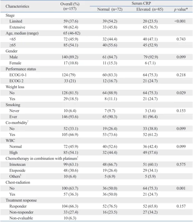
Table 1. Baseline Characteristics
Characteristics Overall (%)
(n=157) Serum CRP
Normal (n=72) Elevated (n=85) p value*
Stage
Limited 59 (37.6) 39 (54.2) 20 (23.5) <0.001
Extensive 98 (62.4) 33 (45.8) 65 (76.5)
Age, median (range) 65 (46-82)
<65 72 (45.9) 32 (44.4) 40 (47.1) 0.743
≥65 85 (54.1) 40 (55.6) 45 (52.9)
Gender
Male 140 (89.2) 61 (84.7) 79 (92.9) 0.099
Female 17 (10.8) 11 (15.3) 6 (7.1)
Performance status
ECOG 0-1 124 (79) 60 (83.3) 64 (75.3) 0.218
ECOG 2 33 (21) 12 (16.7) 21 (24.7)
Weight loss
No 128 (81.5) 64 (88.9) 64 (75.3) 0.029
Yes 29 (18.5) 8 (11.1) 21 (24.7)
Smoking
Never 10 (6.4) 7 (9.7) 3 (3.6) 0.153
Ever 146 (93.6) 65 (90.3) 81 (96.4)
Co-morbidity†
No 52 (33.1) 19 (26.4) 33 (38.8) 0.099
Yes 105 (66.9) 53 (73.6) 52 (61.2)
WBC
Normal 72 (45.9) 40 (52.6) 36 (42.4) 0.099
High 85 (54.1) 32 (44.4) 49 (57.6)
Chemotherapy in combination with platinum‡
Irinotecan 99 (63.1) 48 (66.7) 51 (60.1) 0.575
Etoposide 48 (30.6) 19 (26.4) 29 (34.1)
Others§ 10 (6.4) 5 (6.9) 5 (5.9)
Chest-radiation
No 100 (63.7) 36 (50.0) 64 (75.3) 0.001
Yes 57 (36.3) 36 (50.0) 21 (24.7)
Treatment response
Responder 104 (66.3) 52 (76.5) 52 (65.8) 0.157
Non-responder 33 (27.4) 16 (23.5) 27 (34.2)
Non-evaluable 10 (6.3)
ECOG, Eastern Cooperative Oncology Group; CRP, C-reactive protein; WBC, white blood cell count.
*Chi-squre test between normal CRP and elevated CRP group.
†Co-morbidity, hypertension, diabetes mellitus, cerebrovascular disease, ischemic heart disease, asthma, chronic obstructive lung dis- ease, liver cirrhosis, and end stage renal disease (severity was not specified).
‡Platinum, cisplatin or carboplatin.
§Others, ifosfamide, topotecan, or belotecan.
65.8%; p=0.157). With a median follow-up duration of 9.3 months (range, 0.3-62.7), median PFS was 9.3 months [95% CI, 7.6-11.1], and median OS was 13.7 months (95%
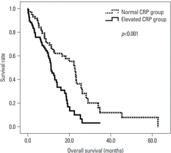
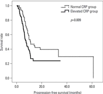
CI, 8.9-18.6). The median PFS and OS in the normal CRP group was significantly longer than with high CRP group (PFS, 11.0 months vs. 7.5 months; p=0.009; OS, 22.5 months vs. 11.2 months, p<0.001) (Figs. 1 and 2). Addition- ally, the extent of disease (23.0 months vs. 10.2 months, p<0.001), age (19.1 months vs. 12.5 months, p=0.026), per- formance status (18.4 months vs. 5.3 months, p<0.001), and chemo-responsiveness (22.2 months vs. 4.9 months, p<0.001) were statistically significant factors in univariate analysis (Table 2). However, weight loss was not statistical- ly significant for survival in univariate analysis (p=0.17).
In multivariate Cox regression model (Table 3), elevated CRP level was an independent prognostic marker for poor survival (HR=1.8, 95% CI, 1.1-2.9; p=0.014), regardless of extensive disease (HR=3.7; p<0.001) and poor perfor- mance status (HR=2.2; p<0.001).
DISCUSSION
To our best knowledge, this is the first report to evaluate the clinical usefulness of serum CRP for predicting survival of SCLC patients. In the multivariate analysis, pre-treatment serum CRP was revealed as a significant prognostic marker of SCLC, together with other well-known factors, such as stage and performance status.
The mean value of serum CRP prior to treatment was 4.7±7.6 mg/dL; 0.3±0.2 mg/dL in the normal CRP group and 8.4±8.8 mg/dL in the high CRP group. Serum CRP level was significantly associated with the extent of disease (p<0.001), chest radiation (p=0.001) and weight loss (p=0.029), but two groups of extensive disease (limited vs. extensive) and group who received chest radiation or not showed almost same composition.
Tumor response and survival
There was a trend for patients in the normal CRP group to- ward higher response rate to chemotherapy (76.5% vs.
Table 2. Univariate Analysis of Survival
Variables Median survival time (95% confidence interval) p value
Age <65 19.133 (11.868-26.399) 0.025
≥65 12.467 (10.386-14.547)
Performance status ECOG 0-1 18.400 (13.818-22.982) <0.001
ECOG ≥2 5.300 (1.484-9.116)
Stage Limited 23.000 (21.998-24.002) <0.001
Extensive 10.167 (8.106-12.228)
CRP Normal 22.533 (18.918-26.149) <0.001
Elevated 11.167 (9.373-12.960)
CRP, C-reactive protein.
Table 3. Factors Independently Affecting Overall Survival
Variable p value* Hazard ratio (95% confidence interval)
Serum CRP 0.014 1.803 (1.129-2.877)
Performance status <0.001 2.226 (1.427-3.474)
Extent of disease <0.001 3.660 (1.129-2.877)
CRP, C-reactive protein.
*Multivariate analysis by Cox's regression.
Fig. 1. Kaplan-Meier survival curve for overall survival in patients with nor- mal CRP and elevated CRP group. CRP, C-reactive protein.
0.0 0.2 0.4 0.6 0.8 1.0
Survival rate
0.0 20.0 40.0 60.0
Overall survival (months)
Normal CRP group Elevated CRP group p<0.001
Brekel, et al.37 reported that hypermetabolism and weight loss are related to enhanced inflammatory response in SCLC patients. In addition, several studies showed that acute phase response is involved in the pathogenesis of cancer cachex- ia.38,39 We also found that extensive disease was associated with high serum CRP level, possibly demonstrating that large tumor burden is likely to increase inflammatory cytokines, such as IL-1, IL-2, tumor necrosis factor-alpha, and interfer- on-gamma,2,3 which stimulates CRP production.18
In terms of response, the relationship between serum CRP and response was not clear. It is highly possible that pre-treat- ment serum CRP was not sufficient for predicting chemo-re- sponse. Therefore, it is more reasonable to monitor serial serum CRP level, including not only pre-treatment but also during and after the treatment. Milroy, et al.40 demonstrated that acute phase response during chemotherapy might have a potential for predicting chemo-response in SCLC since chemo-sensitive tumors might result in tumor necrosis, there- by inducing an acute phase reaction, and significant reduc- tion in the level of CRP was observed after chemotherapy.41
As for prognosis, elevated serum CRP was associated with reduced OS and PFS, apart from all clinically estab- lished prognosticators. This result suggests that it might be a useful marker to define a subset of patients with bad prog- nosis who require intensive treatment. For example, patients with higher pretreatment CRP within the same stage require more enhanced systemic chemotherapy than lower CRP group patients. In the present study, we found a trend be- tween serum CRP level and survival within each stage: ED with high serum CRP group showed relatively shorter OS In various types of malignancy, clinical decision making
before treatment is generally based on established clinical and histopathologic prognosticators. The knowledge of prog- nostic factors, therefore, is important, so that it allows to clas- sify patients who are candidates for newest intensive treat- ment. Traditionally, two-stage system, performance status, and weight loss have been key prognostic factors in SCLC patients.1,27 Other prognostic variables, such as gender and age, have also been well-known as prognostic factors for SCLC, even though some controversies remain.1,28 Simple biochemical tests or serum markers are also important, and numerous studies showed that NSE, CYFRA 21-1, and LDH are promising prognostic factors.29-36
Serum CRP levels, measurement of which is relatively inexpensive and easy to quantify in daily clinical practice, can be elevated in various acute and chronic benign condi- tions such as cardiovascular disease, type 2 DM, arthritis, inflammatory bowel disease, trauma, and transplant rejec- tion.4,24 In this study, patients with co-morbidity were 66.9%
of all patients, however, we failed to observe any correla- tion between CRP levels and co-morbidity (Table 1). As stat- ed above, there have been many efforts to investigate the relationship between serum CRP and prognosis in several types of cancer. In NSCLC, preoperative CRP level provid- ed prognostic information and was associated with patho- logic tumor size and lympho-vascular invasion.21,23,24 In he- patocellular carcinoma, the correlation of preoperative CRP level with tumor size and portal vein invasion was found, and CRP level was an independent indicator of poor prog- nosis and early recurrence.11 In ovarian cancer, preoperative CRP was an independent prognostic marker associated with stage and postoperative residual tumor mass.18 In renal cell carcinoma (RCC), serum CRP was significantly associ- ated with RCC-specific mortality.15 In esophageal cancer, preoperative high CRP level was associated with tumor progression and poor survival.8-10 Finally, in colorectal cancer, preoperative elevation of CRP level was an indicator of ma- lignant potential of tumors such as liver metastasis, perito- neal carcinomatosis, lymph node metastasis, and vascular invasion, as well as a predictor of poor prognosis.12,14 Until now, however, a few data showed the relationship between CRP and treatment outcomes in small cell lung cancer.
In our study, a positive correlation between CRP and weight loss was observed. This is consistent with previous studies, which showed that systemic inflammatory response is associated with increase in resting energy expenditure and loss of lean tissue in patients with lung cancer: Staal-van den
Fig. 2. Kaplan-Meier survival curve for progression-free survival in patients with normal CRP and elevated CRP group. CRP, C-reactive protein.
0.0 0.2 0.4 0.6 0.8 1.0
Survival rate
0.0 20.0 40.0 60.0
Progression-free survival (months) Normal CRP group Elevated CRP group p=0.009
2004;279:48487-90.
8. Gockel I, Dirksen K, Messow CM, Junginger T. Significance of preoperative C-reactive protein as a parameter of the perioperative course and long-term prognosis in squamous cell carcinoma and adenocarcinoma of the oesophagus. World J Gastroenterol 2006;12:3746-50.
9. Nozoe T, Saeki H, Sugimachi K. Significance of preoperative ele- vation of serum C-reactive protein as an indicator of prognosis in esophageal carcinoma. Am J Surg 2001;182:197-201.
10. Shimada H, Nabeya Y, Okazumi S, Matsubara H, Shiratori T, Aoki T, et al. Elevation of preoperative serum C-reactive protein level is related to poor prognosis in esophageal squamous cell car- cinoma. J Surg Oncol 2003;83:248-52.
11. Hashimoto K, Ikeda Y, Korenaga D, Tanoue K, Hamatake M, Ka- wasaki K, et al. The impact of preoperative serum C-reactive pro- tein on the prognosis of patients with hepatocellular carcinoma.
Cancer 2005;103:1856-64.
12. Crozier JE, McKee RF, McArdle CS, Angerson WJ, Anderson JH, Horgan PG, et al. Preoperative but not postoperative systemic in- flammatory response correlates with survival in colorectal cancer.
Br J Surg 2007;94:1028-32.
13. Nielsen HJ, Christensen IJ, Sørensen S, Moesgaard F, Brünner N.
Preoperative plasma plasminogen activator inhibitor type-1 and serum C-reactive protein levels in patients with colorectal cancer.
The RANX05 Colorectal Cancer Study Group. Ann Surg Oncol 2000;7:617-23.
14. Nozoe T, Matsumata T, Kitamura M, Sugimachi K. Significance of preoperative elevation of serum C-reactive protein as an indica- tor for prognosis in colorectal cancer. Am J Surg 1998;176:335-8.
15. Karakiewicz PI, Hutterer GC, Trinh QD, Jeldres C, Perrotte P, Gallina A, et al. C-reactive protein is an informative predictor of renal cell carcinoma-specific mortality: a European study of 313 patients. Cancer 2007;110:1241-7.
16. Lamb GW, McMillan DC, Ramsey S, Aitchison M. The relation- ship between the preoperative systemic inflammatory response and cancer-specific survival in patients undergoing potentially cu- rative resection for renal clear cell cancer. Br J Cancer 2006;94:
781-4.
17. Miyata Y, Koga S, Nishikido M, Noguchi M, Kanda S, Hayashi T, et al. Predictive values of acute phase reactants, basic fetoprotein, and immunosuppressive acidic protein for staging and survival in renal cell carcinoma. Urology 2001;58:161-4.
18. Hefler LA, Concin N, Hofstetter G, Marth C, Mustea A, Sehouli J, et al. Serum C-reactive protein as independent prognostic variable in patients with ovarian cancer. Clin Cancer Res 2008;14:710-4.
19. Gagnon B, Abrahamowicz M, Xiao Y, Beauchamp ME, MacDon- ald N, Kasymjanova G, et al. Flexible modeling improves assess- ment of prognostic value of C-reactive protein in advanced non- small cell lung cancer. Br J Cancer 2010;102:1113-22.
20. Koch A, Fohlin H, Sörenson S. Prognostic significance of C-reac- tive protein and smoking in patients with advanced non-small cell lung cancer treated with first-line palliative chemotherapy. J Tho- rac Oncol 2009;4:326-32.
21. Hara M, Matsuzaki Y, Shimuzu T, Tomita M, Ayabe T, Enomoto Y, et al. Preoperative serum C-reactive protein level in non-small cell lung cancer. Anticancer Res 2007;27:3001-4.
22. Wilop S, Crysandt M, Bendel M, Mahnken AH, Osieka R, Jost E.
Correlation of C-reactive protein with survival and radiographic response to first-line platinum-based chemotherapy in advanced non-small cell lung cancer. Onkologie 2008;31:665-70.
than ED with low serum CRP group (mean OS for high CRP group vs. low CRP group, 19.1 months vs. 23.2 months;
p=0.062).
The mechanism by which an inflammatory response is evoked by SCLC is not yet clear. One possible mechanism is coexisting pulmonary infection (or obstructive pneumo- nia), which could increase white blood cell counts (WBC), subsequently increasing pro-inflammatory cytokine CRP concentration. In the current study, therefore, we evaluated the correlation between WBC and CRP, however, failed to find any significant correlations (pearson correlation coeffi- cient=0.140; p=0.099). This result might explain why in- fection is not the main stimulus to the increased CRP. The other possibility is that pro-inflammatory cytokines are pro- duced by tumor necrosis or local tissue damage which is caused by the tumor-host cell interaction, however, we are not certain whether those cytokines are produced directly by tumors.
In conclusion, we suggest that pre-treatment serum CRP could be used as a prognostic marker in SCLC patients, and it might complement the prognostic value of stage and per- formance status. However, the association between CRP and survival outcome might require further investigation, and our result should await internal or external validation before be- ing used in clinical practice, because of the limitations inher- ent to retrospective study with a small sample size.
ACKNOWLEDGEMENTS
This study was supported by a faculty research grant of Yon- sei University College of Medicine for 6-2007-0194.
REFERENCES
1. Buccheri G, Ferrigno D. Prognostic factors of small cell lung can- cer. Hematol Oncol Clin North Am 2004;18:445-60.
2. Balkwill F, Mantovani A. Inflammation and cancer: back to Vir- chow? Lancet 2001;357:539-45.
3. Coussens LM, Werb Z. Inflammation and cancer. Nature 2002;420:860-7.
4. Heikkilä K, Ebrahim S, Lawlor DA. A systematic review of the association between circulating concentrations of C reactive pro- tein and cancer. J Epidemiol Community Health 2007;61:824-33.
5. Mantovani A, Allavena P, Sica A, Balkwill F. Cancer-related in- flammation. Nature 2008;454:436-44.
6. Chiang AC, Massagué J. Molecular basis of metastasis. N Engl J Med 2008;359:2814-23.
7. Black S, Kushner I, Samols D. C-reactive Protein. J Biol Chem
32. Pujol JL, Grenier J, Parrat E, Lehmann M, Lafontaine T, Quantin X, et al. Cytokeratins as serum markers in lung cancer: a compari- son of CYFRA 21-1 and TPS. Am J Respir Crit Care Med 1996;
154:725-33.
33. Pujol JL, Quantin X, Jacot W, Boher JM, Grenier J, Lamy PJ.
Neuroendocrine and cytokeratin serum markers as prognostic de- terminants of small cell lung cancer. Lung Cancer 2003;39:131-8.
34. Buccheri G, Ferrigno D; Cuneo Lung Cancer Study Group. Serum biomarkers of non-neuron-endocrine origin in small-cell lung can- cer: a 16-year study on carcinoembryonic antigen, tissue polypep- tide antigen and lactate dehydrogenase. Lung Cancer 2000;30:37- 49.
35. Quoix E, Purohit A, Faller-Beau M, Moreau L, Oster JP, Pauli G.
Comparative prognostic value of lactate dehydrogenase and neu- ron-specific enolase in small-cell lung cancer patients treated with platinum-based chemotherapy. Lung Cancer 2000;30:127-34.
36. Boher JM, Pujol JL, Grenier J, Daurès JP. Markov model and markers of small cell lung cancer: assessing the influence of re- versible serum NSE, CYFRA 21-1 and TPS levels on prognosis.
Br J Cancer 1999;79:1419-27.
37. Staal-van den Brekel AJ, Dentener MA, Schols AM, Buurman WA, Wouters EF. Increased resting energy expenditure and weight loss are related to a systemic inflammatory response in lung can- cer patients. J Clin Oncol 1995;13:2600-5.
38. Martín F, Santolaria F, Batista N, Milena A, González-Reimers E, Brito MJ, et al. Cytokine levels (IL-6 and IFN-gamma), acute phase response and nutritional status as prognostic factors in lung cancer. Cytokine 1999;11:80-6.
39. O’Riordain MG, Falconer JS, Maingay J, Fearon KC, Ross JA.
Peripheral blood cells from weight-losing cancer patients control the hepatic acute phase response by a primarily interleukin-6 de- pendent mechanism. Int J Oncol 1999;15:823-7.
40. Milroy R, Shapiro D, Shenkin A, Banham SW. Acute phase reac- tion during chemotherapy in small cell lung cancer. Br J Cancer 1989;59:933-5.
41. Staal-van den Brekel AJ, Schols AM, Dentener MA, ten Velde GP, Buurman WA, Wouters EF. The effects of treatment with chemo- therapy on energy metabolism and inflammatory mediators in small-cell lung carcinoma. Br J Cancer 1997;76:1630-5.
23. Kasymjanova G, MacDonald N, Agulnik JS, Cohen V, Pepe C, Kreisman H, et al. The predictive value of pre-treatment inflam- matory markers in advanced non-small-cell lung cancer. Curr On- col 2010;17:52-8.
24. Lee JG, Cho BC, Bae MK, Lee CY, Park IK, Kim DJ, et al. Pre- operative C-reactive protein levels are associated with tumor size and lymphovascular invasion in resected non-small cell lung can- cer. Lung Cancer 2009;63:106-10.
25. Micke P, Faldum A, Metz T, Beeh KM, Bittinger F, Hengstler JG, et al. Staging small cell lung cancer: Veterans Administration Lung Study Group versus International Association for the Study of Lung Cancer--what limits limited disease? Lung Cancer 2002;37:271-6.
26. Therasse P, Arbuck SG, Eisenhauer EA, Wanders J, Kaplan RS, Rubinstein L, et al. New guidelines to evaluate the response to treatment in solid tumors. European Organization for Research and Treatment of Cancer, National Cancer Institute of the United States, National Cancer Institute of Canada. J Natl Cancer Inst 2000;92:205-16.
27. Osterlind K, Ihde DC, Ettinger DS, Gralla RJ, Karrer K, Krauss S, et al. Staging and prognostic factors in small cell carcinoma of the lung. Cancer Treat Rep 1983;67:3-9.
28. Paesmans M, Sculier JP, Lecomte J, Thiriaux J, Libert P, Sergysels R, et al. Prognostic factors for patients with small cell lung carci- noma: analysis of a series of 763 patients included in 4 consecu- tive prospective trials with a minimum follow-up of 5 years. Can- cer 2000;89:523-33.
29. Buccheri G, Ferrigno D. Prognostic factors in lung cancer: tables and comments. Eur Respir J 1994;7:1350-64.
30. Bremnes RM, Sundstrom S, Aasebø U, Kaasa S, Hatlevoll R, Aamdal S; Norweigian Lung Cancer Study Group. The value of prognostic factors in small cell lung cancer: results from a ran- domised multicenter study with minimum 5 year follow-up. Lung Cancer 2003;39:303-13.
31. Jørgensen LG, Osterlind K, Genollá J, Gomm SA, Hernández JR, Johnson PW, et al. Serum neuron-specific enolase (S-NSE) and the prognosis in small-cell lung cancer (SCLC): a combined mul- tivariable analysis on data from nine centres. Br J Cancer 1996;
74:463-7.