• 검색 결과가 없습니다.

Computed Tomographic Angiogram of an Anterior Communicating Artery Aneurysm Causing Acute Retrobulbar Optic Neuropathy: A Case Report

N/A
N/A
Protected

Academic year: 2022

Share "Computed Tomographic Angiogram of an Anterior Communicating Artery Aneurysm Causing Acute Retrobulbar Optic Neuropathy: A Case Report"

Copied!
3
0
0

로드 중.... (전체 텍스트 보기)

전체 글

(1)

366

Korean J Ophthalmol 2011;25(5):366-368 http://dx.doi.org/10.3341/kjo.2011.25.5.366 pISSN: 1011-8942 eISSN: 2092-9382

Case Report

Computed Tomographic Angiogram of an Anterior Communicating Artery Aneurysm Causing Acute

Retrobulbar Optic Neuropathy: A Case Report

Jee Ho Chang

1

, Dong-Kyu Lee

1

, Bum Tae Kim

2

, Young-Hoon Ohn

1

1

Department of Ophthalmology, Soonchunhyang University Bucheon Hospital, Soonchunhyang University College of Medicine, Bucheon, Korea

2

Department of Neurosurgery, Soonchunhyang University Bucheon Hospital, Soonchunhyang University College of Medicine, Bucheon, Korea

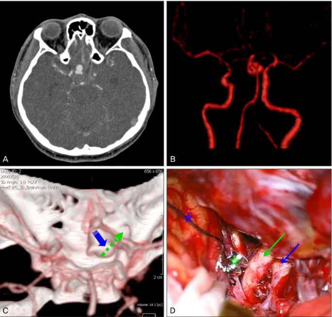
Three-dimensional computed tomographic (3D-CT) angiography is a widespread imaging modality for intracranial vascular lesions. However, 3D-CT angiograms of an anterior communicating artery aneurysm associated with acute retrobulbar optic neuropathy have not been previously described. We present 3D-CT angiograms of an aneurysm of the anterior communicating artery that caused subarachnoid hemorrhage and vision loss in a 39-year old man. The 3D-CT angiograms were consistent with findings identified directly during surgery.

Key Words: Intracranial aneurysm, Retrobulbar optic neuropathy, Three-dimensional computed tomographic angiogram

ⓒ2011 The Korean Ophthalmological Society

This is an Open Access article distributed under the terms of the Creative Commons Attribution Non-Commercial License (http://creativecommons.org/licenses /by-nc/3.0/) which permits unrestricted non-commercial use, distribution, and reproduction in any medium, provided the original work is properly cited.

Received: January 30, 2009 Accepted: June 8, 2010

Reprint requests to Young-Hoon Ohn, MD, PhD. Department of Ophthalmology, Soonchunhyang University Bucheon Hospital, #1174 Jung-dong, Wonmi-gu, Bucheon 420-767, Korea. Tel: 82-32-621-5053, Fax: 82-32-621-5433, E-mail: [email protected]

Three-dimensional computed tomographic (3D-CT) an- giography has recently gained popularity for diagnosing in- tracranial vascular disease [1]. However, to our knowledge 3D-CT angiographic images of an anterior communicating artery aneurysm associated with acute retrobulbar optic neu- ropathy have never been reported. Here, we report a case of a ruptured anterior communicating artery aneurysm causing acute retrobulbar optic neuropathy that was examined by 3D-CT angiography.

Case Report

A 39-year-old man presented to the emergency department of Soonchunhyang University Bucheon Hospital with dis- orientation and severe headache. Brain CT scans demon- strated subarachnoid hemorrhage. 3D-CT angiography of the brain (Somatom Sensation 16; Siemens, Forchheim, Germany) revealed a large aneurysm arising from the junction of the right anterior cerebral and the anterior communicating ar- teries that was oriented downward (Fig. 1A-1C). This finding prompted emergent neurosurgical intervention, during which the aneurysmal neck was clipped successfully (Fig. 1D). On

the fifth postoperative day, the patient’s mental status had im- proved and he complained of right eye vision loss. On oph- thalmologic examination, the patient’s visual acuity was no light perception (NLP) OD and 1.0 OS and his right pupil was nonresponsive. In retrospect, the patient recalled notic- ing a visual field defect encroaching from the inferior field in the right eye 10 days before admission, but he thought it was a manifestation of headache. The results of external, motility, and slit lamp examinations were all normal. The results of di- lated fundus examinations were normal in both eyes, and the optic nerves in particular appeared normal. After three months, the vision in the patient’s right eye remained NLP, and atrophy of the right optic nerve had occurred.

Discussion

With recent advances of CT and magnetic resonance imag- ing, the diagnosis and treatment of cerebrovascular disease have become more accurate and effective. Acute retrobulbar optic neuropathy due to an anterior communicating artery aneurysm is rare, and to our knowledge 3D-CT angiographic images of this condition have never been reported.

Although sudden unilateral visual loss combined with signs

of retrobulbar optic nerve dysfunction suggests a diagnosis

of optic neuritis, especially when associated with normal

looking discs, vascular and compressive lesions may be con-

sidered in certain cases. Diagnosis was straightforward in the

present case. The patient presented with altered mental status

(2)

JH Chang, et al. CT Angiogram of Ruptured Aneurysm

367

A B

C D

Fig 1. Preoperative brain three-dimensional (3D) computed tomographic angiography images. (A) Enhanced axial view shows an aneurysm arising from the junction between the right anterior cerebral artery (ACA) and anterior communicating artery (AComA).

(B) Vessel subtractional view shows a large aneurysm in the right ACA at the AComA junction oriented primarily downward. (C) 3D views show the large aneurysm arising from the junction of the right ACA and AComA, oriented primarily downward (blue ar- row) toward the right optic nerve (green arrow). (D) Intra-operative view shows the right optic nerve (green arrow), internal carotid artery (blue arrow) and frontal lobe (blue star). After clipping, the ruptured aneurysm with its clip (green star) can be seen arising from the junction between the right ACA and AComA and the pinkish optic nerve (green arrow).

and headache. Conventional CT revealed vascular pathology, and the choice of advanced vascular imaging modality was left to the physician. Although conventional digital sub- traction angiography (DSA) is considered the most sensitive tool for detection of intracranial aneurysms, we did not per- form DSA because it is an invasive study. Furthermore, con- sidering the potential delays in time required to assemble the angiography team and perform the angiogram, CT angiog- raphy is often faster and is comparably accurate [2].

Magnetic resonance angiogram is another option, and it has advantages for the evaluation of cerebrovascular lesions.

However, in our case, CT angiography was perceived to be faster and less likely to induce claustrophobia [3].

The choice of treatment modality is also worthy of

discussion. Although endosaccular occlusion of the intra-

cranial aneurysm was considered in the present case, urgent

decompression of the aneurysm could not be achieved by

this treatment because of the volume effect of the coils. In

our patient, immediate decompression was mandatory and

emergent surgical decompression with clipping of the

aneurysm was identified as the treatment most likely to res-

cue vision [4].

(3)

Korean J Ophthalmol Vol.25, No.5, 2011

368

Unfortunately, despite successful surgical decompression, the patient’s vision did not recover. There are several ex- planations for monocular visual loss due to a ruptured anterior communicating artery aneurysm, including direct bleeding into the optic nerve, direct compression by the aneurysm [5], and interruptions of blood flow to arteries supplying posterior portions of the optic nerve as a result of either the subarachnoid hemorrhage itself, subsequent vasospasm, or the surgical clipping of the aneurysm [6]. The previous rec- ognition of an inferior visual field defect 10 days before the attack suggests that direct compression of the optic nerve by the aneurysm was contributory.

Conflict of Interest

No potential conflict of interest relevant to this article was reported.

Acknowledgements

We thank Dr. Nancy J. Newman (Director, Section of Neuro- Ophthalmology, Department of Ophthalmology, Emory

University, Atlanta, GA, USA) for her critical review and proofreading of this manuscript.

References

1. Lovblad KO, Altrichter S, Viallon M, et al. Neuro-imaging of cerebral ischemic stroke. J Neuroradiol 2008;35:197-209.

2. Alberico RA, Patel M, Casey S, et al. Evaluation of the circle of Willis with three-dimensional CT angiography in patients with suspected intracranial aneurysms. AJNR Am J Neuroradiol 1995;16:1571-8.

3. Thijs V. Imaging techniques for acute ischemic stroke: nice gadgets or essential tools for effective treatment? Neuroradiology 2010;52:169-71.

4. Craenen G, Brown SM, Freedman KA, et al. Rapid, painless unilateral vision loss in a 37-year-old healthy woman. Surv Ophthalmol 2004;49:343-8.

5. Chan JW, Hoyt WF, Ellis WG, Gress D. Pathogenesis of acute monocular blindness from leaking anterior communicating ar- tery aneurysms: report of six cases. Neurology 1997;48:680-3.

6. Hara N, Mukuno K, Ohtaka H, Shimizu K. Ischemic optic neu-

ropathy associated with subarachnoid hemorrhage after rupture

of anterior communicating artery aneurysm. Ophthalmologica

2003;217:79-84.

수치

Fig 1. Preoperative brain three-dimensional (3D) computed tomographic angiography images

참조

관련 문서

Chapter 15 Analysis of Qualitative Data Part V Communicating with Others Chapter 16 Writing the Research Report and the Politics

Cerebral Cerebral Cerebral angiography Cerebral angiography angiography angiography shows shows shows shows feeding feeding feeding artery feeding artery

In an effort to survey the level of communication that children feel in communicating with their mothers, 'Parents Facilitated Communication Test' was used..

The objective of this study is to analyze the shear bond strength of orthodontic buttons according to light tip distance and optic fiber diameter when an extended optic

Time series of vertical cross section of potential vorticity and wind vector calculated by Case 1 along the A-A' line indicated at Fig.. Same

Generally, in case that we predict the vibration by blasting, we compute it on the basis of the charge weight per delay but in case of shocking

- A few meters of water can create hydrate plug that blocks a pipeline or subsea equipment.. North Sea Plug Case History.. • 16inch, 22mile pipeline

At the end of the study, a reevaluation of each study case was performed with the same questionnaire. The result shows there has been a meaningful result in the group