교신저자 : 김영희, 부산시 금정구 장전동 부산대학교 자연과학대학 분자생물학과
(Tel: 82-51-510-2526, E-mail: [email protected])
∙접수 2009/11/27 ∙수정 2009/12/02 ∙채택 2009/12/05 Otolaryngology & Dermatology 2009;22(3) : 11-19
Inhibitory Effect of Dendrobium moniliforme on NO and IL-1β Production in LPS-stimulated Macrophages
Ga-young Park1,a)·Chang Hwan Bae1,a)·Sun Young Park1)·Ji-Hee Kim1)· Woo Shin Ko2)·YoungHee Kim1)
1)Department of Molecular Biology, College of Natural Sciences, Pusan National University
2)Department of Oriental Medicine, College of Oriental Medicine, Dongeui University
a)These authors have contributed equally to this work.
LPS로 자극된 대식세포에서 석곡의 NO 및 IL-1β 생성 억제 효과
박가영·배창환·박순영·김지희·고우신·김영희
석곡은 난초과의 여러해살이풀 Dendrobium moniliforme의 지상부를 건조한 것으로 예로부터 養胃生津, 滋陰除 熱 등의 효능이 있어 해열, 진통의 작용과 위액분비 촉진, 혈압강하의 작용이 있는 것으로 알려져 있다. 본 연구에서 는 석곡의 항염증 작용 기전을 알아보기 위하여 석곡 열수추출물을 대식세포주에 처리하여 NO 및 IL-1β의 생성에 미치는 영향을 조사하였다. LPS로 자극된 대식세포주 RAW264.7 세포에서 석곡은 NO 및 IL-1β 생성과 iNOS 단백 질 발현을 저해하였으며, LPS에 의해서 활성화되는 ERK, p38, JNK 효소의 활성을 현저히 억제하였다. 이 결과들로 보아 석곡의 항염증 작용이 MAPK 활성 저해로 인한 NO 및 IL-1β 생성의 억제 때문인 것으로 사료된다.
Key words : Dendrobium moniliforme; Nitric oxide (NO); Inducible nitric oxide synthase (iNOS);
Extracellular signal-regulated kinase (ERK); c-Jun N-terminal kinase (JNK); p-38
I. Introduction
The stems of Dendrobium moniliforme (DM) have been used in traditional Chinese and Korean medicine as a Yin tonic to nourish the
stomach, promote the production of body fluid and reduce fever. DM is known to produce a variety of secondary metabolites, such as alkaloids1), sesquiterpens2,3), and phenanthrens4). But little is known about the mechanism responsible for anti-inflammatory effects of DM.
Macrophages are immune cells usually dispersed throughout the body. They are
particularly important in innate immunity as they are among the first cells responding to microbial infection. They detect pathogenic substances through pattern-recognition receptors and subsequently initiate and regulate inflammatory response5) using a wide range of soluble pro-inflammatory mediators.
Lipopolysaccharide (LPS) is one of the most powerful activators of macrophages, and macrophages and monocytes induced by LPS are known to be activated through the production of inflammatory mediators, such as NO and other free radicals, in addition to numerous cytokines including TNF-α , IL-1β and IL-6. These cytokines lead to a variety of responses including the activation of Th1 response, increased expression of adhesion molecules on vascular endothelial cells6), the induction of acute-phase response proteins by the liver7), and are generally involved in the development of inflammatory diseases8). Thus, the inhibition of excessive production of these cytokines can be employed as criteria to evaluate anti-inflammatory effects of drugs.
Nitric oxide (NO) is a free radical with multiple effects on various organ systems. The most prominent physiological actions of NO as a biological mediator include cGMP-dependent vasodilation, neural communication, host defense, inflammation, immune suppression and blood clotting9). NO is produced in physiological and pathophysiological conditions by NO synthase (NOS), and inducible NOS (iNOS) is induced by inflammatory cytokines and/or bacterial LPS in various cell types including macrophages. A large amount of
NO, particularly synthesized by iNOS, induces an inflammatory response to inhibit the growth of invading microorganisms and tumor cells. This strong inflammatory response to foreign cells could also cause further damage for the neighboring cells and tissues of the host10). Therefore isozyme specific inhibitors of NOS are essential for therapeutic purposes and drugs that specifically inhibit iNOS could be useful in treating diseases mediated by NO overproduction11).
In the present study, we investigated the effects of DM on LPS-induced inflammatory response and further explored the possible mechanisms of this inhibition by DM in the macrophages. Our results provide a molecular basis for understanding the inhibitory effects of DM on endotoxin-mediated inflammation.
II. Materials and methods 1. Preparation of extract
The stems of DM were purchased from a local herb store, Kwang Myoung Dang (Busan, Korea) in April 2005. DM was identified and authenticated by Professor Woo Shin Ko who specializes in traditional Chinese herb medicine in College of Oriental Medicine, Dongeui University (Busan, Korea). A voucher specimen (number DM-05-04) has been deposited at the Department of Molecular Biology, Pusan National University, Busan, Korea. The dry stems (300 g) were extracted with distilled
water at 100℃ for 4 h. The extract was filtered through 0.45 ㎛ filter and freeze-dried (yield, 8 g) and kept at 4℃. The dried extract was dissolved in phosphate buffered saline (PBS) and filtered through 0.22 ㎛ filter before use.
2. Cell culture
Murine macrophage RAW 264.7 cells were maintained in Dulbecco's modified Eagle's medium supplemented with 10% FBS at 37℃
in an atmosphere of 5% CO2. 3. Cell viability assay
The cytotoxicity of DM was assessed using the microculture tetrazolium (MTT)-based colorimetric assay12). The remaining cells after Griess reaction were used for MTT assay. 5㎕
of MTT solution (5 ㎎/㎖) was added to each well (final concentration is 62.5 ㎍/㎖). After incubation for 3 h at 37℃ and 5% CO2, the supernatant was removed and the formed formazan crystals in viable cells were solubilized with 150 ㎕ of DMSO. The absorbance of each well was then read at 570
㎚ using microplate reader.
4. Measurement of nitrite concentration
NO synthesis in cell cultures was measured by a microplate assay method, as described13). To measure nitrite, 100 ㎕ aliquots were removed from conditioned medium and incubated with an equal volume of the Griess
reagent (1% sulfanilamide/0.1% N-(1-naphthyl) -ethylenediamine dihydrochloride/2.5% H3PO4) at room temperature for 10 min. Nitrite concentration was determined by measuring the absorbance at 540 ㎚ with a Vmax 96-well microplate spectrophotometer (Molecular Devices, Menlo Park, CA). The sodium nitrite was used as a standard.
5. ELISA
RAW 264.7 cells were incubated with various concentrations of DM for 1 h, and then LPS (1 ㎍/㎖) was treated. Following 6 h incubation, IL-1β levels were quantified in culture media by ELISA Kit (Komabiotech, Korea) according to the manufacturer's instructions.
6. Western blot analysis
The cells were washed with phosphate buffered saline (PBS) three times and scraped off and lysed with lysis buffer (1% Triton X-100, 1% deoxycholate). Protein concentration of lysates was determined and equal amounts of protein were separated electrophoretically using 10% SDS-PAGE (sodium dodecyl sulfate-polyacrylamide gel electrophoresis), and then the gel was transferred to 0.45 μm nitrocellulose paper. The blot was incubated with anti-iNOS and α-tubulin antibody (Santa Cruz Biotechnology), anti-JNK, phospho-JNK, p38, phospho-p38, ERK, phospho-ERK antibody (Cell Signaling Technology), and secondary antibody and then detected by the
enhanced chemiluminescence detection system according to the recommended procedure (Amersham Co.).
7. Statistical analysis
All results were expressed as means ± SE.
Each experiment was repeated at least three times. Statistical significances were compared between each treated group and analyzed by the paired Student's t-test using SPSS 14 for windows. Data with p < 0.05 were considered statistically significant.
III. Results
1. Effect of DM on cell viability in macrophages
To examine the effect of DM on cell viability, we performed MTT assay. RAW 264.7 macrophage cells were treated with LPS in the presence or absence of DM. DM did not decrease cell viability but rather protected cells from the toxicity of LPS(Fig. 1).
2. Effect of DM on NO synthesis and on iNOS expression in macrophages
Next, we examined the effect of DM on NO synthesis. RAW 264.7 cells were incubated with DM for 1 h and stimulated with 1 ㎍/㎖
LPS for 24 h. The amount of NO released into culture medium was measured by the method of Griess. Whereas LPS-treated cells
Fig. 1. Effect of DM on cell viability. RAW 264.7 cells were incubated with various concentration of DM in the presence of 1
㎍/㎖ of LPS for 24 h. Then cell viability was measured by MTT assay according to Materials and methods. Data represent the relative viability to LPS-alone group and are expressed as the means ± S.E. of three independent experiments.
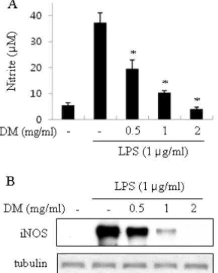
Fig. 2. `Effect of DM on the NO secretion and iNOS expression in macrophages. (A) RAW 264.7 cells were incubated with various concentrations of DM for 1 h and then stimulated with 1 ㎍/㎖ LPS for 24 h at 37℃. The amount of nitrite released was measured by the method of Griess. Data are expressed as the means ± S.E. of four individual experiments, performed in duplicate. * p < 0.05 vs
LPS-alone group. (B) RAW 264.7 cells were treated as described above and equal cytosolic extracts were analyzed by Western blotting with anti-iNOS antibody. Western blot detection of α -tubulin was estimated protein-loading control for each lane.
produced a large amount of NO, DM suppressed NO release into culture supernatant in a dose-dependent manner (Fig. 2A). When macrophages were treated with 2 ㎎/㎖ of DM, the level of NO released was decreased to basal level. To determine whether the decreased nitric oxide synthesis is correlated with iNOS expression, we analyzed the amount of iNOS by Western blot analysis. RAW 264.7 cells were treated with DM as mentioned above. The level of iNOS was dramatically reduced by DM in a dose-dependent manner(Fig. 2B).
3. Inhibitory effect of DM on pro- inflammatory cytokine production in macrophages
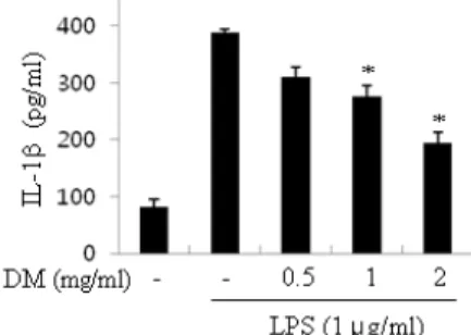
To investigate the anti-inflammatory effect of DM, we examined the effect of DM on proinflammatory cytokines production. RAW 264.7 macrophage cells were challenged with LPS in the presence or absence of DM and the level of IL-1β in the medium was measured. Whereas LPS-treated cells produced a large amount of IL-1β, DM suppressed the release of IL-1β into culture supernatant in a dose-dependent manner(Fig. 3).
Fig. 3. Suppression of IL-1β release by DM. RAW 264.7 cells were incubated with various concentrations of DM for 1 h, and then LPS (1 ㎍/㎖) was treated for 6 h. IL-1β in the cultured supernatant were measured by ELISA kit. Values are means ± S.E. of three independent experiments. * p < 0.05 vs LPS-alone group.
4. Inhibitory effect of DM on LPS-induced MAPK activities
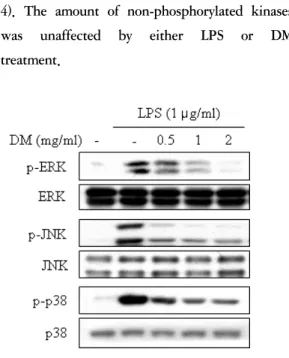
Because activations of MAPKs such as extracellular signal-regulated kinases (ERK), p38, c-Jun NH2-terminal kinase (JNK) are known to participate in the regulation of LPS-induced proinflammatory cytokine and NO production14-16), we investigated the effects of DM on the activation of ERK, JNK and p38 in LPS-stimulated macrophages. Activation of MAPKs requires phosphorylation of serine and threonine residues. RAW 264.7 cells were treated with indicated concentrations of DM for 1 h and then stimulated with 1 ㎍/㎖ LPS for 15 min. Using immunoblot analysis with anti-phospho-specific antibody, we found that DM suppressed LPS-induced activation of ERK, JNK and p38 in dose-dependent manners (Fig
4). The amount of non-phosphorylated kinases was unaffected by either LPS or DM treatment.
Fig. 4. Effect of DM on ERK, p38 and JNK activity in LPS-stimulated macrophages. RAW 264.7 cells were treated with indicated concentrations of DM for 1 h and stimulated with 1 ㎍/㎖ LPS for 15 min. Equal amount of cell extracts was analyzed by western blotting with anti-phospho-ERK, anti-phospho-p38 or anti-phospho-JNK antibody. Western blot detection of non-phosphorylated kinases was estimated protein-loading control for each lane.
IV. Discussion
The application of medicinal herbs dates back to the beginning of civilization, and interestingly, medicinal herbs are still routinely used by most of world's population. Over time, many information has been accumulated by using these herbs in practice, and it is experientially proved that these herbs have the
effect of pain alleviation, life extension and disease prevention. The stems of DM have been used in traditional Chinese and Korean medicine as a Yin tonic to nourish the stomach, promote the production of body fluid and reduce fever. Recently, denbinobin, a 1,4-phenanthrenequinone, has been reported to inhibit nuclear factor-kappaB and induce apoptosis via reactive oxygen species generation in human leukemic cells17). However, the underlying mechanism of anti-inflammatory effects has remained to be characterized so far. In this study, DM significantly inhibited LPS-induced NO production and iNOS expression in macrophages without appreciable cytotoxic effects (Fig. 1, 2). The expression of iNOS and the release of large amounts of NO by macrophages are considered to play a significant role in the pathogenesis of various inflammatory disorders. Inhibition of iNOS activity in macrophages may thus represent an interesting target to treat various diseases associated with increased NO production. In fact, administration of the selective inhibitors of iNOS has been reported to attenuate osteoarthritis18), periodontitis19), experimental autoimmune myocarditis20), multiple sclerosis21), shock22).
Several inflammatory cytokines, particularly TNF-α, IL-1β and IL-6, are known to play key roles in the induction and perpetuation of inflammation in macrophages. These cytokines play pivotal roles in the induction of the innate immune response as well as in determining the magnitude and nature (Th1 vs Th2) of the acquired immune response23). We
demonstrated that DM exhibit anti-inflammatory function by inhibiting the production of NO and IL-1β in macrophages and these results suggest that DM acts as anti-inflammatory agent influencing proinflammatory cytokine and NO.
MAPKs are a family of serine/threonine protein kinases that are an important part of intracellular signaling pathways, connecting extracellular signals to intracellular regulatory proteins. MAPK family members ERK and p38 MAPK are known to participate in the regulation of LPS-induced proinflammatory cytokine production14-16). The third signal transduction pathway of the MAPK family is the JNK pathway, which is also activated primarily by cellular stress and cytokines, and its downstream targets include transcription factors important in cytokine expression24,25). Therefore, the activations of ERK, p38 and JNK are used as a hallmark of LPS-induced signal transduction in macrophages. Thus we investigated the effects of DM on MAPKs phosphorylation in macrophages stimulated with LPS for 15 min, and it was found that ERK, p38 and JNK phosphorylations were suppressed by DM in a dose-dependent manner. These results suggest that DM inhibits LPS-induced NO and cytokine production by down-regulating the phosphorylations of ERK, p38 and JNK.
In conclusion, we demonstrated that DM inhibited NO release and iNOS expression in LPS-stimulated macrophages, and that these effects are mediated by inhibition of the activity of ERK, p38 and JNK. Our finding
could help us to understand the molecular mechanism of anti-inflammatory action of DM, although it was examined in vitro. Further research is therefore obviously required to determine its anti-inflammatory properties against inflammatory diseases such as septic shock, arthritis, inflammatory bowel disease (IBD) and rheumatoid arthritis.
Acknowledgements
This work was supported by Pusan National University Research Grant.
References
1. Chen, YL, Zhang M, Hua YF, He GQ.
Studies on polysaccharide alkaloids and minerals from Dendrobium moniliforme (L.) Sw. Zhongguo Zhong Yao Za Zhi 2001;26:709-10.
2. Zhao, C, Liu Q, Halaweish F, Shao B, Ye Y, Zhao W. Copacamphane, picrotoxane, and alloaromadendrane sesquiterpene glycosides and phenolic glycosides from Dendrobium moniliforme. J Nat Prod 2003;66:1140-3.
3. Zhao, W, Ye Q, Dai J, Martin MT, Zhu J. allo-aromadendrane- and picrotoxane-type sesquiterpenes from Dendrobium moniliforme. Planta Med 2003;69:1136-40.
4. Lin, TH, Chang SJ, Chen CC, Wang JP, Tsao LT. Two phenanthraquinones from
Dendrobium moniliforme. J Nat Prod 2001;64:1084-6.
5. Medzhitov, R, Janeway CA,Jr. Innate immunity: the virtues of a nonclonal system of recognition. Cell 1997;91:295-8.
6. Luscinskas, FW, Gimbrone MA,Jr.
Endothelial-dependent mechanisms in chronic inflammatory leukocyte recruitment.
Annu Rev Med 1996;47:413-21.
7. Diehl, S, Rincon M. The two faces of IL-6 on Th1/Th2 differentiation. Mol Immunol 2002;39:531-6.
8. Mahida, YR. The key role of macrophages in the immunopathogenesis of inflammatory bowel disease. Inflamm Bowel Dis 2000;6:21-33.
9. Moncada, S, Palmer RM, Higgs EA. Nitric oxide: physiology, pathophysiology, and pharmacology. Pharmacol Rev 1991;43:
109-42.
10. MacMicking, J, Xie QW, Nathan C. Nitric oxide and macrophage function. Annu Rev Immunol 1997;15:323-50.
11. Southan, GJ, Szabo C. Selective pharmacological inhibition of distinct nitric oxide synthase isoforms. Biochem Pharmacol 1996;51:383-94.
12. Kim, YH, Ko WS, Ha MS, Lee CH, Choi BT, Kang HS, et al. The production of nitric oxide and TNF-alpha in peritoneal macrophages is inhibited by Dichroa febrifuga Lour. J Ethnopharmacol 2000;
69:35-43.
13. Schmidt HHHW, Kelm M. Determination of Nitrite and Nitrate by the Griess Reaction. Methods in Nitric Oxide
research: John Wiley & Sons Ltd., 1996:491-7.
14. Xiao, ZY, Zhou WX, Zhang YX, Cheng JP, He JF, Yang RF, et al. Inhibitory effect of linomide on lipopolysaccharide-induced proinflammatory cytokine tumor necrosis factor-alpha production in RAW264.7 macrophages through suppression of NF-kappaB, p38, and JNK activation.
Immunol Lett 2007;114:81-5.
15. Lee, JC, Kumar S, Griswold DE, Underwood DC, Votta BJ, Adams JL.
Inhibition of p38 MAP kinase as a therapeutic strategy. Immunopharmacology 2000;47:185-201.
16. Guha, M, Mackman N. LPS induction of gene expression in human monocytes. Cell Signal 2001;13:85-94.
17. Sanchez-Duffhues, G, Calzado MA, de Vinuesa AG, Appendino G, Fiebich BL, Loock U, et al. Denbinobin inhibits nuclear factor-kappaB and induces apoptosis via reactive oxygen species generation in human leukemic cells.
Biochem Pharmacol 2009;77:1401-9.
18. Pelletier, JP, Jovanovic D, Fernandes JC, Manning P, Connor JR, Currie MG, et al.
Reduced progression of experimental osteoarthritis in vivo by selective inhibition of inducible nitric oxide synthase.
1998;41:1275-86.
19. Lohinai, Z, Benedek P, Feher E, Gyorfi A, Rosivall L, Fazekas A, et al. Protective effects of mercaptoethylguanidine, a selective inhibitor of inducible nitric oxide synthase, in ligature-induced periodontitis
in the rat. 1998;123:353-60.
20. Shin, T, Tanuma N, Kim S, Jin J, Moon C, Kim K, et al. An inhibitor of inducible nitric oxide synthase ameliorates experimental autoimmune myocarditis in Lewis rats.
1998;92:133-8.
21. Hua, LL, Liu JS, Brosnan CF, Lee SC.
Selective inhibition of human glial inducible nitric oxide synthase by interferon-beta: implications for multiple sclerosis. 1998;43:384-7.
22. Levy, B, Valtier M, de Chillou C, Bollaert PE, Cane D, Mallie JP. Beneficial effects of L-canavanine, a selective inhibitor of inducible nitric oxide synthase, on lactate metabolism and muscle high energy phosphates during endotoxic shock in rats.
1999;11:98-103.
23. Netea, MG, van der Meer JW, van Deuren M, Kullberg BJ. Proinflammatory cytokines and sepsis syndrome: not enough, or too much of a good thing? Trends Immunol 2003;24:254-8.
24. Kyriakis, JM, Avruch J. Sounding the alarm: protein kinase cascades activated by stress and inflammation. J Biol Chem 1996;271:24313-6.
25. Ciallella, JR, Saporito M, Lund S, Leist M, Hasseldam H, McGann N, et al.
CEP-11004, an inhibitor of the SAPK/JNK pathway, reduces TNF-alpha release from lipopolysaccharide-treated cells and mice.
Eur J Pharmacol 2005;515:179-87.