• 검색 결과가 없습니다.

Assessment of ASPECTS from CT Scans using Deep Learning

N/A
N/A
Protected

Academic year: 2021

Share "Assessment of ASPECTS from CT Scans using Deep Learning"

Copied!
7
0
0

로드 중.... (전체 텍스트 보기)

전체 글

(1)

1. INTRODUCTION

The diagnosis and management of acute ische- mic stroke has become a major concern in the medical field since stroke is responsible for 5% of deaths annually [1]. Non-contrast computed tomo- graphic (CT) images are most widely used in the diagnosis of stroke because of its fast scan time and low-cost assessment of affected ischemic area.

The Alberta Stroke Program Early Computed

Tomographic Score (ASPECTS) [2] is a quantita- tive and clinically validated method to measure the extent of ischemic signs on brain CT scans. Scoring early ischemic changes on CT scans remains a challenge, particularly for clinicians with minimal experience. Therefore, an automated ASPECTS scoring system that offers objective assessment and decision-making support is necessary.

Convolutional neural networks (CNNs) have produced state-of-the-art results for image classi-

Assessment of ASPECTS from CT Scans using Deep Learning

Trinh Le Ba Khanh

†+

, Byung Hyun Baek

††+

, Seul Kee Kim

†††

, Luu-Ngoc Do

††††

, Woong Yoon

†††††

, Ilwoo Park

††††††

, Hyung-Jeong Yang

†††††††

ABSTRACT

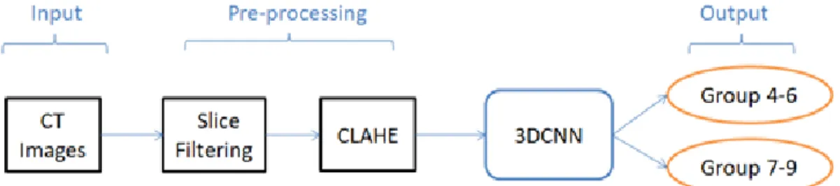
Alberta Stroke Program Early Computed Tomographic Scoring (ASPECTS) is a 10-point CT-scan score designed to quantify early ischemic changes in patients with acute ischemic stroke. However, an assessment of ASPECTS remains a challenge for neuroradiologists in stroke centers. The purpose of this study is to develop an automated ASPECTS scoring system that provides decision-making support by utilizing binary classification with three-dimensional convolutional neural network to analyze CT images. The proposed method consists of three main steps: slice filtering, contrast enhancement and image classification. The experiments show that the obtained results are very promising.

Key words: Deep Learning, Three-dimensional Convolutional Neural Network, CT Scans, ASPECTS, Ischemic Stroke.

※ Corresponding Author : Hyung Jeong Yang, Ilwoo Park, Address: (61186) Yongbong-ro 77, Buk-gu, Gwangju, Korea, (61469) Jebongro 42, Dong-gu, Gwangju, Korea, TEL : +82-62-530-3436, +82-62-220-5744, FAX : +82- 62-530-3439, E-mail : [email protected], ipark@jnu.

ac.kr

(+: Equal contribution)

Receipt date : Mar. 8, 2019, Revision date : Apr. 26, 2019 Approval date : May 8, 2019

†††

Dept of Electronics and Computer Engineering, Chon- nam National University, Gwangju, South Korea (E-mail : [email protected])

†††

Department of Radiology, Chonnam National Univer- sity Hospital, Gwangju, South Korea

(E-mail : [email protected])

†††

Department of Radiology, Chonnam National Univer- sity Hwasun Hospital, Hwasun, South Korea (E-mail : [email protected])

†††††††

Chonnam University Research Institute of Medical Sciences, Chonnam National University, Gwangju, South Korea (E-mail : [email protected])

†††††††

Department of Radiology, Chonnam National University Medical School and Hospital, Gwangju, South Korea (E-mail : [email protected])

†††††††

Department of Radiology, Chonnam National University Medical School and Hospital, Gwangju, South Korea

††††††

Dept of Electronics and Computer Engineering, Chonnam National University, Gwangju, South Korea

※This work was supported by a National Research Foundation of Korea (NRF) grant funded by the Korean government (MSIP)(NRF-2017R1A2B4011409 and No.

2017R1C1B5018396) and grants from Chonnam National

University (Grant Number: 2018-3426) and Chonnam

National University Hospital Biomedical Research Institute

(CRI18019-1).

(2)

fication and segmentation [3]. These networks are composed of layers that can learn representations of data with multiple levels of abstractions. Deep learning approaches can save time and effort by extracting features by themselves since the fea- tures that compose these layers are learned from data and do not need to be designed by a human.

CNNs have shown great potential for medical ap- plications such as brain tumor segmentation [4], liver tumor segmentation [5], pancreas segmenta- tion [6] and in computer-aided diagnostic applica- tions [7]. Based on the outstanding performance of the deep learning approach, CNNs are ideal for an- alyzing CT data.

More recently, CNN deep learning techniques have been applied to lesion segmentation of acute ischemic stroke with diffusion-weighted imaging (DWI) [8]. In the previous study two CNN net- works were combined to develop their model. The first CNN network was an ensemble of two DeconvNets (EDD Net), and the second was a multi-scale convolutional label evaluation net (MUSCLE Net) that evaluated the results from the EDD Net to remove potential false positives. In re- cent years, evidences have been accumulating that automated ASPECTS methods based on machine learning are comparable to expert readings of ASPECTS [9, 10, 11].

In this study, we develop a system for automati- cally assessing ASPECTS based on deep learning by utilizing a binary classification with a 3DCNN on CT data. The proposed system uses a three-di- mensional CNN (3DCNN) [12] to extract in- formation and predict the ASPECTS of CT images.

The results showed 70% accuracy in quantifying ASPECTS.

The remainder of the paper is organized as the follows. In the second section, we present our pro- posed approach for the automatic assessment.

Comprehensive experiments on a CT dataset are used to validate the effectiveness of our system in Section 3. Finally, in section 4, the conclusion and

future works are presented.

2. THE PROPOSED METHOD

In this section, we present automatic assessment of ASPECTS using deep learning and data aug- mentation. ASPECTS is a 10-point scoring system for measuring the early ischemic changes in pa- tients with anterior circulation stroke, where “10”

is given to a patient with the least degree of cere- bral ischemia and “1” to the patient with the high- est degree of cerebral ischemia [2]. ASPECTS were divided into four groups; Groups 1 through 4 consists of patients with scores of 1–3, 4–6, 7–

9, and a score of 10, respectively. In our research, because the collected number of patients with ASPECTS of ≤ 3 or equal to 10 is limited, we fo- cus on two groups with scores of 4–6 and 7–9.

Fig. 1 shows an example of CT images from the two groups.

The CT data varied in terms of spatial resolution and the number of slices. The number of slices ranges from 18 to 39. Since original CT data con- tain a lot of information, it would be very useful if we could exclude the uninformative slices and use only the slices that are informative for ASPECTS. Only the CT slices containing an area with a middle cerebral artery and its major branch, which are informative for ASPECTS, are included, and the cranial and caudal sections of the brain that are non-informative for ASPECTS are removed.

Fig. 2 shows examples of informative and non-in- formative slices for assessment of ASPECTS.

(a) (b)

Fig. 1. (a) Group 4–6, (b) Group 7–9.

(3)

After down- or up-sampling the remaining data, each CT sample consist of 17 slices, each slice has size of "80 × 80 × 3", therefore CT sample have fi- nal resolution of "17 × 80 × 80 × 3".

Due to the drastic difference in image contrast between skull and soft tissue, that is brain, in head CT images, the contrast of brain is not optimally set. A contrast limited adaptive histogram equal- ization (CLAHE) [13] is used in this study to en- hance the contrast within the soft tissues of the brain CT images. Fig. 3 shows an example of a

CT image after CLAHE is applied.

Each CT sample has a final resolution of 17 × 80 × 80 ×3, which is as a sequence of images. An augmentation step is applied to each slice of the CT sample. The data used for training is aug- mented by a three-degree rotation to the left and right as shown in Fig. 4.

In this study, we use a 3DCNN for classification of ASPECTS. The model consists of four blocks of convolution and pooling layers with two fully connected layers attached. These are followed by two dropout layers. The first block and the second block have one convolution layer with kernel size of 3 × 3 × 3. The third block and fourth block have two convolution layers with kernel size of 3 × 3 × 3 and 2 × 2 × 2, respectively. The number of feature maps in each convolution block is 32, 64, 128, 256, respectively.

We train 3DCNN after the preprocessing and augmentation steps. The input of the model is a CT images with size of 17 × 80 × 80 × 3 and the

(a) (b) (c)

Fig. 2. (a) Informative slice, and (b)–(c) Non-informative slices.

(a) (b)

Fig. 3. (a) Original Image, (b) Image after CLAHE is applied.

(a) (b) (c)

Fig. 4. (a) Preprocessed Image, (b) Rotation Right, (c) Rotation Left.

(4)

output of the model is one of two groups with scores of 4–6 and 7–9 in which a CT images be- long to. Fig. 5 shows the process of the proposed method. Our model uses Adam optimizer with a constant learning rate of 10

-6

and a batch size of 16.

3. EXPERIMENTAL RESULTS

267 brain CT datasets corresponding to the same number of patients were collected from Chonnam National University Hospital. It consists of 95 and 172 datasets for two groups with scores of 4–6 and 7–9, respectively. The datasets are divided in- to training (75%) and testing (25%). After aug- mentation, the total number of the training dataset is 597. Table 1 provides details on the amount of data utilized in each group. The results are eval- uated by accuracy and area under the curve (AUC) from a receiver operating characteristic curve (ROC).

We evaluated the effects of preprocessing and augmentation on the performance of our model.

The performance was improved when using slice filtering and CLAHE for preprocessing. The pro- posed model obtains better generalization by ap- plying data augmentation to acquire much varia- tion of data. Table 2 shows the effect of pre-

processing and augmentation on our 3DCNN model.

We also evaluated the proposed model with oth- er pre-trained models such as VGG16 [14], In- ceptionV3 [15]. During the training, the weight of pre-trained models was frozen, only the weight of either long short term memory (LSTM) layer or multi-layer perceptron (MLP) was adjusted. The lower accuracy of pre-train models shows that the pre-train models failed to capture the appropriate features of CT images. We also compared the pro- posed model to the long-term recurrent convolu- tional network (LRCN) model [16], which was op- timized for fully sequential data. The LRCN failed to capture the correct features of 3D data, resulting in a lower performance compared to the 3DCNN models. The results of testing are shown in Table 3.

Table 1. Datasets for both training and testing Group

4–6 Group

7–9 Total

Training (with

augmentation) 213 484 597

Testing 24 44 68

Table 2. Preprocessing and Data Augmentation Preprocessing Data

Augmentation Accuracy

No No 64.71%

Slice Filtering,

CLAHE No 69.12%

Slice Filtering,

CLAHE Yes 70.59%

Table 3. Performances of various CNN structures

Model Accuracy AUC score

VGG16+MLP 66.18% 0.567

InceptionV3+LSTM 64.71% 0.612

LRCN 67.65% 0.640

3DCNN (proposed) 70.59% 0.670

Fig. 5. Block diagram of the proposed method.

(5)

4. CONCLUSION

This study classified CT images into two cate- gories of ASPECT by applying a deep learning neural network, 3DCNN. We proposed a model for automatic assessment of ASPECTS by utilizing binary classification with a 3DCNN using CT data and data augmentation. We applied slice filtering to filter out non-informative slices, and the quality of CT images was enhanced using CLAHE. The augmentation was also applied to improve the training efficiency. Our goal is to extend this meth- od to improve the accuracy of automatic ASPECTS assessment system with a combination of im- proved preprocessing steps and fusion model. In the future study, we will extend our method to oth- er areas of brain imaging modalities.

REFERENCE

[ 1 ] R. Feng, M. Badgeley, J.D. Mocco, and E.K.

Oermann, “Deep Learning Guided Stroke Management: A Review of Clinical Applica- tions,” J ournal of NeuroInterventional Surg- ery, Vol. 10, No. 4, pp. 358-362, 2018.

[ 2 ] J.H. Pexman, P.A. Barber, M.D. Hill, R.J.

Sevick, A.M. Demchuk, M.E. Hudon, et al.,

“Use of the Alberta Stroke Program Early CT Score (ASPECTS) for Assessing CT Scans in Patients with Acute Stroke,” American Journal of Neuroradiology, Vol. 22, No. 8, pp.

1534-1542, 2001.

[ 3 ] S.W. Park and D.Y. Kim, “Comparison of Image Classification Performance in Convolu- tional Neural Network according to Transfer Learning,” Journal of Korea Multimedia Society, Vol. 21, No. 12, pp. 1387-1395, 2018.

[ 4 ] M. Havaei, A. Davy, D. Warde-Farley, A.

Biard, A. Courville, Y. Bengio, et al., “Brain Tumor Segmentation with Deep Neural Net- works,” Medical Image Analysis, Vol. 35, No.

1, pp. 18-31, 2017.

[ 5 ] W. Li, F. Jia, and Q. Hu, “Automatic Segmen-

tation of Liver Tumor in CT Images with Deep Convolutional Neural Networks,” J ournal of Computer and Communications, Vol. 3, No.

11, pp. 146-151, 2015.

[ 6 ] H.R. Roth, A. Farag, L. Lu, E.B. Turkbey, and R.M. Summers, “Deep Convolutional Net- works for Pancreas Segmentation in CT Imaging,” Proceedings of Society of Photo- graphic Instrumentation Engineers 9413, Medical Imaging 2015: Image Processing, pp. 94131G, 2015.

[ 7 ] H.C. Shin, H.R. Roth, M. Gao, L. Lu, Z. Xu, I. Nogues, et al., “Deep Convolutional Neural Networks for Computer-Aided Detection:

CNN Architectures, Dataset Characteristics and Transfer Learning,” IEEE Transactions on Medical Imaging, Vol. 35, No. 5, pp. 1285- 1298, 2016.

[ 8 ] L. Chen, P. Bentley, and D. Rueckert, “Fully Automatic Acute Ischemic Lesion Segmenta- tion in DWI Using Convolutional Neural Networks,” NeuroImage: Clinical, Vol. 15, No. 1, pp. 633-643, 2017.

[ 9 ] C. Herweh, P.A. Ringleb, G. Rauch, S. Gerry, L. Behrens, M. Mohlenbruch, et al., “Perform- ance of e-ASPECTS Software in Comparison to that of Stroke Physicians on Assessing CT Scans of Acute Ischemic Stroke Patients,”

International J ournal of Stroke, Vol. 11, No.

4, pp. 438-445, 2016.

[10] S. Nagel, D. Sinha, D. Day, W. Reith, R.

Chapot, P. Papanagiotou, et al., “e-ASPECTS Software is Non-inferior to Neuroradiologists in Applying the ASPECT Score to Computed Tomography Scans of Acute Ischemic Stroke Patients,” International J ournal of Stroke, Vol. 12, No. 6, pp. 615-622, 2017.

[11] J. Hampton-Till, M. Harrison, A.L. Kuhn, O.

Anderson, D. Sinha, S. Tysoe, et al., “Auto-

mated Quantification of Stroke Damage on

Brain Computed Tomography Scans: e-

ASPECTS,” European Medical Journal Neu-

(6)

rology, Vol. 3, No. 1, pp. 69-74, 2015.

[12] D. Tran, L. Bourdev, R. Fergus, L. Torresani, and M. Paluri, “Learning Spatiotemporal Features with 3D Convolutional Networks,”

P roceedings of the 2015 IEEE International Conference on Computer Vision, pp. 4489- 4497, 2015.

[13] S.M. Pizer, E.P. Amburn, J.D. Austin, R. Cro- martie, A. Geselowitz, T. Greer, et al., “Adap- tive Histogram Equalization and Its Varia- tions,” Computer Vision, Graphics, and Image Processing, Vol. 39, No. 3, pp. 355-368, 1987.

[14] K. Simonyan and A. Zisserman, Very Deep Convolutional Networks for Large-Scale Image Recognition, https://arxiv.org/abs/1409.1556

(accessed Jan., 10, 2019).

[15] C. Szegedy, V. Vanhoucke, S. Ioffe, J. Shlens, and Z. Wojna, “Rethinking the Inception Architecture for Computer Vision,” Proceed- ings of the 2016 IEEE Conference on Compu- ter Vision and Pattern Recognition, pp. 2818- 2826, 2016.

[16] J. Donahue, L.A. Hendricks, M. Rohrbach, S.

Venugopalan, S. Guadarrama, K. Saenko, et

al., “Long-term Recurrent Convolutional Net-

works for Visual Recognition and Descrip-

tion,” Proceedings of the 2015 IEEE Confer-

ence on Computer Vision and Pattern Re-

cognition, pp. 2625-2634, 2015.

(7)

Trinh Le Ba Khanh

He received the B.S from Ho Chi Minh City University of Tech- nology, Vietnam National Uni- versity, Ho Chi Minh City, Viet- nam in 2018. He is currently a M.S student at Dept. of Elec- tronics and Computer Engineer- ing, Chonnam National University, South Korea. His main research interests include Computer Vision and Deep Learning

Byung Hyun Baek

He received his B.S., M.S. and Ph. D. degrees from Chonnam National University, Korea. He is currently a Clinical Assistant Professor in Deparment of Radiology at Chonnam National University Hospital. His re- search interests include acute stroke, neuro- intervention, and neuroradiology.

Seul Kee Kim

He received his B.S., M.S. and Ph. D from Chonnam National University, Korea. He is cur- rently an associate professor at department of radiology, Chon- nam National University Medical School, Korea. His main re- search interests include brain tumor, ischemic stroke, and artificial intelligence.

Luu-Ngoc Do

He received the B.S, M.S and Ph.D from Chonnam National University, South Korea. He is currently an academic research professor at Medical Science In- stitute, Chonnam National Uni- versity, South Korea. His main research interests include Image Processing, Deep Learning, Machine Learning and Bioinformatics.

Woong Yoon

He received his B.S., M.S. and Ph. D. degrees from Chonnam National University, Korea. He has been a Professor in the Department of Radiology at Chonnam National University Hospital. His research interests include acute stroke, neurointervention, and neuroradiology.

llwoo Park

He received the B.S. and Ph.D.

degrees in Bioengineering from University of California Berke- ley, USA in 2004 and 2010, respectively. He worked in the Department of Radiology and Biomedical Imaging at Univer- sity of California San Francisco, USA from 2011 to 2016 as a postdoctoral scholar and an assistant pro- fessional researcher. Since 2017, he has been a pro- fessor in the Department of Radiology at Chonnam National University, College of Medicine. His research interests include advanced magnetic resonance imag- ing technologies and deep learning applications in medical imaging.

Hyung-Jeong Yang

She received her B.S., M.S. and

Ph.D. from Chonbuk National

University, Korea. in 1991, 1993,

and 1998. She worked in the

Department of Computer Sci-

ence at Carnegie Mellon Uni-

versity, USA as a special re-

searcher from 2003 to 2005. She joined Chonnam

Naitonal University, South Korea as a professor. Her

main research interests include multimedia data min-

ing, pattern recognition, artificial intelligence, e-

Learning, and e-Design.

수치

Fig. 1 shows an example of CT images from the two groups.
Fig. 4. (a) Preprocessed Image, (b) Rotation Right, (c) Rotation Left.
Table 1. Datasets for both training and testing Group 4–6 Group7–9 Total Training (with augmentation) 213 484 597 Testing 24 44 68

참조

관련 문서

*1st Author, Department of International Trade and Business, Kangwon National University, South Korea. ** Coauthor, Department of International Trade and Business,

Department of Naval Architecture and Ocean Engineering, Seoul National University.. Naval Architecture

Department of Naval Architecture and Ocean Engineering, Seoul National University of College

Department of Naval Architecture and Ocean Engineering, Seoul National University of College of

Department of Nuclear Engineering

Department of Mechanical and Aerospace Engineering Seoul National

When users download a web page, transfer a file, send/read email, or involve the network in almost any interaction, they want their transaction to complete in the shortest

Department of Naval Architecture and Ocean Engineering, Seoul National University of College