Molecular Pathology of Lung Cancer: Current Status and Future Directions
Mee Sook Roh, M.D., Ph.D.
Department of Pathology, Dong-A University College of Medicine, Busan, Korea
The rapid development of targeted therapies has enormously changed the clinical management of lung cancer patients over the past decade; therefore, molecular testing, such as epidermal growth factor receptor (EGFR) gene mutations or anaplastic lymphoma kinase (ALK) gene rearrangements, is now routinely used to predict the therapeutic responses in lung cancer patients. Moreover, as technology and knowledge supporting molecular testing is rapidly evolving, the landscape of targetable genomic alterations in lung cancer is expanding as well. This article will summarize the current state of the most commonly altered and most clinically relevant genes in lung cancer along with a brief review of potential future developments in molecular testing of lung cancer.
Keywords: Lung Neoplasms; Biological Markers; Molecular Diagnostic Techniques
factor receptor (EGFR) gene mutations or anaplastic lympho- ma kinase (ALK) gene rearrangements has been associated with dramatic response rates and improved progression-free survival (PFS)3-7, therefore, molecular testing is now routinely used to guide clinical care of lung cancer patients to predict one’s therapeutic response1.
Moreover, as the technology and knowledge supporting mo- lecular testing is rapidly evolving, the landscape of targetable genomic alterations in lung cancer is expanding2,8-11. However, the challenge of identifying biologically relevant driver muta- tions from the vast majority of passenger mutations remains.
To date, introduction of next-generation sequencing (NGS) technology offers the ability to detect high-throughput, mul- tiple genetic alterations in both constitutional and cancer genomes12. Such modern, but costly, technologies have not only contributed to our understanding of lung cancer biology, but have also provided the impetus for technical advances that may improve our ability to accurately discover the cancer genome13,14.
This article will summarize the current state of the most commonly altered and most clinically relevant genes in lung cancer and potential future developments in molecular test- ing of lung cancer will be briefly reviewed.
Copyright © 2014
The Korean Academy of Tuberculosis and Respiratory Diseases.
All rights reserved.
Introduction
Cancer is a disease of the cellular genome, and therefore lung cancers are understandably characterized by abundant genetic diversity. Information about genetic alterations and protein expression level is considered alongside histology in order to better comprehend the pathogenesis of lung cancer1. Recent advances in the field of molecular-based cancer biol- ogy revealed that approximately 60% of adenocarcinomas and 20% of squamous cell carcinomas have an identified gene signature, and a finding that has led to the successful develop- ment and approval of targeted therapies2. The use of targeted agents in lung cancer patients harboring epidermal growth
Address for correspondence: Mee Sook Roh, M.D., Ph.D.
Department of Pathology, Dong-A University College of Medicine, 32 Daesingongwon-ro, Seo-gu, Busan 602-714, Korea
Phone: 82-51-240-2833, Fax: 82-51-243-7396 E-mail: [email protected]
Received: Jun. 21, 2014 Revised: Jun. 27, 2014 Accepted: Jul. 4, 2014
cc It is identical to the Creative Commons Attribution Non-Commercial License (http://creativecommons.org/licenses/by-nc/3.0/).
Current Status
1. Novel molecular targets
Although EGFR mutations and ALK fusions are well- characterized molecular targets in non-small cell lung cancer (NSCLC), activating alterations in a variety of potential on- cogeneic driver genes have also been identified in NSCLC.
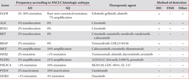
Table 1 summarizes a variety of genes involved in targeted treatment of NSCLC along with their respective histologic subtypes and the method of detection. However, it is impor- tant to recognize that not all potentially targetable biomarkers have been standardized outside of clinical trials. A deeper understanding of the molecular alteration of lung cancer may ultimately lead to personalized treatment strategies, which will improve care for those patients most likely to benefit, and spare the cost and morbidity associated with failed treatment interventions.
1) EGFR: EGFR encodes a transmembrane tyrosine kinase with an extracellular ligand-binding domain and an intracel- lular tyrosine kinase domain15. Activating somatic mutations are present in exons 18−21 of the tyrosine-kinase domain and deletions in exon 19 and the L858R point mutation in exon 21 occur in 90% of all EGFR mutations. They are associated with a response rate of approximately 70% to EGFR-tyrosine kinase inhibitor (TKI) therapy16. EGFR mutations are primarily seen in adenocarcinoma. Therefore, EGFR mutation analysis is the best predictive marker for the use of EGFR-TKI therapy in NSCLC with an adenocarcinoma component, but gender, ethnicity, and smoking status are unsuitable for use as triage
for mutation analysis. The most common mechanism of resis- tance to EGFR-TKI is the T790M gatekeeper mutation, caused by a single-base substitution of C to T, at nucleotide 236917.
2) ALK: The rearrangement results from a short inversion in chromosome 2p, whereby ALK signaling is activated by the creation of oncogenic fusions of the intron 10 of ALK gene within an upstream partner intron 13 of echinoderm micro- tubule associated protein-like 4 (EML4)6. More recently, less than 1% of ALK rearrangements cases have different partner genes including kinesin family member 5B (KIF5B), TFG, and KLC-111,18. ALK rearrangements occur in approximately 4% of lung adenocarcinoma patients, usually young, non-smokers with clinically advanced disease6-8. The United States Food and Drug Administration approved the ALK Break Apart fluorescence in situ hybridization (FISH) Probe Kit (Abbott Molecular, Des Plaines, IL, USA) as a companion diagnostic for targeted therapy with crizotinib in lung cancers1.
3) ROS1: ROS1 is a proto-oncogene located on chromo- some 6q22 that encodes a transmembrane tyrosine kinase receptor of the insulin receptor family19. ROS1 gene rear- rangements are known oncogenic drivers in NSCLC, and several fusion partners have been identified, including CD74, SLC34A2/NaPi2b, and FIG9,19. ROS1 fusions are present in about 2% of NSCLC cases and are often seen in young, non- smokers with adenocarcinoma, a population similar to those with ALK-rearranged NSCLC9,19. There is in vitro and early clinical evidence that lung cancers with ROS1 rearrangements are sensitive to TKIs including crizotinib9,11,19.
4) RET: RET is a proto-oncogene located on chromosome
Table 1. A variety of genes involved in targeted treatment of non-small cell lung cancer along with their respective histologic subtypes and the method of detection2,8,9,11
Gene Frequency according to NSCLC histologic subtype
Therapeutic agent Method of detection
AD SQ IHC FISH Other
EGFR 10−40% mutation Rare non-canonical mutation, 7% amplification
Erlotinib, gefitinib, afatinib ± – +
ALK 4% translocation 0% Crizotinib + + +
ROS1 2% translocation 0% Crizotinib ± + –
RET 2% translocation 0% Crizotinib, sunatinib, sorafenib, vandetanib, cabozatinib
– + –
BRAF 2% mutation 0% Vemurafenib, GSK2118436 ± – +
MET 5% amplification 10% amplification Cabozantinib, crizotinib, rilotumumab + + – HER2 2% mutation <1% mutation Trastuzumab, afatinib, dacomitinib, neratinib – – + FGFR1 3% amplification 21% amplification AZD4547, brivanib, S49076, ponatinib – + –
PIK3CA <2% mutation 10% mutation BKM120, GDC-0941, XL-147 – – +
PTEN 2% inactivation 10% inactivation Vandetanib + – –
DDR2 <1% mutation 4% mutation Dasatinib – – +
NSCLC: non-small cell lung cancer; AD: adenocarcinoma; SQ: squamous cell carcinoma; IHC: immunohistochemistry; FISH: fluorescence in situ hybridization; Other: other molecular methods for the detection of genetic alteration including mutation and indels.
10q11.2 that encodes a receptor tyrosine kinase involved in neural crest development20. Translocations resulting in fu- sions with several partners have been reported in lung cancer, including multiple variants of KIF5B-RET (the most com- mon type), CCDC6-RET (PTC1), NCOA4-RET (PTC3), and TRIM33-RET2. RET fusions are known to occur in the young, non-smokers with adenocarcinoma, but no other driver mutations, and can be targeted with TKIs such as sunitinib, sorafenib, vandetanib, and cabozantinib2,9,11,20.
5) BRAF: BRAF encodes a serine/threonine protein kinase that is the downstream effector protein of KRAS and activates the mitogen-activated protein kinase (MAPK) signal trans- duction pathway involved in the regulation of cell prolifera- tion and survival11,21. In contrast to melanoma, about 50% of the mutations are non-V600E mutations such as L596R and G468A. It is unknown whether V600E BRAF mutations func- tion as driver mutations and other mutations as passenger mutations2. Non-V600E mutations have been associated with current or former smokers, while V600E mutations appear to be more common in female, non-smokers11,21. BRAF-mutated NSCLC cases have been reported to respond to vermu- rafinib2,11.
6) MET: MET is a proto-oncogene located on chromosome 7q21−q31 that encodes a transmembrane tyrosine kinase receptor for its ligand hepatocyte growth factor (HGF)22. Mu- tations in MET are rare, but a high MET gene copy number has been detected in 1−11% of NSCLC cases. High gene copy numbers are more common in squamous cell carcinoma than adenocarcinoma and are often associated with high MET protein expression and poor prognosis9,11. MET ampli- fication that drives and maintains the phosphatidylinositol 3-kinases (PI3K)/AKT pathway, bypassing EGFR blockade by TKIs, has emerged as one of the critical events for secondary resistance to EGFR-TKIs in patients with EGFR-mutated lung adenocarcinoma23. A number of therapeutic agents targeting the MET/HGF pathway are in clinical development, including cabozantinib, crizotinib, and rilotumumab (antagonistc anti- bodies against HGF)9.
7) HER2: HER2 is member of the HER (EGFR) family of ty- rosine kinase receptors. HER2 mutations are detected mainly in exon 20 in approximately 1−2% of NSCLC cases, predomi- nantly in adenocarcinomas in non-smoking women11. HER2- mutated adenocarcinoma cases have been reported to re- spond to trastuzumab and afatinib24.
8) FGFR1: FGFR1 is a membrane-bound tyrosine-kinase receptor involved in the regulation of cell proliferation and angiogenesis through activation of the MAPK and PI3K path- ways9,25. FGFR1 amplification occurs more frequently in squa- mous cell carcinoma (21%) than in adenocarcinoma (3%)9,25. The novel FGFR inhibitor ponatinib may be effective in pa- tients with FGFR1 overexpression and the results of ongoing trials are pending25.
9) PIK3CA: PI3K protein family are intracellular lipid ki-
nases and the main catalytic subunit, the p110α isoform, is en- coded by the PIK3CA9. PIK3CA mutations mostly involve the catalytic domain and have been identified in approximately 1−3% of NSCLCs, particularly in squamous cell carcinomas9,26. Trials with PI3K inhibitors in combination with chemotherapy and other targeted agents are ongoing.
10) PTEN: PTEN encodes a lipid and protein phosphatase on chromosome 10 that plays a significant role in cell cycle progression, apoptosis, growth, proliferation, and migration via negative control of the PI3K/AKT pathway27. PTEN muta- tions and loss of PTEN protein expression are relatively com- mon in squamous cell carcinoma9. The TKI vandetanib has shown efficacy against EGFR mutation-positive lung cancer cell lines showing a loss of PTEN28.
11) Discoidin domain receptor 2 (DDR2): DDR2 en- codes a membrane-bound receptor tyrosine kinase that binds collagen and is involved in the regulation of cell proliferation and survival2,29. Mutations of DDR2 gene occur in 3.8% of squamous cell carcinomas and are associated with oncogenic activity that may respond to dasatinib29.
2. Present strategy for molecular testing
The molecular targets as described above now define the characteristics of NSCLC. However, EGFR mutation testing and ALK rearrangement status are the only two molecular makers considered the standard-of-care of NSCLC treatment in daily practice currently. The development of new therapeu- tic agents has led accurate histologic subtyping and molecular predictive testing to become mandatory in NSCLC cases.
1) Histologic subtyping: Histologic subtyping is still im- portant in triaging tumor samples for appropriate molecular testing, as pathologists become more important in the man- agement of patients with lung cancer. It has become clinically relevant to distinguish between adenocarcinoma and squa- mous cell carcinoma, as the chemotherapy regimen differs for these histologic subtypes1. Because minimally invasive procedures must be employed to obtain diagnostic material in more than 85% of NSCLC cases, an accurate but tissue- sparing approach from the limited cytology or small biopsy specimen is necessary1. Pathologists should try to classify further poorly differentiated NSCLC with the application of a panel of immunohistochemical markers, such as thyroid tran- scription factor-1 and napsin A to identify an adenocarcinoma component, and p63/p40 to identify a squamous cell carci- noma component30.
2) Molecular guidelines for testing of EGFR and ALK:
Several guidelines for molecular testing have been published in the past couple of years1,31,32. Recently, the College of Ameri- can Pathologists/International Association for the Study of Lung Cancer/Association for Molecular Pathology guidelines provide the first standardized evidence-based approach for performing molecular testing to on select patients with lung
cancer for EGFR or ALK TKI therapy that is multidisciplinary and multicontinental in scope1. The details have been de- scribed elsewhere. Briefly, adenocarcinoma is a basis for con- ducting molecular testing. Specimens from either primary or metastatic lesions are equally suitable for testing. For limited specimens, EGFR testing should be prioritized first and ALK testing should be prioritized second over other molecular markers. The guidelines encourage EGFR and ALK testing of biopsies at the time of diagnosis for patients presenting with early stage lung cancer. Alternatively, if testing is not per- formed in these early stage cancers, the guidelines encourage the retaining of cancer tissue for future biomarker testing. Mu- tation analysis, using a validated method with sufficient per- formance characteristics, such as direct sequencing, pyrose- quencing, and peptide nucleic acid polymerase chain reaction (PCR) clamping, is recommended for EGFR mutation testing.
A wide range of sample types, including cytology specimens and fixatives (formalin-fixed paraffin-embedded [FFPE], fresh, frozen, and alcohol) are allowable for EGFR mutation testing.
FISH assays using dual-labeled break-apart probes are recom- mended for ALK gene rearrangement testing1.
Future Directions
1. Future development of molecular testing
Advances in genomic technology such as NGS and mul- tiplex PCR assays have now made it possible to analyze the genomic landscape of lung cancer tissues comprehensively.
A detailed understanding of the complex genetic pathways responsible for the initiation and maintenance of malignant transformation may lead to the identification of vulnerabili- ties in cancers, and in turn may enable identification of novel therapeutic targets.
1) NGS: NGS offers simultaneous sequencing of thousands to millions of short nucleic acid sequences in a massively par- allel way12. These modern but costly technologies have been applied to whole genome sequencing and whole exome se- quencing for the discovery of mutations and polymorphisms, transcriptome sequencing for the quantification of gene expression, small ribonucleic acid sequencing for microRNA profiling, large-scale analysis of DNA methylation, and chro- matin immunoprecipitation mapping of DNA-protein interac- tion. NGS can detect chromosomal rearrangements and gene copy number alterations at a very high resolution33. It can be performed on FFPE and freshly collected tissue specimens and on small fine-needle aspiration biopsies.
Moreover, targeted NGS is beginning to be implemented in clinical laboratory practice. It is applied to detect individual mutations in cancer-related genes that may assist in cancer diagnosis, have prognostic value, or be used for prediction of response to targeted therapy8,13. Targeted NGS assays require
no prior knowledge of the mutations present in a patient’s tu- mor and are not limited to an evaluation of comparatively few
‘hot spots’ in which specific mutations are known to occur13. However, NGS is not clinically applicable as of today. The implementation of NGS technology in a clinical laboratory is complex and requires significant expertise in clinical, techni- cal, cost-benefit, and the bioinformatics aspects of sequenc- ing. Careful ethical consideration must also be given to the design of control arms in clinical trials of biomarker-selected patients8,9.
2) Multiplex PCR: Multiple PCRs that evaluates known ‘hot spot’ mutations by multiplex PCR involve the simultaneous amplification of two or more cDNA/DNA targets in a single reaction vessel with uniquely labelled probes for each target34. A number of multiplex PCR-based assays area available, in- cluding SNaPshot (Applied BIosystems, Foster City, CA, USA) and Sequenome MassARRAY (Sequenom Inc., San Diego, CA, USA)9. Multiplex PCR has been used to differentiate me- tastases from synchronous primary tumors in patients with multiple lung masses, and has the advantage of needing only a small sample of tumor compared with conventional tests, but it is restricted to codons previously determined as mutation hotspots, and is unable to detect chromosomal rearrange- ments or determine gene copy number9.
2. The Cancer Genome Atlas (TCGA) Project
More recently, systematic cancer genomics projects have applied emerging technologies to the analysis of specific tumor types including the TCGA project35. TCGA’s principal aims are to generate, quality control, merge, analyze, and in- terpret molecular profiles at the DNA, RNA, protein, and epi- genetic levels for hundreds of clinical tumors from various tu- mor types and their subtypes35. By the end of 2015, the TCGA Network plans to have achieved the ambitious goal of analyz- ing the genomic, epigenomic, and gene expression profiles of more than 10,000 specimens from 25 different tumor types including lung cancer. TCGA has already identified both com- mon and unique mutation spectra and pathway activation in 183 lung adenocarcinomas36 and 178 squamous cell carcino- mas37, two major histologies in NSCLC. Exonic regions of the 183 adenocarcinoma cases contained 77,736 somatic variants corresponding to a median of 8.1 mutations/megabase (MB) and a mean of 11.9 mutations/MB (range, 0.04−117.4 muta- tions/MB)36, whereas a total of 48,690 non-silent mutations with a mean of 228 non-silent and 360 total exonic mutations per tumor, corresponding to a mean somatic mutation rate of 8.1 mutations/MB and media of 8.4/MB in squamous cell car- cinoma37. Those rates are higher than rates observed in other TCGA projects including acute myelogenous leukemia (0.56/
MB), breast carcinoma (1.0/MB), ovarian cancer (2.1/MB), glioblastoma multiforme (2.3/MB), and colorectal carcinoma (3.2/MB).
1) Adenocarcinoma: Statistical driver analysis yielded pre- viously reported and novel lung adenocarcinoma genes and these included lung adenocarcinoma genes with non-synony- mous mutation frequencies consistent with previous reports:
TP53 (50%), KRAS (27%), EGFR (17%), STK11 (15%), KEAP1 (12%), NF1 (11%), BRAF (8%), and SMAD4 (3%). In total, 25 genes were shown to have a statistically significant number of mutations in lung adenocarcinoma. Unexpectedly, U2AF1, RBM10, and ARID1A mutations were identified. One of the most significantly mutated genes in this lung adenocarcinoma cohort was U2AF1, which had non-synonymous mutations in 3% of cases. U2AF1 mutation may confer tumorigenic capa- bility independent of known proliferations-sustaining driver genes and patients with U2AF1 mutations had significantly reduced PFS. RBM10, encoding an RNA-binding protein, was mutated in 7% of cases and its mutations co-occurred with those in known lung adenocarcinoma oncogenes. ARID1A, encoding a key protein in the SW1/SNF chromatin-remodel- ing complex, was mutated in 8% of cases and showed a signifi- cant accumulation of nonsense substitutions and frameshift indels36.
2) Squamous cell carcinoma: Lung squamous cell car- cinoma is characterized by complex genomic alterations. In total, 18 genes were shown to have a statistically significant number of mutations including mutation of TP53 in nearly all specimens. Significantly altered pathways by mutation or somatic copy number alterations included NFE2L2/KEAP1 in 34%, SOX2/TP63/NOTCH1 pathways in 44%, one of three components of the PI3K/AKT pathway (PIK3CA, PTEN or AKT3) in 47%, and CDKN2A (a known tumor suppressor gene that encodes the INK4A/p16 and ARF/p14 proteins)/
RB1 in 72% of cases, providing evidence of common dysfunc- tion in response to oxidative stress, squamous cell differentia- tion, apoptotic signaling, and/or cell cycle control, respec- tively. Previously unreported loss-of-function mutations were seen in the HLA-A class I major histocompatibility gene, sug- gesting a possible role for genotypic selection of patients for immunotherapies. Lung squamous cell carcinomas also share many alterations in common with head and neck squamous cell carcinomas without evidence of human papillomavirus infection, suggesting that the biology of these two diseases may be similar37.
Conclusion
In light of the myriad new biomarkers and targeted agents, multiplex testing strategies will be invaluable in identifying the appropriate patients for each therapy and enabling targeted agents to be channeled to the patients most likely to gain benefit. As further studies with sensitive multigene assays are completed, one might expect that the data will provide a key comprehensive means of identifying somatic alterations in
entire lung cancer genomes or exomes and will ultimately in- form clinical decision-making.
Conflicts of Interest
No potential conflict of interest relevant to this article was reported.
Acknowledgements
This work was supported by the National Research Founda- tion of Korea (NRF) grant funded by the Korea government (Ministry of Science, ICT & Future Planning) [grant number:
2013R1A1A3007362].
References
1. Lindeman NI, Cagle PT, Beasley MB, Chitale DA, Dacic S, Giaccone G, et al. Molecular testing guideline for selection of lung cancer patients for EGFR and ALK tyrosine kinase in- hibitors: guideline from the College of American Pathologists, International Association for the Study of Lung Cancer, and Association for Molecular Pathology. J Thorac Oncol 2013;8:
823-59.
2. Thunnissen E, van der Oord K, den Bakker M. Prognostic and predictive biomarkers in lung cancer. A review. Virchows Arch 2014;464:347-58.
3. Lynch TJ, Bell DW, Sordella R, Gurubhagavatula S, Okimoto RA, Brannigan BW, et al. Activating mutations in the epider- mal growth factor receptor underlying responsiveness of non- small-cell lung cancer to gefitinib. N Engl J Med 2004;350:
2129-39.
4. Mok TS, Wu YL, Thongprasert S, Yang CH, Chu DT, Saijo N, et al. Gefitinib or carboplatin-paclitaxel in pulmonary adenocar- cinoma. N Engl J Med 2009;361:947-57.
5. Sequist LV, Yang JC, Yamamoto N, O’Byrne K, Hirsh V, Mok T, et al. Phase III study of afatinib or cisplatin plus pemetrexed in patients with metastatic lung adenocarcinoma with EGFR mutations. J Clin Oncol 2013;31:3327-34.
6. Soda M, Choi YL, Enomoto M, Takada S, Yamashita Y, Ishi- kawa S, et al. Identification of the transforming EML4-ALK fu- sion gene in non-small-cell lung cancer. Nature 2007;448:561- 6.
7. Shaw AT, Kim DW, Nakagawa K, Seto T, Crino L, Ahn MJ, et al. Crizotinib versus chemotherapy in advanced ALK-positive lung cancer. N Engl J Med 2013;368:2385-94.
8. Dacic S, Nikiforova MN. Present and future molecular testing of lung carcinoma. Adv Anat Pathol 2014;21:94-9.
9. Shames DS, Wistuba II. The evolving genomic classification of lung cancer. J Pathol 2014;232:121-33.
10. Cagle PT, Allen TC, Olsen RJ. Lung cancer biomarkers: pres- ent status and future developments. Arch Pathol Lab Med 2013;137:1191-8.
11. Cooper WA, Lam DC, O’Toole SA, Minna JD. Molecular biol- ogy of lung cancer. J Thorac Dis 2013;5 Suppl 5:S479-90.
12. Bentley DR, Balasubramanian S, Swerdlow HP, Smith GP, Milton J, Brown CG, et al. Accurate whole human genome se- quencing using reversible terminator chemistry. Nature 2008;
456:53-9.
13. Summerer D. Enabling technologies of genomic-scale se- quence enrichment for targeted high-throughput sequencing.
Genomics 2009;94:363-8.
14. Glenn TC. Field guide to next-generation DNA sequencers.
Mol Ecol Resour 2011;11:759-69.
15. Prenzel N, Fischer OM, Streit S, Hart S, Ullrich A. The epider- mal growth factor receptor family as a central element for cellular signal transduction and diversification. Endocr Relat Cancer 2001;8:11-31.
16. Dearden S, Stevens J, Wu YL, Blowers D. Mutation incidence and coincidence in non small-cell lung cancer: meta-analyses by ethnicity and histology (mutMap). Ann Oncol 2013;24:
2371-6.
17. Pao W, Miller VA, Politi KA, Riely GJ, Somwar R, Zakowski MF, et al. Acquired resistance of lung adenocarcinomas to ge- fitinib or erlotinib is associated with a second mutation in the EGFR kinase domain. PLoS Med 2005;2:e73.
18. Takeuchi K, Choi YL, Togashi Y, Soda M, Hatano S, Inamura K, et al. KIF5B-ALK, a novel fusion oncokinase identified by an immunohistochemistry-based diagnostic system for ALK- positive lung cancer. Clin Cancer Res 2009;15:3143-9.
19. Bergethon K, Shaw AT, Ou SH, Katayama R, Lovly CM, Mc- Donald NT, et al. ROS1 rearrangements define a unique mo- lecular class of lung cancers. J Clin Oncol 2012;30:863-70.
20. Kohno T, Ichikawa H, Totoki Y, Yasuda K, Hiramoto M, Nam- mo T, et al. KIF5B-RET fusions in lung adenocarcinoma. Nat Med 2012;18:375-7.
21. Marchetti A, Felicioni L, Malatesta S, Grazia Sciarrotta M, Guetti L, Chella A, et al. Clinical features and outcome of pa- tients with non-small-cell lung cancer harboring BRAF muta- tions. J Clin Oncol 2011;29:3574-9.
22. Sadiq AA, Salgia R. MET as a possible target for non-small- cell lung cancer. J Clin Oncol 2013;31:1089-96.
23. Engelman JA, Zejnullahu K, Mitsudomi T, Song Y, Hyland C, Park JO, et al. MET amplification leads to gefitinib resistance in lung cancer by activating ERBB3 signaling. Science 2007;
316:1039-43.
24. Mazieres J, Peters S, Lepage B, Cortot AB, Barlesi F, Beau- Faller M, et al. Lung cancer that harbors an HER2 mutation:
epidemiologic characteristics and therapeutic perspectives. J
Clin Oncol 2013;31:1997-2003.
25. Dutt A, Ramos AH, Hammerman PS, Mermel C, Cho J, Sharif- nia T, et al. Inhibitor-sensitive FGFR1 amplification in human non-small cell lung cancer. PLoS One 2011;6:e20351.
26. Spoerke JM, O’Brien C, Huw L, Koeppen H, Fridlyand J, Brachmann RK, et al. Phosphoinositide 3-kinase (PI3K) path- way alterations are associated with histologic subtypes and are predictive of sensitivity to PI3K inhibitors in lung cancer preclinical models. Clin Cancer Res 2012;18:6771-83.
27. Abdulkareem IH, Blair M. Phosphatase and tensin homo- logue deleted on chromosome 10. Niger Med J 2013;54:79-86.
28. Takeda H, Takigawa N, Ohashi K, Minami D, Kataoka I, Ichi- hara E, et al. Vandetanib is effective in EGFR-mutant lung can- cer cells with PTEN deficiency. Exp Cell Res 2013;319:417-23.
29. Hammerman PS, Sos ML, Ramos AH, Xu C, Dutt A, Zhou W, et al. Mutations in the DDR2 kinase gene identify a novel therapeutic target in squamous cell lung cancer. Cancer Dis- cov 2011;1:78-89.
30. Travis WD, Brambilla E, Noguchi M, Nicholson AG, Geisinger KR, Yatabe Y, et al. International Association for the Study of Lung Cancer/American Thoracic Society/European Respira- tory Society international multidisciplinary classification of lung adenocarcinoma. J Thorac Oncol 2011;6:244-85.
31. Shim HS, Chung JH, Kim L, Chang S, Kim WS, Lee GK, et al.
Guideline recommendations for EGFR mutation testing in lung cancer: proposal of the Korean Cardiopulmonary Pa- thology Study Group. Korean J Pathol 2013;47:100-6.
32. Kim H, Shim HS, Kim L, Kim TJ, Kwon KY, Lee GK, et al.
Guideline recommendations for testing of ALK gene rear- rangement in lung cancer: a proposal of the Korean Cardio- pulmonary Pathology Study Group. Korean J Pathol 2014;48:
1-9.
33. Thomas A, Rajan A, Lopez-Chavez A, Wang Y, Giaccone G.
From targets to targeted therapies and molecular profiling in non-small cell lung carcinoma. Ann Oncol 2013;24:577-85.
34. Persson K, Hamby K, Ugozzoli LA. Four-color multiplex re- verse transcription polymerase chain reaction--overcoming its limitations. Anal Biochem 2005;344:33-42.
35. Cancer Genome Atlas Research Network, Weinstein JN, Collisson EA, Mills GB, Shaw KR, Ozenberger BA, et al. The Cancer Genome Atlas Pan-Cancer analysis project. Nat Gen- et 2013;45:1113-20.
36. Imielinski M, Berger AH, Hammerman PS, Hernandez B, Pugh TJ, Hodis E, et al. Mapping the hallmarks of lung adeno- carcinoma with massively parallel sequencing. Cell 2012;150:
1107-20.
37. Cancer Genome Atlas Research Network. Comprehensive genomic characterization of squamous cell lung cancers. Na- ture 2012;489:519-25.