Introduction
Inflammatory diseases of the respiratory tract including allergic rhinitis, asthma, and rhinosinusitis are among the most prevalence conditions to affect the general population
1,2. Many steroids including budesonide (BUD), fluticasone (FP), and mometasone fuorate (MF) have been used to treat rhi- nosinusitis
3and asthma
4. Excessive mucin secretion is a hall- mark of the pathogenesis of several airway diseases, including asthma and rhinosinusitis. Overexpression of MUC5AC and MUC5B has reported in chronic rhinosinusitis
5,6. In addition to an increase in MUC5AC and MUC5B, MUC2 overexpres- sion was described in asthma
7. Despite wide use of steroids, their role is still controversial in many pulmonary conditions, especially regarding to mucin secretion. BUD suppressed
Mometasone Furoate Suppresses PMA-
Induced MUC-5AC and MUC-2 Production in Human Airway Epithelial Cells
Orapan Poachanukoon, M.D.
1, Sittichai Koontongkaew, D.D.S., Ph.D.
2, Paopanga Monthanapisut, M.Sc.
2and Napaporn Pattanacharoenchai, B.Sc.
21
Department of Pediatrics, Faculty of Medicine, Thammasat University (Rangsit Center), Pathum Thani,
2Oral Biology Laboratory, Faculty of Dentistry, Thammasat University (Rangsit Center), Pathum Thani, Thailand
Background: Mucus hypersecretion from airway epithelium is a characteristic feature of airway inflammatory diseases.
Tumor necrosis factor α (TNF-α) regulates mucin synthesis. Glucocorticoids including mometasone fuorate (MF) have been used to attenuate airway inflammation. However, effects of MF on mucin production have not been reported.
Methods: Effects of MF and budesonide (BUD) on the phorbol-12-myristate-13-acetate (PMA)–induction of mucin and TNF-α in human airway epithelial cells (NCI-H292) were investigated in the present study. Confluent NCI-H292 cells were pretreated with PMA (200 nM) for 2 hours. Subsequently, the cells were stimulated with MF (1–500 ng/mL) or BUD (21.5 ng/mL) for 8 hours. Dexamethasone (1 μg/mL) was used as the positive control. Real-time polymerase chain reaction was used to determine MUC2 and MUC5AC mRNA levels. The level of total mucin, MUC2, MUC5AC, and TNF-α in culture supernatants were measured using enzyme-linked immunosorbent assay.
Results: MF and BUD significantly suppressed MUC2 and MUC5AC gene expression in PMA-stimulated NCI-H292 cells. The inhibitory effects of the two steroid drugs were also observed in the production of total mucin, MUC2 and MUC5AC proteins, and TNF-α.
Conclusion: Our findings demonstrated that MF and BUD attenuated mucin and TNF-α production in PMA-induced human airway epithelial cells.
Keywords: Mometasone Fuorate; Budesonide; Mucins; MUC2 Protein, Human; MUC5AC Protein, Human; Tumor Necrosis Factor-Alpha; Epithelial Cells
Copyright © 2017
The Korean Academy of Tuberculosis and Respiratory Diseases.
All rights reserved.
Address for correspondence: Orapan Poachanukoon, M.D.
Department of Pediatrics, Faculty of Medicine, Thammasat University (Rangsit Center), Praholyothin Road, Klongluang, Pathum Thani 12120, Thailand
Phone: 66-2-926-9669, Fax: 66-2-516-3771 E-mail: [email protected] Received: Oct. 5, 2015
Revised: Nov. 24, 2015 Accepted: Jun. 15, 2016
cc It is identical to the Creative Commons Attribution Non-Commercial License (http://creativecommons.org/licenses/by-nc/4.0/).
interleukin-1β up-regulated MUC2 and MUC5AC synthesis in human airway epithelial cells (NCI-H292)
8. FP repressed MUC5AC mRNA expression or MUC5AC protein production in NCI-H292 stimulated with transforming growth factor α
9. However, a previous study in chronic rhinosinusitis patients with nasal polyposis demonstrated that intranasal FP had no effect on MUC5AC gene and protein expression in nasal pol- yps
10. Although many articles show that MF exhibits the high potency compared to other steroid drugs, there appear to be no reports of the effects of MF on mucin production
11.
Tumor necrosis factor α (TNF-α) has been implicated in the pathophysiologic mechanisms of airway inflammatory diseases, including chronic rhinosinusitis
12and asthma
13. However, the effect of steroids on TNF-α is not clear. BUD sig- nificantly reduced lipopolysaccharide (LPS)-induced TNF-α production from lymphocytes
14. MF suppressed the release of TNF-α in nasal lavage of patients with allergic rhinitis
15. In contrast, TNF-α expression in nasal polyps of patients with chronic rhinosinusitis was unaffected by FP
10.
Therefore, the aim of our study was to investigate the effect of MF on mucin and TNF-α production in human airway epithelial cells, compared to that of BUD. NCI-H292 cells was used a model for studies of mucin synthesis. Phorbol- 12-myristate-13-acetate (PMA) was used as an inflammatory stimulant. Our findings show that MF and BUD can down- regulate MUC5AC and MUC2 gene induction and mucin pro- tein production by PMA. The inhibitory effect of MF and BUD on TNF-α production was also demonstrated in the study.
Materials and Methods
1. Chemicals and reagents
All chemicals and reagents used in the study were obtained from Sigma-Aldrich (St. Louis, MO, USA) unless otherwise specified. Mometasone fuorate monohydrate (Nasonex) was obtained from Merck Sharp & Dohme Corp. (Kenilworth, NJ, USA). The drug was dissolved in distilled water. BUD and dexamethasone (DEX) were dissolved in dimethyl sulfoxide (DMSO). The final concentration of DMSO in the medium was less than 0.1%.
2. Maximum non-toxic dose of MF and BUD
Cytotoxicity of MF and BUD was measured using the 3-(4,5-dimethylthiazol-2-yl)-2,5-diphenyltetrazolium bromide (MTT) colorimetric assay. NCI-H292 cells were maintained in RPMI 1640 medium supplemented with 10% fetal calf serum, penicillin (100 U/mL) and streptomycin (100 μg/mL) at 37
oC in a humidified atmosphere of 95% air and 5% CO
2. NCI-H292 cells were grown until they reached 80% confluence, they were then plated into 96-well culture plates at a density of
5,000 cells/well in a total volume of 100 μL and allowed to at- tach and grow for 24 hours. The supernatant in each well was then replaced with RPMI 1640 medium containing various concentrations of MF (0, 1, 10, 50, and 100 μg/mL) and BUD (0 μg/mL, 4.3 μg/mL [10 nM], 21.5 μg/mL [50 nM], 43 μg/
mL [100 nM], 86 μg/mL [200 nM], and 172 μg/mL [400 nM]).
After 72 hours of incubation, 100 μL of MTT (Sigma-Aldrich) was added to each well. After 4 hours incubation, the super- natant was removed and 150 μL DMSO (Sigma-Aldrich) was added to each well. Samples were then shaken for 15 minutes to dissolve the dark blue crystals. The absorbance of each well was read at 570 nm using an enzyme-linked immunosorbent assay (ELISA) microplate reader (Sunrise; Tecan, Salzburg, Austria). All experiments were performed in triplicate. Cell cytotoxicity was calculated as in Eq. (1).
Percent of growth=[(Ca–Ta)/Ca]×100 ··· (1) , where Ca is the absorbance of untreated cells (control) and Ta is the absorbance of treated cells.
3. Experimental design
NCI-H292 cells were maintained in the complete RPMI 1640 medium as mentioned before. When cultures were confluent, the cell were incubated with RPMI 1640 medium containing 0.5% fetal calf serum for 24 hours, after which they were rinsed with phosphate buffered saline (PBS) and exposed to the indicated concentrations of BUD or MF for 2 hours before exposed to PMA (200 nM) for 8 hours. In case of controls, the cells were incubated with medium alone for the same times. DEX has been reported to inhibit mucin expres- sion in NCI-H292 cells
9. Therefore, it was used as a positive control for inhibition.
Conditioned media were collected for the determination of total mucin, MUC2, MUC5AC, and TNF-α levels. Total RNA was extracted for measuring the mRNA expression of MUC2 and MUC5AC genes by real-time reverse transcription poly- merase chain reaction (RT-PCR).
4. Real-time quantitative RT-PCR
Total RNA was isolated from cells using the Trizol reagents
(Gibco-BRL, Rockville, MD, USA) according to the manufac-
turer’s instruction. cDNA was synthesized from 1 μg of RNA
using the Primer RT-PCR Premix (2×; Genet BIO, Daejeon,
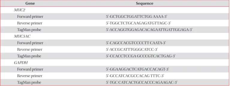
Korea). Real-time RT-PCR TagMan assays were used to deter-
mine MUC2 and MUC5AC expression. Sequences of primers
and TagMan probes (Table 1) were designed and used as
reported previously
16,17. Real-time PCR was performed using
an IQ5 Multicolor Real-time PCR Detection System (Bio-Rad
Laboratories, Hercules, CA, USA) with gene-specific primers
and FastStart Universal Probe Master (Rox; Roche Diagnos-
tic GmbH, Mannheim, Germany). RNA expression levels were calculated using the comparative cycle threshold (Ct) method
18. Quantification of relative mRNA expression levels was determined and normalized to a constitutive expression of glyceraldehyde 3-phosphate dehydrogenase mRNA.
5. Immunoassay of MUC2 and MUC5AC proteins MUC2 levels in conditioned media were measured by sandwich ELISA
19. Briefly, a monoclonal anti-human MUC2 (sc 59956; Santa Cruz Biotechnology, Santa Cruz, CA, USA) at 1:5,000 in a blocking buffer (1% gelatin in PBS-Tween) was in- cubated at 4
oC overnight in a 96-well plate. After washing with PBS-Tween, the plate was blocked with the blocking buffer at 37
oC for 1 hour. Following washing with PBS-Tween, samples were added to each well and the plate was incubated at 37
oC for 2 hours. After washing with PBS-Tween, the plate was incubated with horseradish peroxidase (HRP)–conjugated Helix pomatia (5 μg/mL in the blocking buffer) for 1 hour at 37
oC. Color was developed with tetramethybenzidine (TMB) solution, and the reaction was stopped by adding 0.5 M hydro- chloric acid. Absorbance was read at 450 nm.
MUC5AC protein was determined by ELISA
20. Each sample was incubated at 37
oC in a 96-well plate until dry. After wash- ing with PBS-Tween, the plate was blocked with a blocking solution (1% gelatin in PBS-Tween) at 37
oC for 1 hour. After incubation with monoclonal anti-human MUC5AC (clone 45M1; Neomarkers, Fremont, CA, USA) at 37
oC for 2 hours, the plate was washed and incubated with HRP-conjugated goat anti mouse antibody (Pierce, Rockford, IL, USA) at 37
oC for 2 hours. TMB solution was added to develop color. After the reaction is stopped, the absorbance of each well was read at 450 nm using an ELISA microplate reader (Sunrise;
Tecan). The expression of MUC2 and MUC5AC proteins was presented as absorbance values (arbitrary unit) since there are no commercial standards available for human MUC2 and MUC5AC at present.
6. Total mucin determination
An enzyme-linked lectin assay was used to determine the mucin-like glycoprotein secretion as previously described
19. Briefly, wells of a 96-well plate were coated with samples or standard (bovine submaxillary gland mucin, diluted in sodium carbonate buffer [0.05M, pH 9.6]; Sigma-Aldrich).
The plate was incubated overnight at 4
oC, washed with PBS containing 0.05% Tween 20 (PBS-Tween), and blocked with a blocking buffer (1% gelatin in PBS-Tween) for 2 hours at 37
oC.
After washing with PBS-Tween, the plate was incubated with 5 μg/mL HRP-conjugated Helix pomatia lectin in a blocking buffer for 2 hours at 37
oC. Following washing with PBS-Tween, the plate was incubated with the peroxidase substrate, TMB solution, at room temperature. The reaction was stopped after 10 minutes by the addition of 0.5 M hydrochloric acid. The absorbance of each well was read at 450 nm using an ELISA microplate reader (Sunrise; Tecan). The concentration of total mucin was calculated by comparison with standards.
7. TNF-α measurement
TNF-α levels in culture supernatant were determined by High Sensitive ELISA Kit (eBioscience, San Diego, CA, USA), according to the manufacturer’s recommendations.
Table 1. Primer and probe sequences used in real-time RT-PCR
Gene Sequence
MUC2
Forward primer 5'-GCTGGCTGGATTCTGG AAAA-3'
Reverse primer 5'-TGGCTCTGCAAGAGATGTTAGC-3'
TagMan probe 5'-ACCAGGTGGAGACACAGAATTGATTGGAGA-3'
MUC5AC
Forward primer 5'-CAGCCACGTCCCCTT CAATA-3'
Reverse primer 5'-ACCGCATTTGGGCATCC-3'
TagMan probe 5'-CCACCTCCGA GCCCGTCACTGAG-3'
GAPDH
Forward primer 5'-GGAAGGACTCATGACCACAGT-3'
Reverse primer 5'-GCCATCACGCCACAG TTTC-3'
TagMan probe 5'-TGC CATCACTGCCACCCAGAAGAC-3'
RT-PCR: reverse transcription polymerase chain reaction; GAPDH: glyceraldehyde 3-phosphate dehydrogenase.
8. Data analysis
Statistical analyses were conducted using GraphPad Prizm 5 (GraphPad Software, San Diego, CA, USA). Data were pre- sented as mean±standard error of the mean from three inde- pendent experiments. Statistical comparisons among groups were performed by one-way ANOVA, followed by Dunnett’s post hoc test. Significance was set to p≤0.05.
Results
1. Maximum non-toxic dose of MF and BUD
To test the potential cytotoxicity of MF and BUD at different concentrations, we used MTT assays to examine the viability of NCI-H292 cells and the results are shown in Figure 1. NCI- H292 cells were exposed to MF (0–100 μg/mL) or BUD (0–172 μg/mL) for 72 hours. MF (Figure 1A) had little effect on the viability of NCI-H292 cells when the concentration was 1 μg/
mL; however, when the concentration was higher than 1 μg/
mL, cell viability decreased with increased MF concentrations.
When the concentration was above 10 μg/mL, cell viability markedly decreased to less than 20%. There was no cytotoxic of BUD to NCI-H292 cells at concentrations up to 172 μg/mL (Figure 1B). The maximum non-toxic dose of MF and BUD provided the basis for the dose selection for further experi- ments in our study.
2. MF and BUD inhibit PMA-induced MUC2 and MUC5AC gene expression
PMA (200 nM) increased the expression of MUC2 and MU- C5AC gene expression in NCI-H292 cells (Figures 2, 3). This A
100
80
60
40
20
Cellviability(percentofgrowth)
0
Contro l
MF 1
g/m L
MF
10
g/mL MF
50
g/mL MF
100
g/mL
B
150
100
50
Cellviability(percentofgrowth)
0
0 4.3 21.5 43 86 172
BUD ( g/mL)
Figure 1. Effects of MF and BUD on cell viability. NCI-H292 cells were treated with MF (A) and BUD (B) at indicated concentra- tions for 72 hours. Cell viability was determined by the MTT as- say and results are presented as percent of the control (untreated group). Values are expressed as mean±SEM of three independent experiments. MF: mometasone fuorate; BUD: budesonide; MTT:
3-(4,5-dimethylthiazol-2-yl)-2,5-diphenyltetrazolium bromide;
SEM: standard error of the mean.
10
8
6
4
2 MUC2mRNA/GAPDHmRNA (foldovercontrol)
0
Contro l
PMA 200
nM
PMA+M F1ng/mL
PMA+MF 10
ng/mL
PMA+MF 50
ng/mL
PMA+MF 500
ng/mL
PMA+BUD 21.5
ng/mL
PMA+DEX 1
g/m L
*
*
*
* *
*
*
Figure 2. Glucocorticoids inhibit PMA-induced MUC2 gene expres- sion. NCI-H292 cells were pretreated with or without MF, BUD, or DEX at indicated concentrations for 2 hours. Subsequently, the cells were stimulated with PMA for 8 hours. Total RNA was isolated and MUC2 mRNA levels were analyzed by real-time polymerase chain reaction. Values are expressed as mean±SEM of three independent experiments. *p<0.05, compared to PMA-stimulated cells in the absence of glucocorticoids. PMA: phorbol-12-myristate-13-acetate;
MF: mometasone fuorate; BUD: budesonide; DEX: dexamethasone;
SEM: standard error of the mean; GAPDH: glyceraldehyde 3-phos-
phate dehydrogenase.
effect of PMA was significantly inhibited by pretreatment of cells with BUD (21.5 μg/mL). MF (1–500 ng/mL) exhibited inhibitory effects on the expression of MUC2 and MUC5AC genes (p<0.05).
3. MF and BUD inhibit MUC2 and MU5AC protein expression
To verify the effect of MF and BUD on mucin production, the effects of these steroids on MUC2 and MUC5AC protein expression were evaluated in NCI-H292 cells stimulated by PMA. We demonstrated that MF at 1–500 ng/mL suppressed MUC2 and MUC5AC induced by PMA in a dose-dependent manner (p<0.05) (Figures 4, 5). The stimulating effect of PMA was significantly inhibited with BUD at 21.5 μg/mL (p<0.05).
4. Inhibitory effects of MF and BUD on total mucin production
Treatment NCI-H292 cells with PMA stimulated total mu- cin production (Figure 6). This confirms that we can use PMA as an inflammatory stimulant in our model. BUD (21.5 μg/
mL) and MF (1–500 ng/mL) significantly inhibited the up- regulatory effect of PMA (p<0.05).
2.5
2.0
1.5
1.0
0.5
MUC5ACmRNA/GAPDHmRNA (foldovercontrol) 0
Contro l
PMA 200
nM
PMA+M F1ng/mL
PMA+MF 10
ng/mL
PMA+MF 50
ng/mL
PMA+MF 500
ng/mL
PMA+BUD 21.5
ng/mL
PMA+DEX 1
g/m L
*
*
*
*
* *
*
Figure 3. MF and BUD inhibit PMA-induced MUC5AC gene ex- pression. NCI-H292 cell were pretreated with or without MF, BUD, or DEX at indicated concentrations for 2 hours before stimulation with PMA for 8 hours. Total RNA was isolated and MUC5AC mRNA levels were analyzed by real-time polymerase chain reaction.
Values are expressed as mean±SEM of three independent experi- ments. *p<0.05, compared to PMA-stimulated cells in the absence of glucocorticoids. MF: mometasone fuorate; BUD: budesonide;
PMA: phorbol-12-myristate-13-acetate; DEX: dexamethasone;
SEM: standard error of the mean; GAPDH: glyceraldehyde 3-phos- phate dehydrogenase.
1.5
1.0
0.5
MUC2(foldovercontrol)
0
Contro l
PMA 200
nM
PMA+M F1ng/mL
PMA+MF 10
ng/mL
PMA+MF 50
ng/mL
PMA+MF 500
ng/mL
PMA+BUD 21.5
ng/mL
PMA+DEX 1
g/m L
*
*
*
*
* *
*
Figure 4. MF and BUD inhibit PMA-induced MUC2 synthesis. NCI- H292 cells were pretreated with indicated glucocorticoids for 2 hours.
Subsequently, the cells were stimulated with PMA for 8 hours. The level of MUC2 protein in culture supernatants was determined using ELISA. Data shown are mean±SEM of three independent experi- ments. *p<0.05, compared to PMA-stimulated cells in the absence of glucocorticoids. MF: mometasone fuorate; BUD: budesonide; PMA:
phorbol-12-myristate-13-acetate; ELISA: enzyme-linked immunosor- bent assay; SEM: standard error of the mean; DEX: dexamethasone.
4
3
2
1
MUC5AC(foldovercontrol)
0
Contro l
PMA 200
nM
PMA+M F1ng/mL
PMA+MF 10
ng/mL
PMA+MF 50
ng/mL
PMA+MF 500
ng/mL
PMA+BUD 21.5
ng/mL
PMA+DEX 1
g/m L
* *
* *
*
*
*
Figure 5. MF and BUD inhibit PMA-induced MUC5AC production.
NCI-H292 cells were pretreated with indicated glucocorticoids for 2 hours. Subsequently, the cells were stimulated with PMA for 8 hours. The level of MUC5AC protein in culture supernatants was determined using ELISA. Data shown are mean±SEM of three independent experiments. *p<0.05, compared to PMA-stimulated cells in the absence of glucocorticoids. MF: mometasone fuorate;
BUD: budesonide; PMA: phorbol-12-myristate-13-acetate; ELISA:
enzyme-linked immunosorbent assay; SEM: standard error of the
mean; DEX: dexamethasone.
5. Inhibitory effects of MF and BUD on TNF-α synthesis Figure 7 shows that MF at 1–500 ng/mL inhibited PMA- induced TNF-α production from NCI-H292 cells in a dose- response fashion (p<0.05). BUD at 21.5 μg/mL significantly suppressed the stimulating effect of PMA on TNF-α synthesis in NCI-H292 cells.
Discussion
Previous studies have revealed that normal human bron- chial epithelial (NHBE) and NCI-H292 cells share key com- ponents of the signaling pathways downstream of epidermal growth factor receptor, which are responsible for mucin pro- duction
21. Furthermore, it has been demonstrated that PMA could enhance MUC5AC mRNA and protein expression in both NHBE and NCI-H292 cells
22. Therefore, we assumed that NCI-H292 cells would provide a valid model of mucin produc- tion in normal cells, and thus, focused on investigating the effect of steroid drugs on mucin production using NCI-H292 cells.
Glucocorticoids act via the glucocorticoid receptor, which resides in the cytoplasm. MF exhibits a very high affinity for the glucocorticoid receptor, which is one explanation for the high potency compared to other steroid drugs
11. However, the
effect of this drug on mucin production is still unknown. To investigate this effect, we used PMA, a widely used stimulant known to increase mucin production in airway epithelial cells.
We show that PMA increase the expression of MUC2 and MUC5AC gene and protein expression in NCI-H292 cells. Our findings are in line with previous studies
19,23.
Here, we found that MF inhibited the stimulating effects of PMA on MUC2 and MUC5AC mRNA and protein expression including total mucin synthesis in NCI-H292 cells. To the best of our knowledge, there are no reports about the effect of MF on mucin gene and protein expression from airway epithelial cells. It was reported that mitogen-activated protein kinases and nuclear factor κB pathways play important roles in PMA- induced mucin activation
19,23. Although it is not known exactly how MF suppresses mucin synthesis in airway epithelial cells, it is probably that the suppression occurs somewhere in the pathways.
Previous reports demonstrated that following the dry-pow- der inhaler administration of MF (400 μg); the plasma concen- trations of MF were below the limit of quantification (50 pg/
mL) in most of subjects. The system bioavailability of this drug by this route was estimated to be less than 1%
24,25. However, MF is rapidly and extensively metabolized. Therefore, plasma concentration of MF may not correlate with its clinical effects because the large numbers of active metabolites were formed
800
600
400
Totalmucin(ng/mL) 200
0
Contro l
PMA 200
nM
PMA+M F1ng/mL
PMA+MF 10
ng/mL
PMA+MF 50
ng/mL
PMA+MF 500
ng/mL
PMA+BUD 21.5
ng/mL
PMA+DEX 1
g/m L
* *
* * * *
*
Figure 6. Inhibition of total mucin production by MF, BUD, and DEX. NCI-H292 cells were pretreated with the indicated glucocor- ticoids for 2 hours. The cells were stimulated with PMA and total mucin levels were quantitated by an enzyme-linked lectin assay.
Data shown are mean±SEM of three independent experiments.
*p<0.05, compared to PMA-stimulated cells in the absence of gluco- corticoids. MF: mometasone fuorate; BUD: budesonide; DEX: dexa- methasone; PMA: phorbol-12-myristate-13-acetate; SEM: standard error of the mean.
25
20
15
10
5
TNF-(pg/mL)
0
Contro l
PMA 200
nM
PMA+M F1ng/mL
PMA+MF 10
ng/mL
PMA+MF 50
ng/mL
PMA+MF 500
ng/mL
PMA+BUD 21.5
ng/mL
PMA+DEX 1
g/m L
* *
*
* *
*
*