슬개골에 발생한 관절 내 골연골종
Intra-Articular Osteochondroma of the Patella
김지환 • 남승오 • 변영수 • 조훈식 • 유현승
대구파티마병원 정형외과
골연골종은 원발성 골 종양 중 가장 빈도가 높은 양성 종양 중의 하나이다. 그러나 관절 내, 특히 슬관절에 생기는 골연골종은 매우 드물며, 슬개골과 골교를 형성하는 골연골종의 경우는 더욱 그러하다. 저자들은 58세 여자 환자의 슬개골에 발생한 관절 내 골연골종을 경험하였기 에 문헌 고찰과 함께 보고하는 바이다.
색인단어: 관절 내 골연골종, 슬개골, 슬관절
접수일 2012년 8월 13일 수정일 2012년 9월 18일 게재확정일 2012년 10월 4일
교신저자 김지환
대구시 동구 아양로 99, 대구파티마병원 정형외과 TEL 053-940-7320, FAX 053-954-7417 E-mail [email protected]
Copyright © 2012 by The Korean Orthopaedic Association
“This is an Open Access article distributed under the terms of the Creative Commons Attribution Non-Commercial License (http://creativecommons.org/licenses/by-nc/3.0/) which permits unrestricted non-commercial use, distribution, and reproduction in any medium, provided the original work is properly cited.”
대한정형외과학회지:제 47권 제 6호 2012
골연골종은 가장 흔한 양성 골 종양으로서 주로 장관골의 골간단 에서 호발한다.1,2) 일반적으로 전형적인 골연골종은 장관골의 말 단부위에서 기원하여 관절로부터 멀어지는 양상으로 관찰되는 경우가 많다.2) 관절내, 특히 슬관절에 발생한 경우는 매우 드물어 서 1958년 Jaffe3)가 6예의 슬관절 내 골연골종을 보고한 이래, 현 재까지 국내외에서 14예만이 추가로 보고되었다.4) 또한 슬개골과 골교를 형성하는 골연골종의 경우 국내에 1예만이 보고되어 매 우 희귀하다.5) 저자들은 본원 정형외과에서 좌 슬관절 동통을 주 소로 내원한 58세 여자 환자의 슬개골 하극부에 발생한 슬관절 내 골연골종 1예를 치험하였기에 문헌고찰과 함께 보고하는 바 이다.
증례보고
58세 여자 환자로 약 1년 전부터 간헐적으로 발생한 좌 슬관절 전 방부 통증을 주소로 내원하였다. 최근 몇 달 사이 그 빈도가 잦아 지는 양상이었고 내원 며칠 전 계단에서 무릎을 삐끗한 후 증상 이 더욱 악화되었다. 이학적 소견상 슬관절 전방부 압통을 동반 한 관절주위 삼출이 다소 있었으며, 90도 이상의 슬관절 굴곡 시
전방부 통증으로 인해 굴곡 제한을 보였다. 그러나 슬관절의 불 안정성이나 부종, 열감, 발적 등의 염증 소견은 보이지 않았다. 전 신 및 영양상태는 양호하였으며, 일반 혈액, 혈액 화학 및 요 검사 는 모두 정상이었다. 또한 과거력, 가족력 등에서도 특이 소견은 관찰되지 않았다.
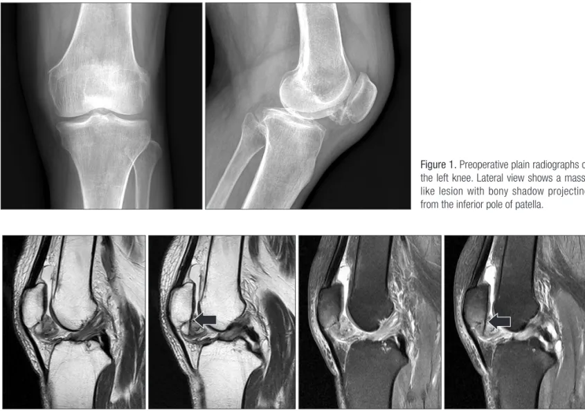
슬관절의 단순 방사선 소견상 전후면상에서 슬개골 하극부에 골 음영을 보이는 종양이 관찰되었다(Fig. 1). 전반적인 대퇴-경 골 상태는 깨끗하여 퇴행성 관절염에 의한 골극으로 보기는 힘 들 것으로 생각되었다. 자기공명영상 소견상 T1 강조영상에서 슬 개골 하극부에 약 3.5×2.5 cm 크기의 비균질성 종양이 슬개골하 지방대(infra-patellar fat pad)의 후상부에서 관찰되었다. 그 중심 부는 골 신호(bone signal)를 보였고, 주위 경계부는 중간 신호 강 도(intermediate signal intensity)를 보였다. T2 강조영상에서 역시 중심부는 골 신호를 보였으나, 주위 경계부는 고신호 강도(high signal intensity)를 보였다(Fig. 2). 중심부에 골을 형성하고 주위 경 계부에는 연골을 형성하는 양성종양인 골연골종 진단하에 수술 을 진행하였다.
슬개골의 전내측에서 종으로 피부를 절개하여 슬관절 내에 도 달하였으며, 수술 소견상 슬개건 후방에서 슬개골 하극부에 부착 된, 연골로 덮힌 골성 종양이 관찰되었다. 슬개골하 지방대, 활액 막, 슬개골 인대와의 유착은 전혀 없는 상태로, 슬개골 하극의 후 내측부와 종양은 골교(bony bridge)를 통해 서로 단단히 부착되어 고정되어 있었다. Chisel을 사용하여 골교를 제거하고 부착부위 에 일부 남은 부분을 Burr를 사용하여 깨끗이 제거하였다(Fig. 3).
절제한 종양은 3.2×2.0×2.0 cm 크기로, 불규칙성 결절상의 연골
슬개골에 발생한 관절 내 골연골종
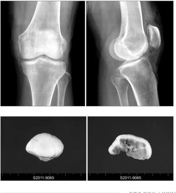
층이 있었고, 내부는 해면골의 소견을 보였다. 수술 후 방사선 소 견상 완전한 골 종양의 제거 소견을 관찰할 수 있었다(Fig. 4).
해부병리학적 소견상 타원형 폴립모양의 골 조직을 절단하여
회백색의 2 mm 직경의 연골막(cartilage cap)을 확인할 수 있었다 (Fig. 5).
현미경적 소견상 가장 바깥층은 섬유성 결체조직으로, 그 심층 Figure 1. Preoperative plain radiographs of the left knee. Lateral view shows a mass- like lesion with bony shadow projecting from the inferior pole of patella.
Figure 2. Magnetic resonance imaging of the lesion sagittal T1, T2-weighted images show cortical and cancellous bony connection between the inhomogenous mass with marginal cartilage shadow and the posteromedial corner of the inferior pole of patella directly (arrow).
Figure 3. In operative field, the mass was rigidly connected with the inferior pole of patella through the bony bridge. There was no connection with infra-patellar fat pad, synovium or patellar tendon. The bony mass was excised clearly, (A) before and (B) after. The cut surface of bony bridge at the posteromedial corner (multi-arrow).
에는 양성 연골 세포층으로, 중심에는 정상 해면골 조직으로 구 성되어 특징적인 골연골종 소견을 나타내었다(Fig. 6).
수술 후 약 2년 추시 결과, 일상 생활 및 보행 시 환측 슬관절의
동통은 완전히 소실되었고, 관절 운동 범위도 정상으로 회복되었 으며, 특별한 합병증도 없었다.
고 찰
골연골종은 골 종양의 약 10-20%, 골 양성 종양의 약 20-50%를 차지하는 가장 빈도가 높은 원발성 골종양이다. 이는 성장판이 위치하는 곳에서 과다 형성 혹은 이상 증식에 의해 발생하는, 신 생물과는 다른 개념의 병변으로서,1,6) 주로 대퇴골 원위부, 경골 근위부, 근위 상완골 등에 호발하고, 장관골의 골간단부에서 골성 융기를 보이며, 반투명의 연골모에 의해 덮여 있다.5) 골연골종은 연골에서 발생하는 모든 골조직에서 발생할 수 있지만 연부 조직 에서 발생하는 경우는 드물며, 관절 내 혹은 관절 주위에서 발생 된 예, 특히 슬관절 내의 슬개골에서 발생한 골연골종은 더욱 드 물다. 특히 저자들이 경험한 슬개골과 골교를 형성하는 골연골종 의 경우 1998년 Sohn과 Kim5)에 의해 국내에 1예만이 보고되었다.
Sohn과 Kim5)은 골연골종의 생성에서 활액막 이형성 또는 외상 등에 의해 형성된 골연골종이 지속적인 자극에 의해 발생한 미란 을 통해 이차적 골교를 형성했을 가능성을 언급하였다. 본 예에 Figure 4. Postoperative plain radiographs show that intra-articular osteochondroma was clearly removed from the inferior pole of patella.
Figure 5. The excised specimen. This consists of ovoid polypoid bony tissue, measuring 3.2×2×2 cm. Cut section shows grayish thickened cartilage, of which cap thickness is 2 mm.
Figure 6. The microscopic finding of the excised mass (H&E, ×20). The typical findings of osteochondroma are seen.
슬개골에 발생한 관절 내 골연골종
서는 자기공명영상 소견상 T1, T2 강조영상 모두에서 슬개골 하 극의 후내측부에서 피질골, 해면골의 신호가 골연골종의 중심부 까지 연결되어 있고, 수술 후 제거한 골연골종의 골교 단면에서 도 뚜렷하게 피질골과 해면골로 구성된 골 소견을 관찰할 수 있 었다. 이런 점에서 본 예의 골연골종은 초기부터 슬개골에서 연 골이형성과 증식, 연골의 변성과 석회화, 연골 내 골화, 골조직 대 치5)의 과정을 통해 형성되었을 것으로 생각된다.
슬관절 내 연골종에 대한 임상증상으로는 슬관절의 동통, 부종, 잠김 현상(locking), 무력감(giving way), 운동 제한 등 다양하게 나 타날 수 있고, 삼출과 활액막 비대가 흔히 동반된다. 진단은 기본 적으로 단순 방사선 검사를 시행하고 관절 조영술, 전산화단층촬 영, 관절경 검사 등이 추가로 이용된다. 단순 방사선 검사에서는 1/3에서 석회화 음영이 보이지 않으므로 골연골종의 위치와 유리 체의 개수 등을 확인하기 위해 관절 조영술 및 전산화단층촬영을 시행해야 한다.7)
감별해야 할 질환으로는 연골육종(chondrosarcoma), 활액막 연골종증(synovial chondromatosis), 색소융모 결절성 활액막염 (pigmented villonodular synovitis), 수지상 지방종(lipoma arbore- scens), 류마티스성 관절염(rheumatoid arthritis), 활액막성 혈관종 (synovial hemangioma) 등이 있다. 이 중 연골육종이 주로 연골로 구성되는 경우에는 감별이 어렵지만, 슬관절 내의 슬개골 하방에 서 발견되고 주위 골 조직을 침범하지 않는 경우 관절 내 연골종 으로 진단할 수 있다. 또한 활액막 연골종증과의 감별은 고립성 활액막 외 병변이라는 점과 관절 내에 유리체를 만들지 않는다는 점 등으로 감별할 수 있다.7,8)
치료는 관절의 동통 및 운동 제한, 미용적 문제, 악성 종양의 가 능성이 있는 경우 단순 변연 절제를 시행하고, 그렇지 않은 경우 경과 관찰을 하는 것이 일반적이다. 현재까지 보고된 예가 많지 않아 그 예후를 단정짓기 어렵지만, 절제술 후에 재발 혹은 악성 종양으로 전환은 보고된 바가 없어 대개 양성 병변으로 간주되고
있다.2,6,9) 또한 수술적 치료에서 종양을 제거할 때 반월상 연골과
부착된 경우는 이를 함께 제거해주면 재발과 악성화는 일어나지 않는 것으로 여겨지고 있다.10)
저자들은 슬개골과 골교를 형성하는 관절 내 골연골종 희귀 1 예를 치험하였기에 문헌 고찰과 함께 보고하는 바이다.
참고문헌
1. Murphey MD, Choi JJ, Kransdorf MJ, Flemming DJ, Gannon FH. Imaging of osteochondroma: variants and complica- tions with radiologic-pathologic correlation. Radiographics.
2000;20:1407-34.
2. Reith JD, Bauer TW, Joyce MJ. Paraarticular osteochondroma of the knee: report of 2 cases and review of the literature. Clin Orthop Relat Res. 1997;(334):225-32.
3. Jaffe HL. Tumors and tumorous conditions of the bones and joints. Philadelphia: Lea & Febiger; 1958. 143-150, 558-567.
4. Han CS, Jeong BO, SO DH. Intra-articular osteochondroma of the knee. J Korean Bone Joint Tumor Soc. 2004;10:147-51.
5. Sohn SW, Kim IS. A case of intraarticular osteochondroma arising from patella. J Korean Orthop Assoc. 1998;33:620-3.
6. Gulati Y, Maheshwari A, Sharma V, Mattoo R, Arora D, Gupta N. Extraskeletal osteochondroma of the thigh: a case report.
Acta Orthop Belg. 2005;71:115-8.
7. Lim Y, Kim ES, Shin JK, Kim BJ. Giant intraarticular osteo- chondroma in the knee joint of 14 years old athlete: report of 1 case. J Korean Knee Soc. 1993;5:218-21.
8. Moon MS, Woo YK, Yang SW. Intra-articular osteochon- droma of the knee: a case report. J Korean Orthop Assoc.
1984;19:735-7.
9. Hagan PF, Schoenecker PL. Para-articular osteochondroma.
Am J Orthop (Belle Mead NJ). 1995;24:65-7.
10. Lee KH, Kang SI, Park CS, Kim MK, Kim MS. Giant intra- articular osteochondroma of the knee: a case report. J Korean Orthop Assoc. 1990;25:973-5.
Intra-Articular Osteochondroma of the Patella
Ji-Hwan Kim, M.D., Seung-Oh Nam, M.D., Young-Soo Byun, M.D., Hun-Sik Cho, M.D., and Hyun Seong Yoo, M.D.
Department of Orthopedic Surgery, Daegu Fatima Hospital, Daegu, Korea
Osteochondroma is one of the most common benign tumors. However, intra-articular occurrence is rare, especially in the knee joint; this is a case of osteochondroma forming bony bridge with the patella. The authors experienced a case of intra-articular osteochondroma of the left patella observed in a 58-year-old female, so we reported it with a literature review.
Key words: intra-articular osteochondroma, patella, knee
Received August 13, 2012 Revised September 18, 2012 Accepted October 4, 2012 Correspondence to: Ji-Hwan Kim, M.D.
Department of Orthopedic Surgery, Daegu Fatima Hospital, 99, Ayang-ro, Dong-gu, Daegu 701-724, Korea
TEL: +82-53-940-7320
FAX: +82-53-954-7417 E-mail: [email protected]Copyright © 2012 by The Korean Orthopaedic Association
“This is an Open Access article distributed under the terms of the Creative Commons Attribution Non-Commercial License (http://creativecommons.org/licenses/by-nc/3.0/) which permits unrestricted non-commercial use, distribution, and reproduction in any medium, provided the original work is properly cited.”
The Journal of the Korean Orthopaedic Association Volume 47 Number 6 2012