A tailgut cyst, also known as retrorectal cystic hamar- toma, is a rare congenital lesion which develops in the retrorectal or presacral space. For the most part, tailgut cysts are found in adult females, but also occur extreme- ly rarely in neonates. To the best of our knowledge, only three cases have been reported to date (1-3). We report the radiologic findings of a tailgut cyst in a neonate, in- cluding the MR, CT and ultrasound results.
Case Report
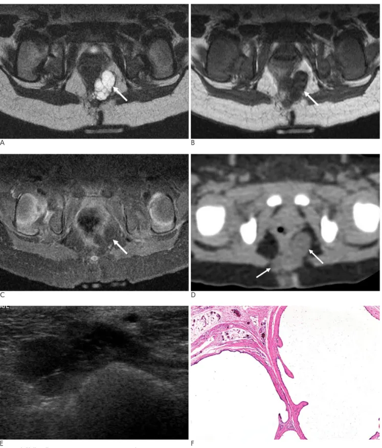
A 23-day-old female infant was referred for an evalua- tion due to a palpable coccygeal mass which was pre- sent since birth. The infant developed to full-term and was delivered by a cesarean section without birth in- jury. Upon a physical examination, a movable, non-ten- der, round mass, measuring 1×2 cm, was palpable cephalad to the anus. An MRI of the spine showed a well-defined, multiloculated cystic mass between the rectum and coccyx, measuring 12×23 mm. The lesion was markedly hyperintense on T2-weighted images (Fig. 1A) and hypointense on T1-weighted images (Fig.
1B). The Gd-enhanced T1-weighted images revealed thin-enhancing septa without a solid component (Fig.
1C). The adjacent fat planes and sacrum were normal and there was no communication between the multiloc- ulated cyst and the thecal sac. A non-enhanced CT re- vealed a lobulating mass of mild hypoattenuation in the retrorectal space (Fig. 1D). Furthermore, no calcification was noted and an ultrasound revealed a lobulating mul- tiseptated cystic mass anterior to the coccyx (Fig. 1E).
The cyst was surgically removed by means of an exci- sion via the posterior sagittal approach. Though the le- sion was attached to the rectum, a complete excision was achieved. The gross specimen consisted of a translucent cystic structure containing a clear aqueous fluid. The microscopic findings revealed that the cystic spaces were lined by ciliated columnar, transitional and squamous cells (Fig. 1F). Also, randomly situated smooth muscles and nerve fibers were present in the ad- jacent cysts and no inflammatory reaction was found.
Discussion
A tailgut cyst arises in the retrorectal space, which is an area bounded by the rectum anteriorly, the sacrum and coccyx posteriorly, the peritoneal reflections superi- orly, the levator ani and coccygeus muscles inferiorly and finally the iliac vessels and ureters laterally (4, 5).
J Korean Radiol Soc 2008;58:177-180
─ 177 ─
Radiologic Features of a Tailgut Cyst in a Neonate:
A Case Report
1Kum Rae Kim, M.D., Won Kyu Park, M.D.
1Department of Radiology, College of Medicine, Yeungnam University Received October 29, 2007 ; Accepted December 20, 2007
Address reprint requests to : Won Kyu Park, M.D., Department of Radiology, College of Medicine, Yeungnam University, 317-1, Daemyungdong, Namgu, Daegu 705-717, Korea.
Tel. 82-53-620-3048 Fax. 82-53-653-5484 E-mail: [email protected]
A tailgut cyst is a rare congenital abnormality located in the retrorectal space and is usually manifested during childhood or adulthood. We report the MR, CT and ultra- sound findings of a tailgut cyst in a 23-day-old neonate.
Index words :Infant, newborn, diseases Congenital abnormalities Sacrococcygeal region
Kum Rae Kim, et al: Radiologic Features of a Tailgut Cyst in a Neonate
─ 178 ─
A B
C D
E F
Fig. 1. A 23-day-old neonate with a tailgut cyst.
A, B. Axial MR images show a multiloculated cystic mass (arrow) in the retrorectal space which is markedly hyperintense on T2- weighted images (A), and hypointense on T1-weighted images (B), respectively.
C. Gd-enhanced T1-weighted image reveals the thin enhancing septa (arrow).
D. A non-enhanced CT scan shows a lobulating mass (arrows) of mild hypoattenuation with well defined borders in the retrorectal space. No calcification is noted.
E. An ultrasound reveals a multiseptated cystic mass (asterisk) abutting the coccyx (Co).
F. The microscopic pathologic finding shows multiloculated cysts (Cy), which are lined by ciliated columnar, transitional and squa- mous cells (Hematoxylin-eosin, ×40).
Early in its development, the embryo possesses a true tail, which develops maximally the 8-mm stage (35 days -gestational age) and usually completely regresses by the 35-mm stage (56 days-gestional age). The anus is formed cephalad to the tail. Because the primitive gut extends into the tail beyond the point at which the anus develops, it is called the tailgut. When the tailgut fails to regress normally, congenital cysts tend to occur in this region (4, 6, 7).
Though reported with respect to a tailgut cyst, the ra- diologic findings of barium enema examination, US, CT and MRI have been primarily described in adults. A bar- ium enema examination shows an extrinsic retrorectal mass (7), whereas the sonographic appearance of this le- sion is a complex retrorectal mass, which is uncharac- teristic of a simple cyst (6). The internal echoes of the cyst are due to the multicystic nature of the mass and the presence of gelatinous material or inflammatory de- bris within the lumen of the lesion. Upon a CT examina- tion, the lesion appeared as a well-marginated presacral low attenuation mass without calcification (6, 8).
However, the focal high attenuation was reported with- in tailgut cyst due to hemorrhaging (8). When a loss of marginal sharpness or invasion to adjacent structures occurs, infection or malignancy should be suspected.
Upon an MRI examination, the tailgut cyst is hypo and markedly hyperintense on T1 and T2-weighted images, respectively (2, 8). The signal characteristics of a tailgut cyst on T1-weighted images may vary depending on the contents of the cyst (8). A hyperintense appearance from T1-weighted images may be caused by either high pro- tein content or hemorrhaging into the cyst.
Upon review of the three cases of previously reported neonatal tailgut cysts, the radiologic findings are similar to the adult cases without complications such as infec- tion, hemorrhaging or malignant transformation. The radiologic findings of our case study are also similar to those of previously reported neonatal tailgut cysts.
Tailgut cysts in neonates should be distinguished from other lesions occuring in the retrorectal space and in the
perianal area. These include teratoma, epidermoid cysts, duplication cysts of rectum and anterior meningo- celes (2, 4, 6). The classification of these masses in the retrorectal region is dependent upon the type of epitheli- um present. However, diverse imaging modalities in- cluding MRI, CT and US can help differentiate tailgut cysts from other cystic masses, in terms of the presence of calcification, fat, hemorrhaging and communication with other structures such as thecal sac.
It is well known that the significance of tailgut cysts is due to morbidity which occurs in unrecognized and in- completely treated lesions. They are prone to infection, recurring formation of fistula and malignant transforma- tions (adenocarcinoma and carcinoid) (5). Thus, com- plete excision is the treatment of choice. Incisional biop- sies are contraindicated because they can lead to fistula formation (2, 4, 5)
In conclusion, tailgut cysts are congenital cysts which develop in the retrorectal space and can cause various morbidities. If a multilocular cystic mass is discovered in the retrorectal space of neonates, tailgut cysts should be included in the differential diagnoses.
References
1. Antao B, Lee AC, Gannon C, Arthur R, Sugarman ID. Tailgut cyst in a neonate with anal stenosis. Eur J Pediatr Surg 2004;14:212-214 2. Oh JT, Son SW, Kim MJ, Kim L, Kim H, Hwang EH. Tailgut cyst
in a neonate. J Pediatr Surg 2000;35:1833-1835
3. Rafindadi AH, Shehu SM, Ameh EA. Retrorectal cystic hamar- toma (tailgut cyst) in an infant: case report. East Afr Med J 1998;75:726-727
4. Hjermstad BM, Helwig E. Tailgut cysts: report of 53 cases. Am J Clin Pathol 1988;89:139-147
5. Killingsworth C, Gadacz TR. Tailgut cyst (retrorectal cystic hamar- toma): report of a case and review of the literature. Am J Surg 2005;71:666-673
6. Jain P, Hawkins S, King A. Tail-gut cyst. Australas Radiol 1997;41:207-210
7. Campbell WL, Wolff M. Retrorectal cysts of developmental origin.
Am J Roentgenol Radium ther Nucl Med 1973;117:307-313
8. Yang DM, Park CH, Jin W, Chang SK, Kim JE, Choi SJ, et al.
Tailgut cyst: MRI evaluation. AJR Am J Roentgenol 2005;184:1519- 1523
J Korean Radiol Soc 2008;58:177-180
─ 179 ─
Kum Rae Kim, et al: Radiologic Features of a Tailgut Cyst in a Neonate
─ 180 ─
대한영상의학회지 2008;58:177-180
신생아에서 발견된 Tailgut Cyst의 영상소견: 증례 보고1
1영남대학교 의과대학 영상의학과학교실
김 금 래・박 원 규
꼬리장 낭종(Tailgut cyst)은 후직장공간에 생기는 드문 기형으로 일반적으로 아동기나 어른에서 발견된다. 우리는 23일 된 신생아에서 발생한 꼬리장 낭종의 초음파, 전산화단층촬영 및 자기공명영상의 소견을 보고한다.