대한내분비외과학회지:제 5 권 제 1 호
□ 증 례 □
Vol. 5, No. 1, June 2005
29
서 론
갑상선 수질암(medullary thyroid carcinoma)은 칼시토닌을 분비하는 neural crest 기원의 C-cell 종양으로, 전 갑상선암 의 1.2∼10%을 차지하는 비교적 드문 질환이며, 갑상선 여 포세포 기원의 암들과 역학적, 생화학적 및 조직학적으로 다른 특징을 보인다.(1)
이 질환의 칼시토닌치는 거의 모든 환자에서 증가되어 있으며, 기저 혈청 칼시토닌치는 종양 크기와 상관관계를 보여 종양이 작을 경우에는 기저 혈청 칼시토닌치는 정상 일 수도 있다.(2) 이와 마찬가지로 갑상선 수질암 환자에서 혈청 CEA (carcinoembryonic antigen)치도 대부분 증가되어 있으나, 이는 다른 갑상선결절 환자나 소화기계 악성종양 환자 등에서도 증가될 수 있어 특이도는 칼시토닌보다 낮 다.(3) 칼시토닌과 CEA의 측정은 진단뿐 아니라 좋은 예후 표지자로 환자의 추적관찰에 이용되고 있다.(4,5) 치료에서 는 방사성 옥소 치료나 항암요법, 방사선 치료에 반응하지 않으므로 조기에 발견하여 갑상선 전절제술 및 전이 부위 의 근치적 수술을 하여야만 완치를 기대할 수 있다.
저자는 1997년에 직장암으로 저위전방절제술 시행 받았 으나, 술 후에 계속된 추적검사에서 혈청 CEA가 12∼20 ng/ml (정상치: 0.0∼5.0 ng/ml)로 이상치를 보였던 50세 환 자가 술 후 8년 후인 2005년에 갑상선 결절로 수술하여 갑 상선 수질암으로 진단된 흥미 있는 예를 경험하였기에 문 헌고찰과 함께 그 증례를 보고하는 바이다.
증 례
환 자: 송○점, 50세, 남자
주소 및 병력: 50세 남자환자가 직장암으로 수술 후에 추 적검사에서 발견된 갑상선 결절을 주소로 내원하었다. 환 자는 항문연에서 4 cm 상방의 직경 2×2 cm인 직장암으로 1997년 5월 22일 근치적저위전방절제술을 시행 받았다. 당 시 병리조직검사 결과에서 TNM 병기는 T1N0M0이었다. 상 기환자는 직장암 수술 후에 3개월에서 12개월 간격으로 혈 청 CEA를 검사하였다(Fig. 1). 2002년 4월에 시행한 대장내 시경에서 항문연 26 cm과 13 cm에 용종절제술을 시행하였
고 CEA혈증으로 인해 재발성 직장암으로 오인된 갑상선 수질암 1예
고신대학교 의과대학 외과학교실 김 정 훈
Medullary Thyroid Cancer Misunderstanding as Recurrent Rectal Cancer due to High Serum CEA Levels
Jeong Hoon Kim, M.D.
The medullary carcinoma of the thyroid gland is relatively rare tumor, accounting about 1.2∼10% of the all thyroid malignancies, which arises from the parafollicular C-cells in thyroid gland. Operation is the only means to cure the patients. Serial concentrations of serum CEA and calcitonin seem to play an important role in the diagnosis and clinical management and also in the therapeutic monitoring of pa- tients with medullary thyroid cancer. In this case, patient with high serum CEA levels after the resection of rectal cancer underwent abdomen CT scan, colonofibroscopy, FDG-PET scan and chest X-ray, but this imaging methods couldn't detect recurrent evidence of rectal cancer. Neck ultrasonog- raphy was performed after 8 years from operation, and fine needle aspiration biopsy was performed for thyroid nodule.
As diagnosed to suspicious medullary carcinoma, patient underwent total thyroidectomy and central compartment neck dissection. Patient diagnosed as medullary thyroid cancer without lymph node metastasis and capsular invasion patho- logically. As well as this case, in patients with high serum CEA levels after definitive surgical resection of gas- trointestinal cancer, if imaging study or FDG-PET scan detect no evidence of recurrence, evaluation of thyroid such as neck ultrasonography and serum calcitonin should be per- formed. (Korean J Endocrine Surg 2005;5:29-31) Key Words: CEA, Medullary thyroid cancer, Rectal cancer 중심 단어: CEA, 갑상선 수질암, 직장암
ꠏꠏꠏꠏꠏꠏꠏꠏꠏꠏꠏꠏꠏꠏꠏꠏꠏꠏꠏꠏꠏꠏꠏꠏꠏꠏꠏꠏꠏꠏꠏꠏꠏꠏꠏꠏꠏꠏꠏꠏꠏꠏꠏꠏꠏꠏꠏꠏꠏꠏꠏꠏꠏ Department of Surgery, Kosin University College of Medicine, Busan, Korea
책임저자 : 김정훈, 부산광역시 서구 암남동 34번지 ꂕ 620-030, 고신대학교병원 외과 Tel: 051-990-6462, Fax: 051-246-6093 E-mail: gskjh@hanafos.com
게재승인일:2005년 5월 30일
2005년 고신대학교 의과대학 연구비 일부를 지원 받았음.
30 대한내분비외과학회지:제 5 권 제 1 호 2005
ꠏꠏꠏꠏꠏꠏꠏꠏꠏꠏꠏꠏꠏꠏꠏꠏꠏꠏꠏꠏꠏꠏꠏꠏꠏꠏꠏꠏꠏꠏꠏꠏꠏꠏꠏꠏꠏꠏꠏꠏꠏꠏꠏꠏꠏꠏꠏꠏꠏꠏꠏꠏꠏꠏꠏꠏꠏꠏꠏꠏꠏꠏꠏꠏꠏꠏꠏꠏꠏꠏꠏꠏꠏꠏꠏꠏꠏꠏꠏꠏꠏꠏꠏꠏꠏꠏꠏꠏꠏꠏꠏꠏꠏꠏꠏꠏꠏꠏꠏꠏꠏꠏꠏꠏꠏꠏꠏꠏꠏꠏꠏꠏꠏꠏꠏ 으며, 각각 관상선종, 만성 대장염으로 진단되었다. 2003년
3월에는 혈청 CEA가 20.1 ng/ml로 이전 검사들보다 많은 증가를 보였으나 대장내시경에서 특이한 소견은 없었으며, 복부컴퓨터단층촬영을 하였으나 재발의 증거는 발견되지 않았다. 2005년 1월 양전자 방출 단층촬영(positron emission tomography, PET)을 하여 특이소견이 없었으나, 경부초음파 에서 갑상선 우엽에 결절이 관찰되어 세침흡인세포검사를 시행하고 갑상선 수질암 의증으로 진단되어 수술 치료를 위해 입원하게 되었다.
기왕력 및 가족력: 특이사항은 없었다.
이학적 소견: 체격은 중등도였고, 영양 상태는 양호했으 며 의식은 명료하였고 혈압, 맥박, 체온은 정상범위였다. 경 부에는 갑상선이 양측으로 3×3×3 cm 정도의 활면을 가졌
고 종괴는 촉지되지 않았다. 측경부 림프절은 촉지되지 않 았고 기타 부위에 이상 소견은 없었다.
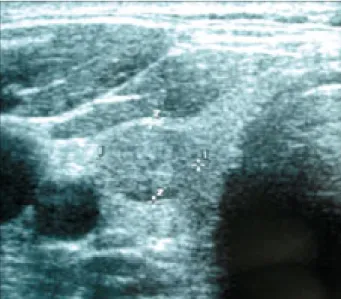
검사소견: 기본적인 혈액검사, 요검사는 정상이었고 간기 능검사, 흉부 X-선 검사, T3, Free T4 및 TSH치도 정상범위 였다. 양전자 방출 단층촬영에서도 특이소견이 없었다. 혈 청 CEA는 15.17 ng/ml이었으며, calcitonin은 107.84 pg/ml (정상치: 0.0∼10.0 pg/ml)이었다. 경부초음파에서 갑상선 우 측엽에 1×0.8 cm의 결절이 관찰되었으며(Fig. 2), 세침흡입 조직검사의 현미경소견에서는 방추형세포들이 다수 보이 며 핵내 봉입체가 관찰되어 갑상선 수질암의증으로 진단되 었다. 경부컴퓨터단층촬영에서 양측 측경부에 림프절비대
Fig. 3. Histologic finding. (A) Round to spinded cells are arranged in clusters, trabecular and acinar pattern with intersecting amyloid deposits (×200). (B) Tumor cells contains uniform, round to oval nuclei with finely granular chromatin, and small and visible nucleoli. cytoplasm is eosinophilic and granular (×400).
Fig. 1. The curve of serum CEA level shows within normal limit
after total thyroidectomy (arrow). Fig. 2. Preoperative neck ultrasonography shows 1×0.8 cm sized well defined, heterogenous nodule at right thyroid.
김정훈:고 CEA혈증으로 인해 재발성 직장암으로 오인된 갑상선 수질암 1예 31 ꠏꠏꠏꠏꠏꠏꠏꠏꠏꠏꠏꠏꠏꠏꠏꠏꠏꠏꠏꠏꠏꠏꠏꠏꠏꠏꠏꠏꠏꠏꠏꠏꠏꠏꠏꠏꠏꠏꠏꠏꠏꠏꠏꠏꠏꠏꠏꠏꠏꠏꠏꠏꠏꠏꠏꠏꠏꠏꠏꠏꠏꠏꠏꠏꠏꠏꠏꠏꠏꠏꠏꠏꠏꠏꠏꠏꠏꠏꠏꠏꠏꠏꠏꠏꠏꠏꠏꠏꠏꠏꠏꠏꠏꠏꠏꠏꠏꠏꠏꠏꠏꠏꠏꠏꠏꠏꠏꠏꠏꠏꠏꠏꠏꠏꠏ 는 관찰되지 않았다.
치료 및 경과와 병리학적 소견: 환자는 내원 다음날인 2005년 2월 16일에 갑상선전절제술과 중앙경부림프절곽청 술을 시행하였으며, 직경 0.9×0.9 cm의 경계가 분명한 종 괴가 우엽 하부에 있었다. 병리소견으로는 방추형세포로 둘러싸인 군집으로 되어있으며, 아밀로이드가 침착되어 있 었고, 세포 내에는 미세한 크로마틴 과립을 가진 균일하고 둥근 핵이 있었으며, 세포질은 호산성의 과립을 포함하고 있어서 갑상선 수질암으로 진단하였다(Fig. 3). 피막침습은 없었고 절제된 림프절 10개 모두에서 전이는 없었다. 수술 다음날 혈액검사는 정상수치였으며, 특별한 합병증 없이 술 후 6일째 퇴원하였다. 2005년 3월 30일 외래에서 시행한 혈액검사에서 혈청 CEA는 3.48 ng/ml이었으며, calcitonin은 2.04 pg/ml이었다. 퇴원 후 6개월에 걸쳐 본원 외과에서 관 찰 중이며 건강한 상태이다.
고 찰
CEA는 180 kDa 분자량의 단백질로 1965년 Gold와 Free- man에 의해 처음 발표되었다. 이후 혈청 CEA는 많은 형태 의 악성종양에서 증가되었으나 특히 대장과 직장암에서 술 전 종양의 진행정도를 예상하며, 술 후 재발 감시에 많은 도움을 주고 있다. Wang등(6)의 후향적 연구에서는 대장항 문악성종양 환자들의 술 후 Dukes병기에 따른 술 전 혈청 CEA는 진행정도가 증가함에 따라 25∼82%의 이상치를 보 였다. 또한 대장직장암에서 술 전에 혈청 CEA가 증가된 환 자들은 근치적 수술을 받은 직후에 대부분 정상 수치를 보이 게 되며, 추적검사에서 증가되었을 경우는 재발을 강력히 의 심해 볼 수 있다. 본 증례는 술 전에 혈청 CEA가 증가되어 있었으나 술 후에도 감소되지 않고 계속적으로 이상치를 보 였으며, Dehdachti등(7)과 Ledermann등(8)의 연구와 같이 양 전자 방출 단층촬영을 시행하였으나 특이한 소견이 없었다.
Madeddu등(3)은 258명의 갑상선결절로 수술 받은 환자를 대상으로 술 전 혈청 CEA를 후향적으로 조사하였으며, 양 성결절과 악성결절에서 각각 4명과 11명이 이상 수치를 보 였다. 이상 수치를 보인 악성결절에서 3예는 유두암, 2예는 여포암, 3예는 수질암, 2예는 미분화암, 1예는 여포상변이 유두암이었다. 이 연구에서 고CEA혈증이 있는 악성결절 환자 중 전이성 미분화암 2예 외에는 수술 후 혈청 CEA는 정상으로 된 것 같이, 본 증례에서도 역시 갑상선전절제술 시행 후에 혈청 CEA가 의미 있게 감소하여 정상치로 전환 되었다(Fig. 1). 오등(9)도 마찬가지로 갑상선 수질암의 수술 후에 혈청 CEA의 감소와 재발된 경우 대부분이 증가됨을 보고한 바 있다. 이와 같이 갑상선 수질암 뿐만이 아니라 다른 갑상선 결절에서도 혈청 CEA가 증가되어 있을 수 있 으며, 갑상선 수질암의 조기 진단에는 혈청 칼시토닌치의 측 정이 CEA보다 우수하지만, 수술 후 원격 전이 발생이나 예
후 인자로는 혈청 CEA가 더 우수하다는 보고도 있다.(10, 11) 본 증례에서와 같이 직장암 등 소화기계 악성종양 환자 에서 술 후에 추적검사에서 지속적으로 혈청 CEA가 증가 되어 있거나, 정상으로 되었다가 증가된 경우에 대개 복부 컴퓨터촬영이나 복부초음파, 대장내시경, 흉부 X-선 촬영 등으로 재발에 대한 검사를 하고, 상기검사에서 발견되지 않는 환자의 상당수는 양전자 방출 단층촬영에서 재발이 진단되기도 한다.(7) 그러나 이러한 모든 검사에서 재발의 증거를 찾지 못한 경우에는 갑상선결절, 특히 갑상선 수질 암으로 인한 혈청 CEA의 증가를 고려해야 하며, 경부초음 파 등으로 갑상선에 대한 평가가 이루어져야 할 것이다.
REFERENCES
1) Saad MF, Ordonez NA, Rashid RK. Medullary carcinoma of the thyroid: a study of the clinical features and prognostic features in 161 patients. Medicine 1984;63:319-25.
2) Robert F. GAGEL, ANA O. Hoff, Gilbert J. COTE. Medullary carcinoma of the thyroid. In: Lewis E. Braverman, Robert D.
Utiger, editors. The thyroid. 9th ed. Philadelphia: Lippincott Co; 2005. p.967-88.
3) Madeddu G, Langer M, Dettori G, Costanza C. Role of serum carcinoembryonic antigen in preoperative diagnosis of cancer in patients with thyroid nodules. Cancer 1980;45:2607-10.
4) Pacini F, Fontanelli M, Fugazzola L, Elisei R, Romei C, Di Cooscio G, et al. Routine measurement of serum calcitonin in nodular thyroid diseases allows the preoperative diagnosis of unsuspected sporadic medullary thyroid carcinoma. J Clin Endocrinol Metab 1994;78:826-9.
5) Rieu M, Lame M-C, Richard A, Lissak B, Sambort B, Vuong- Ngoc P, et al. Prevalence of sporadic medullary thyroid carci- noma: the importance of routine measurement of serum calci- tonin in the diagnostic evaluation of thyroid nodules. Clin Endocrinol 1995;42:453-60.
6) Wang WS, Lin JK, Chiou TJ, Liu JH, Frank S-Fan, Chueh CY, et al. Preoperative CEA level as an independent prognostic factor in colorectal cancer. Jpn J Clin Oncol 2000;30:12-6.
7) Flanagan FL, Dehdashti F, Ogunbiyi OA, Kodner IJ, Siegel BA. Utility of FDG-PET for investigating unexplained plasma CEA elevation in patients with colorectal cancer. Ann Surg 1998;227:319-23.
8) Ledermann J, Arulampalam. Asymptomatic patient with an increasing concerntrationof CEA. Lancet Oncol 2001;2:172.
9) 오승근, 김지수. 갑상선수질암. 대한외과학회지 1999;56:49- 58.
10) 임성희. 갑상선 수질암의 진단. 대한내분비학회지 1996;11:7- 10.
11) Saad MF, Fritsche HA, Samaan NA. Diagnosticand prognostic values of carcinoembryonic antigen in medullary carcinoma of the thyroid. J Clin Endocrinol Metab 1984;58:889-94.