Comparisons of Gastric Cancer Treatments: East vs. West
Kai Bickenbach, and Vivian E Strong1
Department of Surgery, University of Medicine and Dentistry of New Jersey-New Jersey Medical School, Newark, NJ,
1Department of Surgery, Memorial Sloan-Kettering Cancer Center, New York, NY, USA
There has been a large amount of speculation concerning the differences in the outcomes in patients who have gastric cancer in the Eastern and Western worlds. The differences in biology, surgical and adjuvant treatment have been used to explain such differences.
There are clear differences observed in the histology (diffuse vs. intestinal), tumor location (proximal vs. distal), environmental exposures, dietary factors and Helicobacter pylori status. A higher incidence of gastric cancer in the East has led to screening programs, and lead- ing to an earlier stage at presentation. Surgical treatment differs in that the extended lymph node dissection is routinely practiced in the Asian countries. Additionally, different adjuvant therapeutic regimens are used in both regions. The purpose of this review is to describe the differences in both presentation and treatment between the East and the West.
Key Words: Gastric Cancer; Gastric adenocarcinoma; Western experience; Eastern experience; Gastric cancer survival
Correspondence to: Kai Bickenbach
Department of Surgery, University of Medicine and Dentistry of New Jersey-New Jersey Medical School, 205, S. Orange Ave, G-1222, Newark, NJ 07103, USA
Tel: +1-973-972-1110, Fax: +1-973-972-3730 E-mail: [email protected]
Received June 8, 2012 Accepted June 11, 2012
Copyrights © 2012 by The Korean Gastric Cancer Association www.jgc-online.org
This is an open-access article distributed under the terms of the Creative Commons Attribution Non-Commercial License (http://creativecommons.org/
licenses/by-nc/3.0) which permits unrestricted noncommercial use, distribution, and reproduction in any medium, provided the original work is properly cited.
Introduction
Although the incidence of gastric cancer has been declining in most industrialized nations over the past two decades, it still remains the second leading cause of cancer related deaths world- wide.(1) The incidence is highest in Japan, Korea, China, Latin America and Eastern Europe. In Western countries like the United States, the incidence is lower, with 21,000 new cases diagnosed each year.(2) Survival outcomes differ considerably between Eastern and Western populations, with better overall survival reported in Eastern series.(3-7) Many authors have sought explanations for this based on either: 1) Stage Migration, 2) Differences in biology, or 3) Differences in treatment. The adoption of screening programs in Japan has led to screening programs which allow for earlier detec- tion in the East.(8) Additionally, there is discrepancy in pathologic
review between Eastern and Western pathologists, with Eastern pa- thologists having a lower threshold for diagnosis of primary tumor than Western pathologists.(9) Differences in biology are clearly seen in the histology (diffuse vs. intestinal), tumor location (proximal vs.
distal), environmental exposures, dietary factors and Helicobacter pylori status. Surgical treatment differs in that the extended lymph node dissection is routinely practiced in Asian countries, leading to greater lymph node retrieval. Whether this leads to stage migration or to a direct therapeutic effect has yet to be resolved. Laparoscopic gastrectomy has also been much more widely adopted in the East compared to the West. Furthermore, adjuvant therapy differs be- tween the two regions.
The purpose of this review is to describe the differences in both presentation and treatment between the East and the West. We will highlight the differences in clinicopathological presentation, discuss the differences in both surgical and adjuvant treatment, and discuss the differences in survival outcomes.
Stage Differences
The incidence of gastric cancer is higher in Eastern countries compared to the West. This has led to the adoption of mass cancer
screening programs in countries like Japan.(8) In Japan, annual screening with a double-contrast barium technique and endoscopy is recommended for persons over the age of 40 years. As such, many series have shown that gastric cancer tends to present with earlier staged lesions than their Western counterparts.(10,11) Based on reports from cancer registries, 53% of gastric cancers in Japan were localized when diagnosed, as opposed to 27% of those in the United States (US). This has been touted as a potential reason for the discrepancy in survival, although several series have dem- onstrated differences in survival when stratified by stage.(12,13) Additionally, there may be some variation in pathologic evalua- tion between the US and Japan.(9,14,15) Japanese pathologists rely heavily on nuclear cytologic and glandular architecture abnormali- ties, whereas Western pathologists require the presence of invasion to diagnose carcinoma. Therefore, it may be that patients in Japan may have less advanced tumors than US patients even within T- stage.
Biology
One major difference between Eastern and Western gastric cancer is the location of the tumor. Western countries have a much higher incidence of tumors located in the proximal third of the stomach.(10-12,16-18) In fact, the incidence of proximal gastric cancer has been increasing steadily in the US, even while the in- cidence of gastric cancer is on the decline.(19,20) Proximal tumors are known to be associated with worse outcomes and could explain the differences between the two regions.(21) However, even when compared by tumor location, there do appear to be survival differ- ences favoring the East.(12)
Differences in histology are seen between the East and West.
There is a higher prevalence of diffuse histology in Western pa- tients.(12,13) The poor prognosis of diffuse and signet ring histol- ogy could explain worse outcomes in the West. Additionally, differ- ences in the baseline characteristics of patients could explain worse outcomes in the US. Patients in the US develop gastric cancer later in life.(12,13,18) One study demonstrated a median difference of 10 years between Korean and US cohorts.(12) Western populations generally have a higher body mass index. Increased obesity has been shown in many series to be associated with increased peri- operative complications in gastric cancer as well as many other tumors.(22,23) The prevalence of comorbidities such as diabetes and an increased use of tobacco in the US could also influence the discrepant outcomes between the East and West.
Differences in Surgical Treatment
1. Endoscopic mucosal resection (EMR)
The advent of early detection of gastric cancer by screening programs in the East has led to an increase in the prevalence of early gastric cancer (ECG) in those regions. EGC is defined as tu- mor invasion confined to mucosa or submucosa irrespective of the lymph node status. Database studies have demonstrated that the in- cidence of lymph node metastases in EGC confined to the mucosa is less than 4.9%, with a 5 year survival of 99%. The risk of lymph node metastases increases to 23.8% with submucosal invasion.(24) Multiple studies in the East have demonstrated the feasibility of EMR for EGC. Uedo et al.(25) reported on 124 Japanese patients with EGC less than 2 cm with no ulceration. All were completely resected and the disease specific survival (DSS) rates were 99% at 5 and 10 years. Kim et al.(26) reported on 514 patients with EGC who were treated at 13 institutions throughout Korea. The local re- currence rate was 6%, however there were no gastric cancer related deaths in these recurrences.
The use of EMR has been widely adopted in the East. It is esti- mated that approximately 50% of Stage IA gastric cancer is treated by EMR in Japan. Indications for EMR include well to moderately differentiated tumors that are confined to the mucosa. Superficially elevated tumors must be less than 2 cm and those that are flat or depressed should be less than 1 cm. There should be no ulceration or scar present and there should be no lymphatic or venous in- volvement.(27) Unlike Japan, EMR has not been as widely adopted in Western countries; however its use is starting to increase.
2. Lymph node dissection
The surgical approach to lymph node dissection has differed between the East and the West. D2 lymphadenectomy, which entails systematic dissection of the perigastric nodes along with the nodes along the celiac artery and its branches, is the standard of care in Japan and Eastern countries. In Western countries, D1 lymph node dissection (perigastric nodes only) is routinely per- formed. Lymph node dissection in the West has been driven by two large European randomized controlled trials which compared D1 to D2 lymphadenectomy (Table 1). Cuschieri et al.(28,29) reported the results of a multi-center randomized controlled trial from the United Kingdom (UK). In it, 400 patients were randomized to either D1 or D2 lymphadenectomy. The results showed a signifi- cantly higher morbidity (46% vs. 28%, P<0.001) and mortality (13%
vs. 6.5%, P=0.04) in the D2 arm. This was mainly driven by distal
pancreatectomy and splenectomy.(28) There was no difference seen in overall survival or disease-specific survival between D1 and D2 lymph node dissection.(29) This study was highly criticized due to contamination and non-compliance. The median number of lymph nodes retrieved was 17 in the D2 group and 13 for the D1.
Additionally, many Eastern surgeons find the mortality rate unac- ceptably high. The second major European study was the Dutch Gastric Cancer trial, in which 711 patients were randomized to ei- ther D2 or D1 lymph node dissection. All surgeons were instructed by an expert from Japan and all D2 lymph node dissections were supervised by a specially trained surgeon. Despite this there was still non-compliance in 36% of D1 and 51% of D2 dissections.(30) As in the UK trial the D2 arm had higher morbidity (43% vs. 25%, P<0.001) and mortality (10% vs. 4%, P=0.004). The survival data has been reported at 3 separate time intervals.(30-32) In the first two pub- lications, there was no difference in overall or disease-specific survival, with a reported 11 year overall survival of 35% in the D2 arm vs. 30% in the D1 arm (P=0.53). Management of gastric cancer in the West was greatly influenced by these outcomes. However, recently the 15 year results of the Dutch trial have been published.
There remains no significant difference in overall survival. How- ever, there is improved loco-regional control in the D2 arm and there is improved gastric cancer mortality in the D2 arm (37% vs.
48%, P=0.01).(32) Whether the 15 year results will influence treat- ment patterns in the US remains to be seen.
In the East, the standard of care is a D2 lymph node dissec- tion. Many Japanese surgeons consider the results of the Dutch and UK trials to have an unacceptably high mortality rate and poor survival rates. Peri-operative mortality in the Japan Clinical Oncol- ogy Group (JCOG) 9501 trial was 0.8% and the 5 year survival was 70%,(33) whereas in the European trials mortality was 4~6% and 5-year survival 33~35%(7,34). Trials in the East have focused on more extensive lymph node dissections than D2. There has been one randomized trial out of Taiwan which compared D1 and D3 lymphadenectomy. In it, 221 patients were randomized at a single institution, in which 3 highly trained surgeons performed all the operations. In their hands, the morbidity rate was higher in the D3 group (17.1% vs. 7.3%, P=0.012) however there was no operative mortality reported in either group.(28) The D3 group had more lymph nodes removed (37.2 vs. 19.4), however there was no dif- ference in number of nodes positive (3.9 vs. 3.4). The study dem- onstrated improved survival with D3 lymphadenectomy (59.5% vs.
53.6%, P=0.041).(35) This study suggests that in the hands of highly experienced surgeons, where the operative mortality is very low, D3 lymphadenectomy is superior to D1.
Two other Japanese trials have examined more extensive Table 1. Summary of lymphadenectomy trails
Trial Number
of patients
randomized Study groups Stratification Morbidity
(%) Mortality
(%) Significance
demonstrated % change identified in trial Western trials
UK trial(28) 400 D1 (n=200)
D2 (n=200)
Institution Nodal status Tumor location
28.0 46.0
6.5 13.0
None 35
33
5-year survival Dutch
trial(30-32)
711 D1 (n=380) D2 (n=331)
Institution 25.0
43.0
4.0 10.0
Yes (survival at 15 years)
37 48 15-year DSS Eastern trials
Wu et al.(35) 221 D1 (n=110) D2 (n=111)
None 7.3
17.1
0.0 0.0
Yes (survival) 53.6 D1 vs. 59.5 D3 5-year survival JCOG 9501(33) 523 D2 (n=263)
D2+PAND (n=260)
T-stage Borrman type Institution
20.9 28.1
0.8 0.8
None 69.2
70.3
5-year survival JCOG 9502(36) 167 LTA (n=85)
TH (n=82)
Stage Borrman type Institution
49.0 34.0
4.0 0.0
None 37.9
52.3
5-year survival UK = United Kingdom; DDS = disease specific survival; JCOG = Japan Clinical Oncology Group; PAND = para-aortic node dissection; LTA = left thoracoabdominal approach; TH = transhiatal approach.
lymphadenectomies. In JCOG 9501, 523 patients were random- ized to D2 lymphadenectomy or D2+Para-aortic node dissec- tion (PAND). The results demonstrated higher morbidity with D2+PAND (28.1% vs. 20.9%, P=0.07), however there was no dif- ference in operative mortality (0.8% in both groups).(33) There was no difference in survival between the two groups, with 5 year survival rates of 70% and 69% (P=0.85). A second Japanese study, JCOG 9502, compared D2 lymphadenectomy with or without me- diastinal nodal dissection. In this trial, 167 patients were random- ized to either a left thoracoabdominal approach with mediastinal lymph node dissection or a trans-hiatal approach. There was no significant difference in post-operative morbidity or mortality. This study failed to demonstrate a survival advantage with the more ex- tensive lymphadenectomy with a 5 year overall of 37.9% vs. 52.3%
(P=0.92).(36)
Differences in Adjuvant Therapy
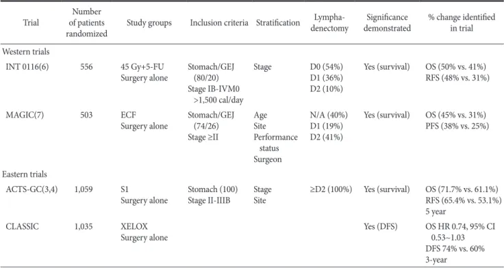
Approaches to multi-modality therapy differ between the East and West. Approaches in the West have been driven by two ran- domized controlled trials that showed a benefit to adjuvant therapy (Table 2). The US Intergroup study (INT) 0116, randomized 556
patients with Stage Ib-IVM0 cancer by the 1988 AJCC staging cri- teria.(6) Tumors were located at the gastroesophageal junction (GEJ) in 20% of patients. Patients were randomized to 45 Gy of radiation and 5-FU/Leucovorin or surgery alone. The study demonstrated improved local control and a clear survival benefit with a 3-year overall survival (OS) of 50% vs. 41% (P=0.005).
The Medical Research Council Adjuvant Gastric Cancer In- fusional Chemotherapy (MAGIC) study from the UK reported a similar survival advantage with the use of peri-operative chemo- therapy.(7) In this trial, patients were randomized to Epirubicin, Cisplatin, and 5-FU for 3 cycles pre-operatively and 3 cycles post- operatively. The patients consisted of adenocarcinoma of the stom- ach (74%), GEJ (15%), and lower esophagus (11%). The results of the trial demonstrated an improved overall and progression free survival in the peri-operative chemotherapy group, with 5-year survivals of 36% and 23% respectively.
These two trials have driven therapy in the West; however there are several criticisms that Eastern investigators have of these trials.
The main criticism is the surgical quality of control in these trials.
In the INT 0116 trial only 10% of patients had a D2 lymphadenec- tomy. In fact, 54% of the patients had a D0 lymphadenectomy.
Similarly, in the MAGIC trial, only 41% of patients had a D2
Table 2. Summary of trials for adjuvant therapy
Trial Number
of patients
randomized Study groups Inclusion criteria Stratification Lympha-denectomy Significance
demonstrated % change identified in trial Western trials
INT 0116(6) 556 45 Gy+5-FU
Surgery alone Stomach/GEJ (80/20) Stage IB-IVM0
>1,500 cal/day
Stage D0 (54%)
D1 (36%) D2 (10%)
Yes (survival) OS (50% vs. 41%) RFS (48% vs. 31%)
MAGIC(7) 503 ECF
Surgery alone Stomach/GEJ (74/26) Stage ≥II
AgeSite Performance
status Surgeon
N/A (40%) D1 (19%) D2 (41%)
Yes (survival) OS (45% vs. 31%) PFS (38% vs. 25%)
Eastern trials
ACTS-GC(3,4) 1,059 S1
Surgery alone Stomach (100) Stage II-IIIB Stage
Site ≥D2 (100%) Yes (survival) OS (71.7% vs. 61.1%) RFS (65.4% vs. 53.1%) 5 year
CLASSIC 1,035 XELOX
Surgery alone Yes (DFS) OS HR 0.74, 95% CI
0.53~1.03 DFS 74% vs. 60%
3-year
INT = US Intergroup study; MAGIC = Medical Reseach Council Adjuvant Gastric Cancer Infusion Chemotheraphy; ACTS-GC = Adjuvant Chemotherapy Trial of S1 in Gastric Cancer; ECF = epirubicin, cisplatin, 5-FU; GEJ = gastroesophageal junction; DFS = disease free survival; OS = overall survival; RFS = recurrence free survival; PFS = progression free survival; HR = hazard ratio; CI = confidence interval.
lymphadenectomy and in 40% of the patients the lymphadenec- tomy was unknown. Japanese surgeons would argue that peri- operative chemotherapy or adjuvant chemoradiation compensates for inadequate surgery. Lending support to this, is the fact that the survival in the surgery alone arm is much lower in both Western trials than is seen in Japanese trials or series. Japanese surgeons would argue that the use of these modalities is unproven in patients with a D2 lymphadenectomy. Additionally, the MAGIC trial in- cluded lower esophageal lesions which confound its results.
In the East, there have been 3 trials which demonstrated a positive effect of adjuvant chemotherapy. In the National Surgical Adjuvant Study for Gastric Cancer (N-SAS-GC) trial 190 patients with T2N1-2M0 cancers were randomized to uracil-tegafur (UFT) for 16 months or observation.(37) The trial was designed to accrue 500 patients, but unfortunately was closed early due to slow accrual.
The study showed a significant survival advantage with adjuvant UFT with 5 year overall survivals of 86% vs. 73% (P=0.017). This trial however has limited statistical power due to its small size.
A second trial from Japan examined the use of adjuvant S1 (tegafur, gimeracil, and oteracil) in Stage II and III gastric cancer.
The Adjuvant Chemotherapy trial of S1 for Gastric Cancer (ACTS- GC) randomized 1,034 patients to 12 months of oral S1 or surgery alone.(3) The surgical quality control was excellent, with all centers performing 100 cases annually and all but 1 patient underwent a D2 or D3 lymphadenectomy.(4) The results of the trial demonstrated an improvement in 5 year overall survival of 71.7% vs. 61.1%. In- terestingly, the survival benefit was more pronounced in Stage II patients.(3)
The CLASSIC trial, a multi-center trial out of South Korea, China and Taiwan, recently presented results at ASCO. In this trial, 1,035 patients with Stage II or III gastric cancer were randomized to XELOX (capecitabine and oxaliplatin) or surgery alone. There was improvement in the XELOX arm with a 3-year DFS of 74%
vs. 60% (P<0.001). There was a trend to improved OS but it did not reach significance (hazard ratio 0.74, 95% confidence interval 0.53~1.03, P=0.0775).
It is difficult to compare the results of the Eastern and West- ern trials, mainly due to the differences in surgical techniques and survival rates. The survival in the surgery alone arms in all of the Eastern trials is greatly improved over the West. Whether this is a result of a more extensive lymphadenectomy or of differences in biology of disease is unknown.
Outcomes
There are clear discrepancies in the outcomes between the East and West which is apparent in the survival differences in the RCTs.
Survival in the surgery alone arms have been 60~70% in Japanese trials as opposed to 30~40% in Western trials.(3,5-7,38) Eastern investigators argue that this is a result of more radical surgery while Western investigators argue that this is a result of earlier detection and differences in biology. There have been many studies that have tried to compare outcomes between Eastern and Western popula- tions. All of these studies are retrospective and the majority of them compare registries. Two trials have evaluated the differences between Asian and Caucasian patients within US in order to as- sess whether there are inherent biologic differences. Schwartz and colleagues evaluated 75 patients treated at a single institution and compared by race.(16) Race was linked to tumor location, extent of operation and tobacco use but was not associated with differences in survival. This study was obviously limited by its size. Theuer et al performed a study on the California Cancer Registry and com- pared outcomes based on race in 2,416 patients.(10,11) They found that Asian patients were more likely to be younger, have lymph node negative, and have tumors located in the antrum. The sur- vival was better in Asian Americans with an OS of 20.9% versus 10.2% (P<0.0001) and on multivariate analysis Asian race was a predictor of survival. Data for TMN stage was not available, so ac- curate comparisons by stage were not performed. A similar study was performed on registry data from 2,043 Canadian patients and demonstrated, however this study showed that ethnicity was not an independent predictor of survival.(39)
Several trials have compared Eastern and Western registries and have demonstrated that Eastern patients have more proximal tumors and lower stage.(18,40) However, even when these differ- ences have been accounted for there still exist survival differences between the two regions.
Memorial Sloan-Kettering Cancer Center (MSKCC) has pub- lished two studies comparing outcomes between the East and the West.(12,13) The advantage of these studies is that it MSKCC is one of the few institutions in the US that routinely performs D2 lymph node dissections. The first study compared patients treated at MSKCC to Japanese patients from Kanagawa Cancer Center and Yokohama City University. Although there was a difference in survival between the two regions, when controlled for by tumor location and T stage there were similar survival results. The sur- vival differences were only present in T1 or T2 tumors in the distal
gastric body or T3 tumors occurring in the middle and proximal third.(13) A second study from MSKCC compared US patients to Korean patients. Contrary to the previous study, Korean patients had an improved survival compared to US patients even when matched by T stage and location. The two cohorts were matched by an internationally validated nomogram for DSS of gastric cancer.
When matched by nomogram, Korean patients exhibited a better DSS than US patients, suggesting an inherent biologic difference.
(12) However, the authors excluded patients who received neoad- juvant therapy to prevent an advantage to the US patients. This left a higher proportion of patients in the Korean cohort that received adjuvant therapy (45% vs. 10%).
Conclusions
There exist clear differences in the both the surgical and adju- vant treatments as well as the long term outcomes in the treatment of gastric cancer between the East and the West. Eastern surgeons perform more radical lymph node dissections, while the practice in the West is driven by the negative results both the UK and Dutch trials. Whether the 15 year results of the Dutch trial will change practices remains to be seen. Western physicians either focus on peri-operative chemotherapy or adjuvant chemoradiotherapy, while those in the East use adjuvant S1.
References
1. Steigman SA, Kunisaki SM, Wilkins-Haug L, Takoudes TC, Fauza DO. Optical properties of human amniotic fluid:
implications for videofetoscopic surgery. Fetal Diagn Ther 2010;27:87-90.
2. Kunisaki C, Makino H, Kimura J, Takagawa R, Kosaka T, Ono HA, et al. Impact of lymphovascular invasion in patients with stage I gastric cancer. Surgery 2010;147:204-211.
3. Sasako M, Sakuramoto S, Katai H, Kinoshita T, Furukawa H, Yamaguchi T, et al. Five-year outcomes of a randomized phase III trial comparing adjuvant chemotherapy with S-1 versus surgery alone in stage II or III gastric cancer. J Clin Oncol 2011;29:4387-4393.
4. Sakuramoto S, Sasako M, Yamaguchi T, Kinoshita T, Fujii M, Nashimoto A, et al; ACTS-GC Group. Adjuvant chemotherapy for gastric cancer with S-1, an oral fluoropyrimidine. N Engl J Med 2007;357:1810-1820.
5. Nashimoto A, Nakajima T, Furukawa H, Kitamura M,
Kinoshita T, Yamamura Y, et al; Gastric Cancer Surgical Study Group, Japan Clinical Oncology Group. Randomized trial of adjuvant chemotherapy with mitomycin, Fluorouracil, and Cytosine arabinoside followed by oral Fluorouracil in serosa- negative gastric cancer: Japan Clinical Oncology Group 9206- 1. J Clin Oncol 2003;21:2282-2287.
6. Macdonald JS, Smalley SR, Benedetti J, Hundahl SA, Estes NC, Stemmermann GN, et al. Chemoradiotherapy after surgery compared with surgery alone for adenocarcinoma of the stom- ach or gastroesophageal junction. N Engl J Med 2001;345:725- 730.
7. Cunningham D, Allum WH, Stenning SP, Thompson JN, Van de Velde CJ, Nicolson M, et al. Perioperative chemotherapy versus surgery alone for resectable gastroesophageal cancer. N Engl J Med 2006;355:11-20.
8. Hanazaki K, Sodeyama H, Wakabayashi M, Miyazawa M, Yokoyama S, Sode Y, et al. Surgical treatment of gastric can- cer detected by mass screening. Hepatogastroenterology 1997;44:1126-1132.
9. Schlemper RJ, Itabashi M, Kato Y, Lewin KJ, Riddell RH, Shimoda T, et al. Differences in diagnostic criteria for gastric carcinoma between Japanese and western pathologists. Lancet 1997;349:1725-1729.
10. Theuer CP. Asian gastric cancer patients at a southern Califor- nia comprehensive cancer center are diagnosed with less ad- vanced disease and have superior stage-stratified survival. Am Surg 2000;66:821-826.
11. Theuer CP, Kurosaki T, Ziogas A, Butler J, Anton-Culver H.
Asian patients with gastric carcinoma in the United States ex- hibit unique clinical features and superior overall and cancer specific survival rates. Cancer 2000;89:1883-1892.
12. Strong VE, Song KY, Park CH, Jacks LM, Gonen M, Shah M, et al. Comparison of gastric cancer survival following R0 resec- tion in the United States and Korea using an internationally validated nomogram. Ann Surg 2010;251:640-646.
13. Noguchi Y, Yoshikawa T, Tsuburaya A, Motohashi H, Karpeh MS, Brennan MF. Is gastric carcinoma different between Japan and the United States? Cancer 2000;89:2237-2246.
14. Willis J, Riddell RH. Biology versus terminology: East meets West in surgical pathology. Gastrointest Endosc 2003;57:369- 376.
15. Lauwers GY, Shimizu M, Correa P, Riddell RH, Kato Y, Lewin KJ, et al. Evaluation of gastric biopsies for neoplasia: differences between Japanese and Western pathologists. Am J Surg Pathol
1999;23:511-518.
16. Schwarz RE, Zagala-Nevarez K. Ethnic survival differences after gastrectomy for gastric cancer are better explained by fac- tors specific for disease location and individual patient comor- bidity. Eur J Surg Oncol 2002;28:214-219.
17. Yao JC, Schnirer II, Reddy S, Chiang S, Najam A, Yu C, et al.
Effects of sex and racial/ethnic group on the pattern of gastric cancer localization. Gastric Cancer 2002;5:208-212.
18. Verdecchia A, Mariotto A, Gatta G, Bustamante-Teixeira MT, Ajiki W. Comparison of stomach cancer incidence and survival in four continents. Eur J Cancer 2003;39:1603-1609.
19. Blot WJ, Devesa SS, Kneller RW, Fraumeni JF Jr. Rising inci- dence of adenocarcinoma of the esophagus and gastric cardia.
JAMA 1991;265:1287-1289.
20. Hansson LE, Sparén P, Nyrén O. Increasing incidence of carci- noma of the gastric cardia in Sweden from 1970 to 1985. Br J Surg 1993;80:374-377.
21. Kattan MW, Karpeh MS, Mazumdar M, Brennan MF. Postop- erative nomogram for disease-specific survival after an R0 re- section for gastric carcinoma. J Clin Oncol 2003;21:3647-3650.
22. Kunisaki C, Makino H, Takagawa R, Sato K, Kawamata M, Kanazawa A, et al. Predictive factors for surgical complications of laparoscopy-assisted distal gastrectomy for gastric cancer.
Surg Endosc 2009;23:2085-2093.
23. Chern H, Chou J, Donkor C, Shia J, Guillem JG, Nash GM, et al. Effects of obesity in rectal cancer surgery. J Am Coll Surg 2010;211:55-60.
24. Hirasawa T, Gotoda T, Miyata S, Kato Y, Shimoda T, Taniguchi H, et al. Incidence of lymph node metastasis and the feasibility of endoscopic resection for undifferentiated-type early gastric cancer. Gastric Cancer 2009;12:148-152.
25. Uedo N, Iishi H, Tatsuta M, Ishihara R, Higashino K, Takeuchi Y, et al. Longterm outcomes after endoscopic mucosal resec- tion for early gastric cancer. Gastric Cancer 2006;9:88-92.
26. Kim JJ, Lee JH, Jung HY, Lee GH, Cho JY, Ryu CB, et al. EMR for early gastric cancer in Korea: a multicenter retrospective study. Gastrointest Endosc 2007;66:693-700.
27. Soetikno R, Kaltenbach T, Yeh R, Gotoda T. Endoscopic mu- cosal resection for early cancers of the upper gastrointestinal tract. J Clin Oncol 2005;23:4490-4498.
28. Cuschieri A, Fayers P, Fielding J, Craven J, Bancewicz J, Joypaul V, et al. Postoperative morbidity and mortality after D1 and D2 resections for gastric cancer: preliminary results of the MRC randomised controlled surgical trial. The Surgical Cooperative
Group. Lancet 1996;347:995-999.
29. Cuschieri A, Weeden S, Fielding J, Bancewicz J, Craven J, Joypaul V, et al. Patient survival after D1 and D2 resections for gastric cancer: long-term results of the MRC random- ized surgical trial. Surgical Co-operative Group. Br J Cancer 1999;79:1522-1530.
30. Bonenkamp JJ, Hermans J, Sasako M, van de Velde CJ, Wel- vaart K, Songun I, et al; Dutch Gastric Cancer Group. Ex- tended lymph-node dissection for gastric cancer. N Engl J Med 1999;340:908-914.
31. Hartgrink HH, van de Velde CJ, Putter H, Bonenkamp JJ, Klein Kranenbarg E, Songun I, et al. Extended lymph node dissection for gastric cancer: who may benefit? Final results of the randomized Dutch gastric cancer group trial. J Clin Oncol 2004;22:2069-2077.
32. Songun I, Putter H, Kranenbarg EM, Sasako M, van de Velde CJ. Surgical treatment of gastric cancer: 15-year follow-up re- sults of the randomised nationwide Dutch D1D2 trial. Lancet Oncol 2010;11:439-449.
33. Sasako M, Sano T, Yamamoto S, Kurokawa Y, Nashimoto A, Kurita A, et al; Japan Clinical Oncology Group. D2 lymphad- enectomy alone or with para-aortic nodal dissection for gastric cancer. N Engl J Med 2008;359:453-462.
34. Redaniel MT, Laudico A, Mirasol-Lumague MR, Gondos A, Pulte D, Mapua C, et al. Cancer survival discrepancies in de- veloped and developing countries: comparisons between the Philippines and the United States. Br J Cancer 2009;100:858- 862.
35. Wu CW, Hsiung CA, Lo SS, Hsieh MC, Chen JH, Li AF, et al.
Nodal dissection for patients with gastric cancer: a randomised controlled trial. Lancet Oncol 2006;7:309-315.
36. Sala FG, Kunisaki SM, Ochoa ER, Vacanti J, Grikscheit TC.
Tissue-engineered small intestine and stomach form from au- tologous tissue in a preclinical large animal model. J Surg Res 2009;156:205-212.
37. Nakajima T, Kinoshita T, Nashimoto A, Sairenji M, Yamaguchi T, Sakamoto J, et al; National Surgical Adjuvant Study of Gas- tric Cancer Group. Randomized controlled trial of adjuvant uracil-tegafur versus surgery alone for serosa-negative, locally advanced gastric cancer. Br J Surg 2007;94:1468-1476.
38. Kinoshita T, Nakajima T, Ohashi Y. Adjuvant chemotherapy with uracil-tegafur (UFT) for serosa negative advanced gastric cancer: results of a randomized trial by national surgical ad- juvant study of gastric cancer. Prog Proc Am Soc Clin Oncol
2005;23:313s-313s.
39. Gill S, Shah A, Le N, Cook EF, Yoshida EM. Asian ethnicity- related differences in gastric cancer presentation and outcome among patients treated at a canadian cancer center. J Clin On- col 2003;21:2070-2076.
40. Bollschweiler E, Boettcher K, Hoelscher AH, Sasako M, Kinoshita T, Maruyama K, et al. Is the prognosis for Japanese and German patients with gastric cancer really different? Can- cer 1993;71:2918-2925.