128
접수:2010년 8월 7일, 수정:2010년 8월 11일, 승인:2010년 8월 24일
책임저자:서정기, 110-740, 서울시 종로구 연건동 28번지, 서울대학교 의과대학 소아과학교실 Tel: 02-2072-3627, Fax: 02-743-3455, E-mail: [email protected]
소아에서 소장형 장중첩증; 자연 정복과 수술적 치료의 비교
서울대학교 의과대학 소아과학교실, *영상의학과교실
박미란ㆍ임미선ㆍ서정기ㆍ고재성ㆍ장주영ㆍ양혜란ㆍ임윤정*ㆍ김우선*
Small Bowel Intussusception in Children: Spontaneous Resolution vs. Surgical Intervention
Mi Ran Park, M.D., Mi Sun Lim, M.D., Jeong Kee Seo, M.D., Jae Sung Ko, M.D., Ju Young Chang, M.D., Hye Ran Yang, M.D., Yoon Joung Lim, M.D.* and Woo Sun Kim, M.D.*
Departments of Pediatrics, *Radiology, Seoul National University College of Medicine, Seoul, Korea Purpose: Intussusception is one of the most common causes of an acute abdomen in infancy. The majority of pediatric cases of intussusception are of the ileocolic type and usually idiopathic. Small bowel intussusception is rarely diagnosed in children, and few cases have been reported. The purpose of this study was to determine the clinical features and causes of small bowel intussusception in children.
Methods: We retrospectively reviewed the clinical and radiologic findings of 21 children with small bowel intussusception who were admitted to Seoul National University Children’s Hospital between March 2005 and January 2010.
Results: The clinical presentation of small bowel intussusception included abdominal pain or irritability (85%), vomiting (23%), fever (14%), bloody stools (14%), and abdominal masses (4%). Six patients required surgical management. Ultrasonography showed that the mean diameter of the lesions and mean thickness of the outer rims were 1.6±0.7 and 1.7±1.8 mm, respectively. Eleven lesions were located in the left abdominal or paraumbilical regions. Children who underwent surgical management were older than children with transient small bowel intussusception (mean age, 51 vs. 109 months). The mean diameter of the lesions and mean thickness of the outer rims were greater in the surgically-managed group.
The location of intussusception was not significantly different between the two groups.
Conclusion: Small bowel intussusception was spontaneously reduced in a large number of pediatric patients. However, sonographic demonstration of larger size, older age, and pathologic lead point warrant surgical intervention. (Korean J Pediatr Gastroenterol Nutr 2010; 13: 128∼133)
Key Words: Small bowel intussusception, Children, Surgical, Spontaneous reduction
서 론
장중첩증은 2세 이하에서 장폐쇄를 유발하는 가장 흔한 원인 중의 하나로 진단과 치료가 적절하게 이루어 지지 않으면 정맥 환류 장애로 장의 괴사나 천공 같은 합병증이 발생할 수 있는 응급질환이다. 주로 심한 복 통, 혈변이나 촉지되는 종괴와 같은 전형적인 증후를 통해 의심하게 된다1). 소아의 장중첩증은 대부분 대장 형(ileocolic type)으로 소장형은 모든 장중첩증의 1.68∼
17% 정도를 차지하는 비교적 드문 유형이다2,3). 과거에 는 소장형 장중첩증이 다른 유형의 장중첩증에 비해 전 형적인 증상이 드물어 진단이 어려운 것으로 알려져 있 지만, 최근 초음파 진단 기술의 발전으로 소장형 장중 첩증의 진단과 함께 그 원인이 되는 질환도 찾는 경우 가 많아졌다4). 소장형 장중첩증은 성인에서와 마찬가 지로 보존적 정복으로 치료가 어려워 수술적인 정복을 고려하는 것으로 알려졌지만 저절로 호전되는 일과성 장중첩 증례도 최근 보고되고 있다5∼7).
저자들은 소아에서 드물게 발생하는 소장형 장중첩 증의 임상양상 및 원인에 대해 알아보고, 수술이 필요 했던 소장형 장중첩증과 일과성 장중첩증의 특징을 비 교하여 향후 임상에서의 치료 결정에 도움이 되고자 한 다.
대상 및 방법
1. 대상2005년 3월부터 2010년 1월까지 서울대병원 소아과 에서 복부 초음파나 컴퓨터 단층 촬영(CT)으로 장중첩 증으로 진단된 176예 중 소장형 장중첩증으로 진단된 21예를 대상으로 임상 양상 및 원인 질환을 후향적으로 조사하였다. 자연 정복된 소장형 장중첩증과 수술적 치 료를 필요로 했던 소장형 장중첩증 환자들의 임상 증상 및 재발 여부를 비교하였다.
2. 방법
임상 양상과 경과 및 원인질환은 후향 의무기록 분석 을 통하여 파악하였다. 소장형 장중첩증은 모두 복부 초음파 검사 또는 복부 CT로 진단되었다. 복부 초음파
검사는 숙련된 소아 방사선 전문의가 LOGIQ 9, GE Medical Systems, Milwaukee, Wis; HDI 3000 and HDI 5000, Advanced Technology Laboratories, Bothell, Wash (5-10-MHz linear array transducers) 등의 기종을 사용하 여 시행하였으며 간, 담관 및 담낭, 비장, 췌장, 신장, 골반 등 전체 복부를 모두 확인하였다. 복부 초음파 검 사에서 횡단 촬영상 도넛소견(doughnut sign or cre- scent-in-doughnut)이나 여러층의 원형 모양, 종단 촬영 상 샌드위치 모양(sandwich sign)이 보일 때 장중첩증으 로 진단했다. 장중첩증의 모양과 위치를 확인한 후 병 변의 전체 두께 및 주변부 저에코성 원형의 두께(가장 바깥벽에서 내강까지의 거리)를 측정하였다. 장이 늘어 나거나 장벽이 두꺼워졌는지, 시작점과 복수 여부 등을 확인하였으며 CT 및 복부 x-ray 검사도 함께 확인하였 다. 자연 정복된 소장형 장중첩증과 수술적 치료를 필 요로 했던 소장형 장중첩증의 임상 양상 및 초음파 소 견을 비교하였다.
3. 통계 분석
모든 통계적 분석은 SPSS 17.0을 통해 수행하였으며 Mann-Whitney U test, Fisher’s exact test를 이용하여 p
<0.05를 유의성이 있는 것으로 판단하였다.
결 과
1. 연령 및 성별분포성별 분포에서 여아 3예와 남아 19예로 연령은 6개 월에서 14세까지 분포하였으며 평균 연령은 5세였다.
2. 임상증상 및 이학적 소견
임상증상은 보챔이나 복통(85%)이 가장 많았고 구토 (57%), 발열(23%), 혈변(14%), 복부종괴(4%) 순서였으 며 증상 발생 후 진단까지 걸린 시간은 1∼30일(평균 3일)이었다. 복부 증상이 없는 경우가 3예였으며 증상 이 있었던 군의 평균 연령은 59±49개월, 증상이 없었던 군의 평균 연령은 117±82개월이었고 증상이 없는 군의 66% (2예)에서 기저 질환이 있었다. 소장형 장중첩증의 위치는 9명에서 오른쪽(우상복부 1예, 우하복부 8예), 8명에서 왼쪽(좌상복부 5예, 좌하복부 3예)이었고 3명 에서 배꼽 주위였으며 1명이 여러 군데에서 관찰되었
Case Age (months) Sex Irritability/Pain Vomiting Bloody stool Abdominal
mass Underlying disorder Management
1 6 M + + − − - SR*
2 17 M + + − − - SR
3 17 M + + − − - SR
4 22 F + + + − - SR
5 27 M + − − − - SR
6 38 M + − − + - SR
7 39 M + + − − - SR
8 51 M + + − − - SR
9 56 M + + − − - SR
10 57 M + − − − - SR
11 57 M + + − − Duplication cyst SR
12 149 M − − − − T-cell lymphoma SR
13 173 F + + − − Peutz-Jeghers SR
14 34 M + − − − Hydronephrosis SR
15 23 M − − − − - SR
16 179 M − − − − DLBL† OP‡ (resection)
17 67 M + − + − Burkitt lymphoma OP (manual)
18 119 M + + − − - OP (manual)
19 19 M + + − − - OP (manual)
20 164 M + + − − Peutz-jeghers OP (resection)
21 107 F + − + − Peutz-jeghers OP (resection)
*SR: spontaneous reduction, †DLBL: diffuse large B cell lymphoma, ‡OP: operation.
Table 1. Clinical Characteristics of Small Bowel Intussusception 다. 초음파 검사에서 횡단 촬영에서 직경은 0.9∼3.6
cm (mean 1.6±0.7 cm)였으며 주변부 저에코성 원형의 두께는 1∼7.8 mm (mean 1.7±1.8 mm)였다.
3. 동반 질환
동반 질환이 있었던 경우는 8예(38%)였으며 Peutz- jeghers 증후군 3예, 림프종 3예, 수신증 1예 및 중복낭 종(duplication cyst) 1예였다. 중복낭종이 있었던 1예는 소장형 장중첩증 진단 당시에는 초음파에서 관찰되었 으나 이후 추적 관찰 시에는 확인되지 않았다. 4예에서 수술적 치료를 시행하였다. 동반 질환이 없는 군의 평 균 연령은 37±29개월이었으며 동반 질환이 있는 군의 평균 연령은 117±57개월로 유의하게 높았다(p=0.001) (Table 1).
4. 정복 방법
6예(28%)에서 수술적 치료가 시행되었으며 15예는 모두 자연 정복되었다(Table 1). 전신 상태가 양호하고 초음파 검사에서 중첩 부위의 장 움직임과 관류가 양호
하며 길이가 짧고 허혈 변화를 보이지 않았던 경우 경 과 관찰하였고 추적 초음파 검사에서 자연 정복을 확인 하였다. 수술을 받았던 소장형 장중첩증은 장의 허혈변 화와 괴사가 동반되어 장 절제술을 시행한 Peutz- jeghers 증후군 2예, 5년 전 담도폐쇄증으로 간 이식술 을 시행받은 병력이 있으며 CT에서 다수의 소장형 장 중첩증을 보였던 1예(조직검사상 Burkitt 림프종으로 진단됨), diffuse large B-cell lymphoma로 항암치료 중으 로 복부팽만이 지속되었던 1예와 기저 질환이 없으나 경과 관찰 중 복통 및 구토가 지속되어 도수 정복술을 시행한 2예였다.
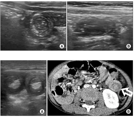
자연 정복된 소장형 장중첩증군과 수술적 치료를 필 요로 했던 소장형 장중첩증군을 비교했을 때 수술 정복 을 시행했던 군의 연령(105±17개월)은 자연 정복된 군 (51±20개월)에 비해 높았으며(p=0.045), 두 군 간의 성 별, 임상 증상이나 재발률의 유의한 차이는 없었다. 초 음파 검사 결과 자연 정복된 소장형 장중첩증(Fig. 1)과 수술적 치료를 필요로 했던 소장형 장중첩증(Fig. 2) 사 이에 위치에는 유의한 차이가 없었고 횡단 직경 및 주
Fig. 2. US (A) and CT scan (B) of a 5-year-old boy with recurrent abdominal pain and melena who underwent sur- gical reduction of small bowel intussusceptions at multiple sites (arrow). Based on bio- psy, he was diagnosed with Burkitt lymphoma.
Fig. 1. Typical benign small bowel intussusceptions (jeju- nojejunal) in a 4 year old boy with abdominal pain. Transe- verse (A) and longitudinal (B) US scan showed doughnut (A) and sandwich signs (B).
Transient SBI (n=15) Surgically managed SBI* (n=6) p value
Clinical features
Male:Female† 13:2 5:1 0.658
Mean age‡ (mo) (range) 51±20 (6 mo∼14 yrs) 109±17 (19 mo∼14 yrs) 0.045 Symptoms
Pain/irritability† 12 (85%) 5 (83%) 0.658
Bloody stool† 1 (6%) 2 (33%) 0.184
Mass† 1 (6%) 0 (0%) 0.714
Bowel ischemia/necrosis† 0 (0%) 2 (33%) 0.071
Recurrence† 3 (18%) 1 (16%) 0.684
Sonographic findings
Location (Right; Left; Middle)† 7; 6; 2 2; 2; 1 (1: multifocal) 0.956 Transverse diameter‡ (cm) 1.6±0.4 (0.9∼2.0) 3.2±0.5 (2.3∼3.5) 0.004 Thickness of outer rim‡ (mm) 1.6±0.8 (1.0∼2.5) 5.8±1.4 (4.4∼7.8) 0.004
Lead point† 1 (6%) 4 (67%) 0.031
Ascites† 1 (6%) 4 (67%) 0.017
Bowel distention† 8 (53%) 5 (83%) 0.221
*SBI: small bowel intussusception, †Fisher’s exact test, ‡Mann-Whitney U-test.
Table 2. Transient Small Bowel Intussusception and Surgically Managed Small Bowel Intussusception 변부 저에코성 원형의 두께는 수술을 했던 군에서 유의
하게 큰 것으로 나타났다(p=0.004). 장중첩증의 시작점
(lead point)과 복수도 수술을 했던 군에서 유의하게 많 이 관찰되었다(Table 2).
고 찰
장중첩증은 영아에서 급성 복통을 유발하는 흔한 원 인으로 대부분 소장-대장형으로 2세 미만에서 흔하게 발생하며 연령대는 3개월에서 1세 정도로 특히 남아에 서 2:1 정도의 비율로 흔하다. 소장형 장중첩증이 발 생하는 평균 연령은 4세 정도로 더 나이가 많은 아이들 에서 발생하는 경향이 있으며 성별과 관계가 없는 것으 로 알려져 있다8). 위치는 왼쪽이 흔하며 Strouse 등9)은 54%에서 공장(jejunum)에서 발생하고 18%는 중간 부 위에서, 29%는 회장(ileum)에서 발생했다고 보고하였 다. 소장형 장중첩증의 증상은 복통, 구토, 보챔이나 발 열같은 비특이적인 증상이 흔한 편으로 혈변, 복부 종 괴와 같은 전형적인 장중첩증의 증상은 비교적 드물어 진단이 어렵고 지연되는 경향이 있다10). 본 증례에서는 위치는 큰 차이를 보이지 않았고 간헐적인 보챔을 동반 한 복통은 3예로 모두 대장형 장중첩증이 함께 있는 경 우였으며 그밖의 경우는 주로 지속적인 복통이 많았고 혈변과 복부 종괴 같은 특징적인 증상은 각각 3예 (14%)와 1예(4%)로 비교적 적은 양상을 보였다. 복부 증상이 전혀 없었던 경우는 3예로 증상이 있었던 군에 비해 평균 연령이 높았고 기저 질환이 있는 경우가 많 았다.
장중첩증의 진단에 있어 초음파 검사는 높은 민감도 (98∼100%)를 보인다2,11∼13). 소장-대장형의 경우 횡단 촬영에서 직경이 3∼5 cm로 비교적 큰 편으로 길이가 길며 근위 횡행 결장이나 간 아래 부위에서 쉽게 발견 되는 경향이 있다11,14,15). 이에 비해 소장형 장중첩증은 횡단 촬영에서 관찰되는 병변의 직경이 2∼3 cm 정도 로 작고 3 cm 미만으로 길이가 짧으며, 회맹부에서 떨 어진 왼쪽이나 배꼽 근처에서 발견되는 경우가 많고, 초음파로 진단되는 정확도가 76.5% 정도인 것으로 나
타났다6,16). 그러나 실제로 소장형 장중첩증을 처음부터
의심하여 초음파 검사를 시행하는 경우는 드물고 대장 형 혹은 다른 질환을 감별하기 위하여 초음파를 시행하 거나, CT나 MRI 등을 시행했다가 우연히 발견되는 경 우가 대부분이다. Doi 등17)은 이러한 현상을 양성 소장 형 장중첩증으로 정의하였다. 소장형 장중첩증으로 진 행될 수 있는 경우로는 장벽이 부어있고 장 운동이 비
정상적이며 이전의 상처로 흉터나 유착이 있는 경우이 며 염증, 항암치료, 출혈이나 이전 수술 병력이 이러한 요인들과 관련이 있는 것으로 알려져 있다6,18). 수술을 시행하게 되는 경우는 진단이 늦어졌거나, 복막 자극 증상, 쇼크나 심한 탈진, 초음파 검사에서 중첩된 장벽 비후, 장 운동성과 관류 감소가 관찰되는 경우에 시행 한다. 국내에서 소장형 장중첩증 15예 중 20%에서 수 술적인 치료가 필요했음을 보고한 바 있으며19), Lee 등14) 은 수술을 시행했던 13명의 장중첩증 증례를 통해 소장 형 장중첩증의 주변부 저음영의 두께가 16 mm 이상인 경우 수술이 필요했다고 보고하였으며 Lim 등20)은 병 변의 두께가 40 mm 이상, 주변부 저음영의 두께가 10 mm 이상 시 수술적 치료가 필요할 수 있다고 하였다.
Kornecki 등6)은 소장형 장중첩증 증례 중 유발 병변이 있어 수술이 필요했던 경우는 매우 낮았다고 보고하였 으나 Ko 등8)은 병변 부위의 직경과 주변부 저음영 두 께가 합병증의 발생을 예측하지 못하며, 수술을 시행한 19예 중 46%에서 합병증이 발생했으므로 빠른 수술적 인 치료가 필요하다고 주장하였다. 본 연구에서는 자연 정복된 군에서 수술적 치료를 했던 군에 비해 병변의 두께 및 주변부 저음영의 두께가 모두 유의하게 얇은 양상을 보였으며 수술적 치료가 필요했던 경우는 28%
정도였다. 특히 동반 질환이 있는 경우에도 수술적인 치료가 필요 없이 회복된 경우가 4예(50%)에서 관찰되 었기 때문에 기질적 원인을 갖더라도 수술적 치료를 결 정하는데 있어 세밀한 검사 및 경과 관찰이 요구된다.
본 연구에서 소아에서 소장형 장중첩증은 비교적 증 상이 비특이적이며 자연 정복되는 경우가 많았다. 그러 나 연령이 높을수록, 초음파에서 관찰되는 병변의 직경 이 클수록, 시작점이 있는 경우에는 수술적인 치료가 필요하였다. 그러므로 소장형 장중첩증이 의심되는 경 우 수술적 치료를 먼저 고려하지 말고 임상 증상과 초 음파 소견을 통해 수술 여부를 판단해야 한다.
요 약
목 적: 장중첩증은 영아기 급성 복통의 흔한 원인 중 하나로 대부분 특발성의 소장-대장형이다. 반면 소장형 장중첩증은 드물며 시작점을 보이는 경우가 있고 수술 적 정복을 필요로하는 경우가 흔하다. 본 연구의 목적
은 소아에서 소장형 장중첩증의 임상 양상과 경과에 대 해 알아보고자 한다.
방 법: 2005년에서 2010년까지 서울대병원에서 소장 형 장중첩증으로 진단받은 21명의 환아들의 임상 양상 및 영상 소견을 후향적으로 분석하였다.
결 과: 임상 양상은 복통 및 보챔(85%), 구토(23%), 발열(14%), 혈변(14%) 및 복부 종괴(4%) 등이었다. 여 섯 명(28%)의 환아에서 수술적 치료가 필요하였다. 초 음파에서 병변의 직경은 1.6±0.7 cm였고 가장자리의 두께는 1.7±1.8 mm였다. 열한 명에서 병변이 왼쪽 복부 혹은 배꼽 주위였다. 수술적 치료가 필요했던 환아들은 자연 정복된 환아들에 비해 평균 연령이 높았다 (109±17개월:51±20개월). 병변의 평균 직경 및 가장 자리의 두께는 수술적 치료가 필요했던 환아들에서 더 큰 소견을 보였고 위치는 두 그룹 간에 차이를 보이지 않았다.
결 론: 소장형 장중첩증은 많은 소아에서 자연 정복 된다. 그러나 초음파에서 크기가 크고, 나이가 많을수 록 또한 병변의 시작점이 있으면 수술적 치료를 고려할 수 있다.
참 고 문 헌
1) DiFiore JW. Intussusception. Semin Pediatr Surg 1999;8:
214-20.
2) Lehnert T, Sorge I, Till H, Rolle U. Intussusception in children-clinical presentation, diagnosis and management.
Int J Colorectal Dis 2009;24:1187-92.
3) Ko SF, Tiao MM, Hsieh CS, Huang FC, Huang CC, Ng SH, et al. Pediatric small bowel intussusception disease:
feasibility of screening for surgery with early computed tomographic evaluation. Surgery 2010;147:521-8.
4) Saxena AK, Seebacher U, Bernhardt C, Hollwarth ME.
Small bowel intussusceptions: issues and controversies related to pneumatic reduction and surgical approach.
Acta Paediatr 2007;96:1651-4.
5) Hur NJ, Ryu MH, Lee DJ, Kwon JH. A clinical observation on children with transient small bowel intus- susception. Korean J Pediatr Gastroenterol Nutr 2000;3:
160-8.
6) Kornecki A, Daneman A, Navarro O, Connolly B, Manson D, Alton DJ. Spontaneous reduction of intus- susception: clinical spectrum, management and outcome.
Pediatr Radiol 2000;30:58-63.
7) Parikh M, Samujh R, Kanojia R, Sodhi KS. Does all small bowel intussuseption need exploration? Afr J Paediatr Surg 2010;7:30-2.
8) Ko SF, Lee TY, Ng SH, Wan YL, Chen MC, Tiao MM, et al. Small bowel intussusception in symptomatic pediatric patients: experiences with 19 surgically proven cases. World J Surg 2002;26:438-43.
9) Strouse PJ, DiPietro MA, Saez F. Transient small-bowel intussusception in children on CT. Pediatr Radiol 2003;
33:316-20.
10) Tiao MM, Wan YL, Ng SH, Ko SF, Lee TY, Chen MC, et al. Sonographic features of small-bowel intussusception in pediatric patients. Acad Emerg Med 2001;8:368-73.
11) del-Pozo G, Albillos JC, Tejedor D, Calero R, Rasero M, de-la-Calle U, et al. Intussusception in children: current concepts in diagnosis and enema reduction. Radiographics 1999;19:299-319.
12) Bisset GS 3rd, Kirks DR. Intussusception in infants and children: diagnosis and therapy. Radiology 1988;168:141- 5.
13) Lim HK, Bae SH, Lee KH, Seo GS, Yoon GS. Assess- ment of reducibility of ileocolic intussusception in child- ren: usefulness of color Doppler sonography. Radiology 1994;191:781-5.
14) Lee HC, Yeh HJ, Leu YJ. Intussusception: the sono- graphic diagnosis and its clinical value. J Pediatr Gastro- enterol Nutr 1989;8:343-7.
15) Verschelden P, Filiatrault D, Garel L, Grignon A, Perreault G, Boisvert J, et al. Intussusception in children:
reliability of US in diagnosis--a prospective study. Radio- logy 1992;184:741-4.
16) Pracros JP, Tran-Minh VA, Morin de Finfe CH, Deffrenne-Pracros P, Louis D, Basset T. Acute intestinal intussusception in children. Contribution of ultrasono- graphy (145 cases). Ann Radiol (Paris) 1987;30:525-30.
17) Doi O, Aoyama K, Hutson JM. Twenty-one cases of small bowel intussusception: the pathophysiology of idiopathic intussusception and the concept of benign small bowel intussusception. Pediatr Surg Int 2004;20:140-3.
18) Kim JH. US features of transient small bowel intus- susception in pediatric patients. Korean J Radiol 2004;5:
178-84.
19) Lee HS, Chung JY, Koo JW, Kim SW, Kim SH. Clinical characteristics of intussusception in children: comparison between small bowel and large bowel type. Korean J Gastroenterol 2006;47:37-43.
20) Lim HT, Park JH, Choi HJ, Kim JS, Shin HK, Gu CH.
Clinical approach of ultrasonography in the diagnosis of intussusception in infant and children. J Korean Pediatr Soc 1994;37:649-54.