■ 접 수 : 2012년 12월 10일, 수정 : 2012년 12월 17일, 채택 : 2013년 1월 5일
■ 교신저자 : 이종수, 서울시 동대문구 회기동 1번지 경희대학교 부속한방병원 한방재활의학과교실 Tel : (02) 958-9299, Fax : (02) 963-4983, E-mail : [email protected]
Ginsenoside Rg3이 흰쥐 척수압박손상의 초기 염증반응에 미치는 영향
정 벌⋅이종수
경희대학교 한의과대학 한방재활의학과교실
Effects of Ginsenoside Rg3 on Early-stage Inflammatory Response in Spinal Cord Compression of Rodents
Beoul Jeong, K.M.D., Jong-Soo Lee, K.M.D.
Dept. of Oriental Rehabilitation Medicine, College of Korean Medicine, Kyung-Hee University
Objectives :
In present study, we investigated the effects of ginsenoside Rg3 on early-stage inflammatory response in spinal cord compression of rodents.
Methods :
Spinal cord injury(SCI) was induced by a vascular clip method(30 g, 5 min) on the spinal cord of mice. Rg3 was treated orally at 1 hour prior to the SCI induction. Messenger ribonucleic acid(mRNA) expression of tumor necrosis factor-α(TNF-α), interleukin-1β(IL-1β), interleukin-6(IL-6) and cyclooxygenase-2(COX-2) was measured by the real-time polymerase chain reaction(RT-PCR). Microglia in the spinal cord tissue, neurophils and COX-2 in the peri-lesion and inducible nitric oxide synthase(iNOS) expression in the ventral horn of SCI induced rats were measured by immunohistochemical stain.
Results :
1. Rg3 significantly reduced the mRNA expression of TNF-α, IL-1β, and COX-2 in the spinal cord tissue compared with SCI group(p<0.05, p<0.01).
2. Rg3 significantly reduced the total number of activated microglia and proportion of phagocytic form in the total activated microglia compared with SCI group(p<0.05, p<0.01).
3. Rg3 significantly reduced myeloperoxidase(MPO) positive neurophil in the peri-lesion compared with SCI group(p<0.05).
4. Rg3 reduced the COX-2 expression in the tissue and motor neurons compared with SCI group.
5. Rg3 significantly reduced iNOS positive motor neurons in the ventral horn compared with SCI group(p<0.01).
Conclusions :
In conclusion, we demonstrated at first that treatment of ginsenoside Rg3 could reduce significantly the levels of inflammatory mediators in a spinal cord compression model of rodents. Therefore, these results suggested that ginsenoside Rg3 may be a useful antimiflamatory therapeutic candidate for SCI.
Key words : Ginsenoside Rg3, Early-stage inflammatory response, Spinal cord injury
Ⅰ. 서 론
척수손상은 기계적 충격이나 생화학적 충격 또는 혈역학적 변화 등이 복합적으로 작용하여 신경학적 장애를 발생시키는 것으로, 직접적인 신경전달로의 장애와 신경세포의 괴사(necrosis) 와 함께 이차적으로 진행되는 신경세포 자연사 (apoptosis)에 의한다
1,2). 전 세계적으로 약 250만 명 이상의 환자가 보고되고 있으며, 평균수명의 증가, 교통수단의 발달 등으로 외부손상에 따른 척수손상이 크게 증가하고 있는 추세이다
3). 척수 손상은 통증 및 운동 마비를 야기하여 환자의 삶 의 질을 저하시키며, 이와 관련된 심리적 불안정 및 경제적 손실 또한 큰 것으로 나타나 치료의 필요성이 대두되고 있다
4).
척수에 손상이 일어날 때 1차 손상은 역학적인 조직의 파괴, 출혈, 손상된 세포로부터의 대사물 질의 분비 등에 의해 일어나며, 이후 2차손상은 척수의 부종, 성장인자, 허혈, 칼슘의 이동, 과산 화물기에 의한 신경조직 손상, 염증반응, cytokine 의 작용, 재관류 등에 의해 발생한다
5). 따라서 약 물적인 치료로 손상의 진행을 막거나 호전을 시 킬 수 있는 여지가 있는 이 2차 손상을 줄이는 방법에 관한 연구가 대두되고 있다
6).
현재까지 허혈성 및 퇴행성의 각종 중추신경 계 질환에 대해 많은 한약물들이 신경세포 보호 또는 염증반응 억제 효과가 있다는 보고가 있었 으나, 이들 연구들은 주로 뇌질환 연구모형들을 통해 연구된 것이다
7-11). 그 중 ginsenoside는 人 蔘 saponin으로부터 추출된 항염증효과와 신경 세포손상 보호효과가 우수한 유효성분으로 약 30 여종이 보고되어 있다
12,13). 특히 ginsenoside Rg3 은 BV-2 microglia에서 cyclooxygenase-2(COX-2) 와 pro-inflammatory cytokine인 tumor necrosis
factor-α(TNF-α)와 interleukin-1β(IL-1β) 및 inducible nitric oxide synthase(iNOS) 발현을 억제하는 효 과와
14,15)국소뇌허혈(focal cerebral ischemia)
16), 대뇌피질 신경세포의 Ca
2+channel-mediated cell death
17), 해마 신경세포의 homocysteine-induced excitotoxicity에 대한 신경세포손상 보호효과
18)등의 많은 연구보고가 있으나, 척수손상에 대한 효과연구는 매우 미흡한 실정이다. 최근 黃連解 毒湯 및 狗脊 등이 척수압박손상 모델에서 신경 세포의 보호에 유효했다는 보고가 있었고
19,20), 특 히 黃連解毒湯은 척수압박손상 모델에서 염증반 응 억제 효과를 나타냈다는 보고가 있었다
21).
이에 저자는 뇌질환 연구모형에서 항염증효과와 신경세포손상 보호효과 등이 실험적으로 보고된 바 있는 ginsenoside Rg3가 척수압박손상의 초기 염증반응에 미치는 효과를 규명하기 위하여 척수 압박손상을 유발한 동물 모델에서 ginsenoside Rg3 투여 후 염증 매개 물질 및 염증 관련 유전 자 발현에 대한 영향을 관찰하여 유의한 결과를 얻었기에 보고하는 바이다.
Ⅱ. 연구방법
1. 재료
1) 동물
본 실험에 사용한 동물은 8주령 25~28 g의
C57BL/6J 수컷 생쥐와 9주령 약 300 g 전후의
Sprague-Dawley계 수컷 흰쥐를 국가공인 동물취
급업체(Nano Biotechnology, Korea)로부터 공급
받았다. 실험동물을 1주 이상의 환경적응 기간을
거치게 하였으며, 자동적으로 항온(21~23 ℃), 항
습(40~60%)이 유지되고 12시간 간격으로 낮과
밤이 교대되는 동일한 실험실 환경에서 무균음수 와 사료를 자유롭게 공급하며 사육하였다. 본 실 험은 경희대학교 실험동물 윤리위원회의 승인을 거친 후 시행하였다.
2) 약물
본 실험에 사용한 약물은 20(s)-ginsenoside Rg3 (Rg3; G3552, LKT Laboratories, USA)이며, 생쥐 와 흰쥐에게 각각 30 mg/kg을 척수압박손상 유 발 1시간 전에 1회 경구투여 하였다.
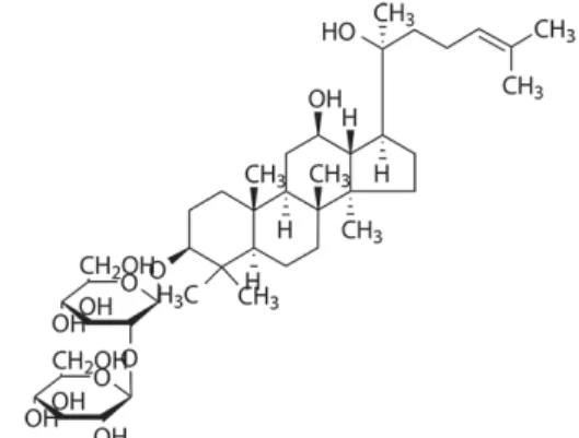
Fig. 1. Chemical structure of 20(s)-ginsenoside Rg3.
2. 방법
1) 실험군 분류
척수압박손상 후 TNF-α, IL-1β, IL-6 및 COX-2 의 messenger ribonucleic acid(mRNA) 발현 측 정을 위해서는 C57BL/6J 수컷 생쥐를 사용하였 고, 면역조직화학염색을 위해서는 Sprague-Dawley 계 수컷 흰쥐를 사용하였으며 각각 실험군은 다 음과 같이 구분하였다. 마취 등 일체의 처치를 가하지 않은 정상군(normal group), 마취와 배부 의 피부절개 및 척추의 천공 과정은 시행하였으 나 압박손상을 가하지 않은 Sham군(sham group),
위와 같이 사전 준비과정에 이어 압박손상을 가 한 대조군(spinal cord injury, SCI group) 및 대 조군과 같이 압박손상을 가하기 전 Rg3을 경구 투여한 군(SCI+Rg3 group)으로 나누었다. 각 군 의 실험동물은 생쥐와 흰쥐 각각 6마리씩 총 48 마리를 사용하였다.
2) 척수압박손상의 유발
척수압박손상의 유발은 실험동물을 pentobarbital (40 mg/kg) 복강주사로 마취한 다음 electronic temperature controller(CMA150, CMA, Sweden) 를 사용하여 정상체온(37±0.5 ℃)이 유지되는 상 태에서, 다음의 과정으로 실시하였다. 배부의 털 을 제거한 다음 피부와 배부 근육들을 절개한 상 태에서, 전기드릴을 사용하여 제 10흉추의 추궁 (vertebral lamina)을 제거하였다. 경막(dura mater) 이 유지되는 상태에서 척수가 노출되도록 하였 다. 이때 전기드릴에 의하여 척수 실질조직이 손 상되지 않도록 하였으며, 실질조직이 손상된 실 험동물은 실험에서 제외하였다. 생쥐의 경우에는 30 g force의 vascular clip(INS14120, Kent Scientific Corporation, USA)으로 척수를 5분간 압박하였 고
22), 흰쥐의 경우에는 척수에 35 g 무게의 원형 쇠막대(지름 약 2.0 mm)를 10분간 올려놓아 약 1.0 mm 정도 척수가 압박되도록 하였다
23). 이후 배부 근육과 피부를 봉합하고 마취에서 깨어나게 하였다.
3) 척수조직의 처리
척수압박손상 4시간 후에 생쥐는 즉시 단두하
고 척수를 분리하여 mRNA 발현량 관찰을 위해
사용하였으며, 흰쥐는 0.05 M phosphate buffered
saline(PBS)과 4% paraformaldehyde로 관류하고
척수를 분리하여 -40 ℃의 dry ice-isophentane
용액으로 동결시키고, cryocut으로 30 μm 두께의 횡단절편으로 제작하여 조직염색에 사용하였다.
4) TNF-α, IL-1β, IL-6 및 COX-2의 mRNA 발현량 측정
생쥐 척수조직으로부터 TNF-α, IL-1β, IL-6 및 COX-2의 mRNA 발현량 측정은 정량적인 real-time polymerase chain reaction(RT-PCR) 방법으로 측 정하였다. 척수압박손상 4시간 후 경추탈구 방법 으로 생쥐를 희생시키고 즉시 척수를 분리하여 압박손상 부위를 기준으로 1 cm 크기의 조직으 로 분리하였다. Trizol(Quiagen, Germany)을 사용 하여 척수조직으로부터 total RNA를 추출하였으 며, cDNA는 iScript cDNA Synthesis Kit(BioRad, USA)를 사용하여 역전사하였다. 희석한 cDNA와 각각 0.5 pMol의 TNF-α, IL-1β, IL-6, COX-2 및 β-actin primer를 혼합한 다음 iQ SYBR Green Supermix kit(BioRad, USA)와 CFX96 REAL-TIME PCR Detection System(BioRad, USA)을 사용하 여 RT-PCR을 수행하였다. 각각의 primer sequence는 Table Ⅰ과 같으며, 정량은 β-actin의 cycle time(Ct) 값과 Normal군의 정량 값으로부터 상대정량하였다.
TNF-α forward 5’-TGA GAA GTT CCC AAA TGG C-3’
reverse 5’-GCT ACA GGC TTG TCA CTC-3’
IL-1β forward 5’-TGA GCA CCT TCT TTT CCT TCA-3’
reverse 5’-TTG TCT AAT GGG AAC GTC ACA C-3’
IL-6 forward 5’-AGA CTT CAC AGA GGA TAC CA-3’
reverse 5’-GCA TCA TCG TTG TTC ATA CA-3’
COX-2 forward 5’-GCT GGC CTG GTA CTC AGT AGG TT-3’
reverse 5’-CGA GGC CAC TGA TAC CTA TTG C-3’
β-actin forward 5’-TTT CCA GCC TTC CTT GGG TAT G-3’
reverse 5’-CAC TGT GTT GGC ATA GAG GTC TTT AC-3’
TNF-α : Tumor Necrotic Factor-α IL-1β : Interleukin-1β
IL-6 : Interleukin 6 COX-2 : Cyclooxygenase-2 Table Ⅰ. Primer Sequences
5) 면역조직화학염색
흰쥐의 척수조직을 0.05 M PBS로 5분간 3회 씻고, 1% H
2O
2에서 10분간 반응시킨 다음 다시 3회 씻어낸 다음 10% normal horse serum(Vectastain, USA)과 bovine serum albumin(Sigma-Aldrich, USA)를 PBS에 섞은 blocking solution에 한 시간 정도 반응시켰다. 이후 PBS로 3회 씻어 낸 후 1차 항체를 처리하였다. 1차항체는 anti-Iba-1(1:250, MCA275EL, Serotec, USA), anti-myeloperoxidase (MPO)(1:250, A-0398, DAKO, USA), anti-iNOS(1:200, 610297, BD, USA) 및 anti-COX-2(1:100, 160106, Cayman Chemical, USA)를 사용하였다. PBS와 Triton X-100을 섞은 용액으로 희석한 후 4 ℃에 서 반응시켰으며, 다음 biotinylated anti-rabbit secondary antibody(1:200, Vector Labolatories, USA)를 실온에서 1시간동안 반응시키고, 조직을 PBS로 씻어내고, avidin-biotin immunoperoxidase 의 방법에 따라 각각 1시간씩 반응시켰다. 다음 0.05% DAB(Sigma-Aldrich, USA) 용액에서 2분간 발색 반응시켰다. 이후 0.5% cresyl violet 또는 0.2% methyl green 용액으로 약하게 배경염색한 다음 탈수, 봉입하여 조직표본을 제작하였다.
6) 조직의 관찰
면역조직화학방법으로 염색된 척수 조직표본
의 압박손상 주변부를 CCD 카메라(DP70, Olympus,
Japan)가 장착된 광학현미경(BX51, Olympus, Japan)
을 사용하여 관찰하였다. 양성반응 세포 수 측정
을 위해서 각각의 영상을 영상분석시스템에 저장
하고, ImageJ software(ver. 1.41, NIH, USA)를
사용하여 양성반응 세포 수를 측정한 다음 일정
면적(10
5μm
2)으로 보정하여 자료로 사용하였다.
3. 통계처리
본 실험에서 얻은 자료는 mean±standard deviation 으로 정리하였으며 SPSS(version 18.0) 통계프로 그램을 이용하여 처리하였다. 측정된 모든 자료 는 student's t-test를 사용하여 p<0.05의 유의수 준으로 검증하였다.
Ⅲ. 결 과
1. TNF-α, IL-1β, IL-6 및 COX-2 mRNA 발현량의 변화
압박손상에 의한 척수조직의 염증반응 변화를 관찰하기 위해서 pro-inflammatory cytokine인 TNF-α, IL-1β 및 IL-6의 mRNA 발현량을 RT-PCR 로 정량적으로 측정하였다. TNF-α mRNA 발현 량은 Normal군의 1.0±0.2 배에 비하여 Sham군은 12.9±3.6 배로 증가하였으며, SCI군은 35.5±6.7 배로 현저한 증가를 나타냈다. 이에 비하여 ginsenoside Rg3 30 mg/kg을 경구투여한 SCI+Rg3군은 15.7±3.1 배로 감소하여 SCI군에 비해 유의성(p<0.05) 있 게 TNF-α mRNA 발현량의 억제를 나타냈다(Fig.
2). IL-1β mRNA 발현량은 Normal군의 0.9±0.4 배에 비하여 Sham군은 7.0±2.4 배로 증가하였으 며, SCI군은 22.3±2.3 배로 현저한 증가를 나타냈 다. 이에 비하여 SCI+Rg3군은 11.1±1.1 배로 감 소하여 SCI군에 비해 유의성 있게 IL-1β mRNA 발현량의 억제를 나타냈다(Fig. 3). IL-6 mRNA 발현량은 Normal군의 1.0±0.2 배에 비하여 Sham군 은 48.5±20.9 배로 증가하였으며, SCI군은 189.1±53.9 배로 매우 현저한 증가를 나타냈다. 이에 비하여 SCI+Rg3군은 130.5±18.6 배로 SCI군에 비해 감소
하였으나 통계적 유의성은 없었다(Fig. 4). 이러한 결과로 ginsenoside Rg3은 pro-inflammatory cytokine 발현을 억제하는 작용이 있는 것을 확 인할 수 있었다.
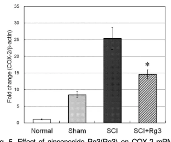
또한 pro-inflammatory cytokine과 함께 주요 한 염증조절 인자인 COX-2의 mRNA 발현량을 측정한 결과, Normal군의 1.1±0.1 배에 비하여 Sham군은 8.5±0.9 배로 증가하였으며, SCI군은 25.4±3.3 배로 현저한 증가를 나타냈다. 이에 비 하여 SCI+Rg3군은 14.6±1.4 배로 감소하여 SCI군 에 비해 유의성 있게 COX-2 mRNA 발현량의 억 제를 나타냈다(Fig. 5). 그러므로 ginsenoside Rg3 은 pro-inflammatory cytokine과 함께 COX-2 발 현을 억제하는 작용이 있는 것을 확인할 수 있었 다.
Fig. 2. Effect of ginsenoside Rg3(Rg3) on TNF-α mRNA expression in the spinal cord tissue of SCI induced mice(n=6).
SCI was induced by a vascular clip method(30 g, 5 min) on the spinal cord of mice. Rg3 was treated orally at 1 hour prior to the SCI induction. TNF-α mRNA expression was measured by the RT-PCR. Rg3 significantly reduced the TNF-α mRNA expression in the spinal cord tissue compared with SCI group.
*p<0.05 compared to the SCI group.
Fig. 3. Effect of ginsenoside Rg3(Rg3) on IL-1β mRNA expression in the spinal cord tissue of SCI induced mice(n=6).
SCI was induced by a vascular clip method(30 g, 5 min) on the spinal cord of mice. Rg3 was treated orally at 1 hour prior to the SCI induction. IL-1β mRNA expression was measured by the real-time PCR. Rg3 significantly reduced the IL-1β mRNA expression in the spinal cord tissue compared with SCI group.
*p<0.01 compared to the SCI group.
Fig. 4. Effect of ginsenoside Rg3(Rg3) on IL-6 mRNA expression in the spinal cord tissue of SCI induced mice.
SCI was induced by a vascular clip method(30 g, 5 min) on the spinal cord of mice. Rg3 was treated orally at 1 hour prior to the SCI induction. IL-6 mRNA expression was measured by the real-time PCR.
Fig. 5. Effect of ginsenoside Rg3(Rg3) on COX-2 mRNA expression in the spinal cord tissue of SCI induced mice.
SCI was induced by a vascular clip method(30 g, 5 min) on the spinal cord of mice. Rg3 was treated orally at 1 hour prior to the SCI induction. COX-2 mRNA expression was measured by the real-time PCR.
*p<0.05 compared to the SCI group.
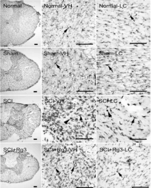
2. 미세아교세포 활성화의 변화
중추신경계 내 염증조절 세포인 미세아교세포 의 활성화 변화를 미세아교세포 표식자인 Iba-1 에 대한 면역조직화학염색 방법을 통해 관찰하였 다. Normal군에서는 척수압박손상 주변부의 회 색질과 백색질 모두에서 작은 세포체와 가늘고 긴 돌기들을 갖은 휴지기 미세아교세포들만이 관 찰되었다(Normal, Normal-VH, Normal-LC, arrows).
Sham군에서는 회색질의 일부 미세아교세포들이
활성화되어 세포질이 커지고 돌기들이 굵어진 것
을 관찰할 수 있었다(Sham, Sham-VH, Sham-LC,
arrows). 이에 비하여 척수에 압박손상을 받은
SCI군에서는 회색질의 거의 모든 미세아교세포
들이 세포질이 매우 커지고 돌기들이 짧고 굵어
진 형태의 activated form(SCI-VH, arrow) 또는
원형의 세포질과 돌기들이 사라진 아메바형태의
phagocytic form(SCI-VH, arrowhead)의 활성화
형태로 변했으며, 백색질의 미세아교세포들도 큰
세포질과 짧고 굵은 돌기의 activated form 활성 화 형태로 변했다(SCI-LC, arrow). 이에 비하여 ginsenoside Rg3 30 mg/kg을 경구투여한 SCI+Rg3 군은 SCI군과 같이 모두 활성화된 미세아교세포 들이 관찰되었으나 회색질에서는 전체적인 활성화 미세아교세포 수의 감소와 함께 phagocytic form 의 활성화 미세아교세포들이 감소하였고(SCI+Rg3, SCI+Rg3-VH, arrows), 백색질에서도 activated form의 미세아교세포가 감소한 것이 관찰되었다 (SCI+Rg3-LC)(Fig. 6).
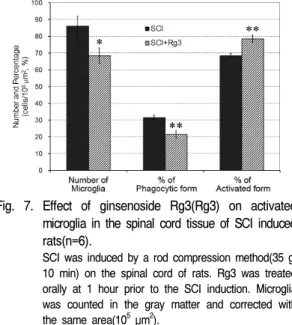
또한 SCI군과 SCI+Rg3군의 회색질에서 활성 화 미세아교세포의 수와 아메바형태의 phagocytic form 및 activated form 미세아교세포의 비율을 측정한 결과, SCI군의 활성화 미세아교세포 수는 86.0±6.0 개/10
5μm
2이었고, SCI+Rg3군은 68.5±4.5 개/10
5μm
2로 SCI군에 비해 유의성(p<0.05) 있게 활성화 미세아교세포 수의 감소가 관찰되었으며, SCI군에서는 phagocytic form이 31.6±1.4%, activated form이 68.4±1.4%인 것에 비해 SCI+Rg3군은 phagocytic form이 21.5±2.1%, activated form이 78.5±2.1%로 SCI군에 비해 유의성(p<0.01) 있게 phagocytic form 활성화 미세아교세포 비율의 감 소가 관찰되었다(Fig. 7). 이러한 결과는 ginsenoside Rg3이 미세아교세포의 활성화를 억제하는 작용 이 있음을 보여주는 것이다.
Fig. 6. Representative sections of Iba-1 immuno-labeled microglia in the spinal cord tissue of SCI induced rats.
SCI was induced by a rod compression method(35 g, 10 min) on the spinal cord of rats. Ginsenoside Rg3 was treated orally at 1 hour prior to the SCI induction.
Normal shows a “resting” form microglia in the ventral horn(VH) and in the lateral column(LC)(arrows). Sham shows some microglia were changed to a “activated”
form which has large cell body and shortened processes in VH and LC(arrows). SCI shows all microglia were changed to “activated” form(arrows) and “phagocytic”
form which is a large ameboid shape without processes (arrowheads) in VH and LC. Ginsenoside Rg3 treated SCI+Rg3 shows a decrease of activated microglia in VH and LC, especially in “phagocytic” form, compared with SCI group(arrows). The scale bars represent 100 μm.
Fig. 7. Effect of ginsenoside Rg3(Rg3) on activated microglia in the spinal cord tissue of SCI induced rats(n=6).
SCI was induced by a rod compression method(35 g, 10 min) on the spinal cord of rats. Rg3 was treated orally at 1 hour prior to the SCI induction. Microglia was counted in the gray matter and corrected with the same area(105μm2).
*p<0.05, †p<0.01 compared to the SCI group.
3. 중성백혈구 침윤의 변화
척수 압박손상 주변부에서 조직손상과 염증반 응을 유발하는 인자인 중성백혈구의 침윤을 중성 백혈구 발현효소인 MPO에 대한 면역조직화학염 색 방법을 통해 관찰하였다. Normal군에서는 MPO 에 양성반응을 보이는 중성백혈구의 침윤이 관찰 되지 않았고(normal), Sham군에서도 중성백혈구 의 침윤은 관찰되지 않았다(sham). 척수에 압박 손상을 받은 SCI군에서는 MPO 양성반응의 중성 백혈구가 압박손상을 받은 주변부에서 관찰되었 으며(SCI, arrows), 그 수를 측정한바 24.3±2.3 개 /10
5μm
2가 관찰되었다. 이에 비하여 ginsenoside Rg3 30 mg/kg을 경구투여한 SCI+Rg3군은 17.7
±1.6 개/10
5μm
2가 관찰되어 SCI군에 비하여 유 의성(p<0.05) 있게 중성백혈구 침윤의 억제를 나 타내었다(SCI+Rg3, arrows)(Fig. 8, 9).
Fig. 8. Representative sections of MPO positive neutrophils in the peri-lesion of SCI induced rats.
SCI was induced by a rod compression method(35 g, 10 min) on the spinal cord of rats. Ginsenoside Rg3 was treated orally at 1 hour prior to the SCI induction.
Normal and Sham do not demonstrate MPO positive neutrophil. Rg3 treated SCI+Rg3 shows a decrease of MPO positive neutrophils compared with SCI group(arrows).
The scale bars represent 100 μm.
Fig. 9. Effect of ginsenoside Rg3(Rg3) on MPO positive neutrophils in the peri-lesion of SCI induced rats (n=6).
SCI was induced by a rod compression method(35 g, 10 min) on the spinal cord of rats. Rg3 was treated orally at 1 hour prior to the SCI induction. MPO positive neutrophils were counted in the peri-lesion and corrected with the same area(105 μm2). Normal and Sham are not represented in the figure because MPO positive neutrophil was not observed. Rg3 significantly reduced MPO positive neurophil in the peri-lesion compared with SCI group. *p<0.05 compared to the SCI group.
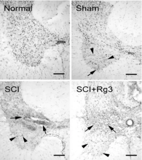
4. 압박손상 주변부에서 COX-2 발현의 변화
척수 압박손상 주변부에서 염증반응 조절인자인 COX-2 발현의 변화를 면역조직화학염색 방법을 통해 관찰하였다. Normal군에서는 척수조직과 전 각의 운동신경세포 모두에서 COX-2가 발현되지 않 았고(normal), Sham군에서는 운동신경세포에서는 COX-2가 발현되지 않았으나(sham, arrowheads), 척수조직의 일부에서 COX-2가 발현된 것이 관찰 되었다(sham, arrow). 척수에 압박손상을 받은 SCI 군에서는 압박손상 주변부에서 매우 강한 COX-2 발현이 관찰되었고(SCI, arrows), 척수 전각의 일 부 운동신경세포에서도 COX-2가 발현되었다(SCI, arrowheads). 이에 비하여 ginsenoside Rg3 30 mg/kg을 경구투여한 SCI+Rg3군은 압박손상 주 변부에서 COX-2의 발현 강도가 미약해졌으며 (SCI+Rg3, arrows), 척수 전각의 운동신경세포에 서는 COX-2 발현을 관찰할 수 없었다(SCI+Rg3, arrowheads)(Fig. 10). SCI군의 운동신경세포에서 만 COX-2 양성반응 세포가 관찰되었으므로 그 수를 측정하여 비교하지는 않았다.
Fig. 10. Representative sections of COX-2 expression in the peri-lesion of SCI induced rats.
SCI was induced by a rod compression method(35 g, 10 min) on the spinal cord of rats. Ginsenoside Rg3 was treated orally at 1 hour prior to the SCI induction.
Normal does not demonstrate COX-2 expression.
Sham shows weak COX-2 expression(arrow), but not in the motor neurons(arrowheads). SCI shows strong COX-2 expression in the tissue(arrows) and motor neurons(arrowheads). Rg3 treated SCI+Rg3 shows a decrease of COX-2 expression in the tissue(arrows) and motor neurons(arrowheads) compared with SCI group. The scale bars represent 200 μm.
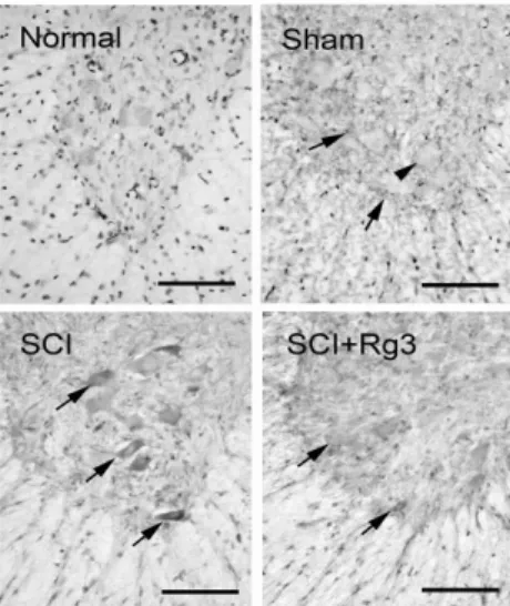
5. iNOS 발현의 변화
척수 전각 운동신경세포에서 염증 관련인자 중
하나인 iNOS의 발현을 면역조직화학염색 방법을
통해 관찰하였다. Normal군에서는 척수 전각과
운동신경세포 모두에서 iNOS가 발현되지 않았고
(normal), Sham군에서는 운동신경세포에서는 iNOS
가 발현되지 않았으나(sham, arrowhead), 척수
전각의 조직에서는 일부 iNOS가 발현된 것이 관
찰되었다(sham, arrows). 척수에 압박손상을 받은
SCI군에서는 척수 전각의 조직은 물론 거의 모든
운동신경세포에서 강한 iNOS 발현이 관찰되었으
며(SCI, arrows), 그 수를 측정한바 12.7±1.1 개
/10
5μm
2가 관찰되었다. 이에 비하여 ginsenoside
Rg3 30 mg/kg을 경구투여한 SCI+Rg3군은 운동신
경세포에서 iNOS 발현의 강도가 미약해졌으며
(SCI+Rg3, arrows), 그 수는 7.8±0.7 개/10
5μm
2로 관찰되어 SCI군에 비하여 유의성(p<0.01) 있
는 iNOS 발현의 억제가 관찰되었다(Fig. 11, 12).
Fig. 11. Representative sections of iNOS expression in the ventral horn of SCI induced rats.
SCI was induced by a rod compression method(35 g, 10 min) on the spinal cord of rats. Ginsenoside Rg3 was treated orally at 1 hour prior to the SCI induction.
Normal does not demonstrate COX-2 expression.
Sham shows weak COX-2 expression(arrows), but not in the motor neurons(arrowhead). SCI shows strong COX-2 expression in the motor neurons(arrows). Rg3 treated SCI+Rg3 shows a decrease of COX-2 expression in the motor neurons(arrows) compared with SCI group.
The scale bars represent 100 μm.
Fig. 12. Effect of ginsenoside Rg3(Rg3) on iNOS positive motor neurons in the ventral horn of SCI induced rats(n=6).
SCI was induced by a rod compression method(35 g, 10 min) on the spinal cord of rats. Rg3 was treated orally at 1 hour prior to the SCI induction. iNOS positive cells were counted in the ventral horn and corrected with the same area(105 μm2). Normal and
Sham are not represented in the figure because iNOS positive cell was not observed.
*p<0.01 compared to the SCI group.
Ⅳ. 고 찰
척수손상의 유병률은 인구 백만 명 당 721명으 로 매년 인구 백만 명 당 30~32명의 새로운 척수 손상이 발생되는 것으로 보고되고 있다
24). 척수 손상 환자의 약 40%는 사지마비, 약 60%는 하반 신마비를 가지고 있으며
25), 환자는 물론 그 가족 들이 느끼는 심리적 불안정과 재활치료에 부담되 는 엄청난 경제적 손실 등을 고려할 때 척수손상 에 대한 유의한 치료법의 개발이 절실하다
26). 최 근 줄기세포 치료법의 개발이 진행되고는 있으나 아직은 초기단계에 머물고 있으며
27,28), axon의 재 생 촉진을 위한 세포이식과 유전공학적 치료법 등의 개발을 위한 몇몇 연구들이 수행되고 있을 뿐이다
29,30).
척수손상은 1차와 2차 손상으로 나눌 수 있는
데, 척수에 1차손상인 기계적인 손상이 일어난
후 수 분에서 수 시간 후부터 복잡한 급성 병태
생리학적 변화에 따른 2차손상이 나타나며, 2차
손상은 보통 24시간 정도에 최고조에 달한다
31).
척수압박손상의 2차손상 과정에서는 microglia의
활성화와 neutrophil의 침윤, cytokine 분비 및
cyclooxigenase-2(COX-2) 발현 등에 의한 염증반응
이 손상을 촉진시킨다. 특히 tumor necrosis factor-α
(TNF-α)를 포함한 염증성 cytokine은 inducible
nitric oxide synthase(iNOS) 발현을 통해 nitric
oxide(NO) 생성을 촉진하여 세포독성 및 조직손
상을 가속화시킨다
5). 1차손상은 발생 즉시 비가
역적인 변화가 일어나므로 치료의 여지가 없지만
2차 손상에 대해서는 진행을 막거나 호전을 시킬
수 있으므로 이에 대한 더욱 활발한 연구가 요구
된다
6,32). 현재까지 현대의학에서는 고용량 스테 로이드를 이용하는 치료 외에 근본적인 치료는 이루어지지 못하고 있으며
33-35), 이와 관련한 한의 학적 연구는 미미한 실정으로, 최근 黃連解毒湯 및 狗脊 등이 척수압박손상 모델에서 신경세포의 보호에 유효했다는 보고가 있으며
19,20), 특히 黃連 解毒湯은 척수압박손상 모델에서 조직손상과 염 증반응 억제 효과를 나타냈다는 보고가 있었다
21).
본 연구에서는 척수압박손상에 의한 2차손상의 원인이 되는 염증반응에 주안점을 두고, 뇌질환 연 구모형에서 항염증효과와 신경세포손상 보호효 과 등이 실험적으로 보고된 바 있는 ginsenoside Rg3가 척수압박손상의 초기 염증반응에 미치는 효과를 규명하기 위해 연구를 설계하였다. 척수 압박손상을 유발한 동물 모델에서 척수조직의 TNF-α, IL-1β, IL-6 및 COX-2 mRNA 발현량을 RT-PCR 방법으로 관찰하였고, 미세아교세포의 활성화, MPO 양성반응의 중성백혈구 침윤, COX-2 와 iNOS 발현을 면역조직화학염색으로 관찰하였 다. 본 연구는 첫째, 척수압박손상 동물 모델에서 의 급성 염증 반응에 대한 ginsenoside Rg3의 억 제 효과를 규명하여 치료에의 응용 가능성을 제 기했다는 데 그 의의가 있으며, 둘째, ginsenoside Rg3가 척수압박손상에서 염증을 억제시키는 중 요한 분자생물학적 기전을 제시했다는 데 있어서 도 큰 의미가 있다고 할 수 있겠다.
Ginsenoside는 人蔘 saponin으로부터 추출된 항염증효과와 신경세포손상 보호효과가 우수한 유효성분으로 약 30여종이 보고되어 있다
12,13). 특히 ginsenoside Rg3은 BV-2 microglia에서 COX-2와 pro-inflammatory cytokine인 TNF-α와 interleukin-1β 및 iNOS 발현을 억제하는 효과와
14,15)국소뇌허 혈(focal cerebral ischemia)
16), 대뇌피질 신경세포 의 Ca
2+channel-mediated cell death
17), 해마 신 경세포의 homocysteine-induced excitotoxicity에
대한 신경세포손상 보호효과
18)등의 많은 연구보 고가 있다. 그러나 척수손상에 대한 효과연구는 매우 드물기에, 본 연구에서 그 척수압박손상에 대한 항염증효과 규명으로 치료에의 응용 가능성 을 시험해 볼 가치가 있다고 판단하였다.
Ginsenoside Rg3의 투여는 압박손상을 받은 척수조직의 TNF-α, IL-1β 및 COX-2 mRNA 발 현량과 미세아교세포 활성화를 유의하게 억제하 였고, 압박손상 주변부에서 MPO 양성반응의 중 성백혈구 침윤을 유의하게 억제하였다. 척수압박 손상 과정에서는 신경계의 손상 후 수 일 이내에 axon의 퇴행성 변화가 일어나는데, 이러한 부위 에서 신경세포의 부산물과 분해 된 수초 등의 제 거를 위해 염증세포들이 활동하게 되는 것이다.
기계적인 손상이 있거나 실험적으로 염증을 유발 하는 cytokine을 주사한 경우 호중성백혈구의 침 윤이 일어나게 되고
36,37), 미세아교세포는 손상 24 시간 후 활성-미세아교세포 수가 증가되고 약 7 일 후 최고조에 달한다
38). 이러한 활성-미세아교 세포가 TNF-α, IL-1, nitric oxide 등을 분비하여 척수의 2차손상을 일으키는 것이다
39,40). 따라서, 위의 결과는 ginsenoside Rg3의 항염증 활성이 이러한 염증 매개 물질의 조절에 의한다는 것을 의미한다.
또한 ginsenoside Rg3은 압박손상 주변부에서 COX-2 발현을 억제하였고, 척수 전각의 운동신 경세포에서 iNOS 발현을 유의하게 억제하였다.
COX-2는 여러 가지 병리적 상태와 관련되며, 염증상
태에서는 prostaglandin을 생산하게 하는 inducible
enzyme으로, 결과적으로 substance P, nitric oxide
등이 분비되어 다양한 병리기전에 관여한다
41,42).
iNOS는 척수손상의 초기에 발현되어 NO를 통해
2차 손상을 유발하고 그 정도를 조절하는 손상
촉진인자이다. 그러므로 척수손상 초기에 iNOS
발현을 억제하는 것이 이후 나타나는 신경학적
증상의 개선과 조직병리학적 소견을 개선하는 데 중요하다고 하겠다
43,44). 따라서 이러한 결과는 ginsenoside Rg3가 척수압박손상에서 염증반응 을 억제하는 기전을 통해서 척수의 2차 손상을 개선하는 효능이 있다는 것을 보여 준다.
이상의 결과들을 종합하면, 본 연구에서는 ginsenoside Rg3가 척수압박손상 동물 모델에서 pro-inflammatory cytokine과 COX-2 발현을 조절 하고 미세아교세포의 활성화를 억제하는 작용을 통 해 척수압박손상의 초기 염증반응에 유의한 효능이 있음을 확인하였다. 이러한 결과는 ginsenoside Rg3가 척수압박손상의 치료에 유효한 대안이 될 수 있음을 암시한다. 다만, 임상에서 척수압박손 상의 치료제로 적용하기 위해서는 더 자세한 기 전을 설명할 수 있는 보다 심도 있는 실험연구 및 임상연구가 필요할 것으로 사료된다.
Ⅴ. 결 론
人蔘의 성분 중 항염증효능과 신경세포손상 보호효과가 우수한 ginsenoside Rg3이 척수압박 손상 초기의 염증반응에 미치는 효과를 규명하기 위해 동물실험을 한 후 다음과 같은 결과를 얻었 다.
1. Ginsenoside Rg3은 압박손상을 받은 척수조 직의 TNF-α, IL-1β 및 COX-2 mRNA 발현 량을 유의하게 억제하였다.
2. Ginsenoside Rg3은 압박손상을 받은 척수조 직의 미세아교세포 활성화를 유의하게 억제 하였다.
3. Ginsenoside Rg3은 압박손상 주변부에서
MPO 양성반응의 중성백혈구 침윤을 유의 하게 억제하였다.
4. Ginsenoside Rg3은 압박손상 주변부에서 COX-2 발현을 억제하였다.
5. Ginsenoside Rg3은 압박손상 주변부 척수 전각의 운동신경세포에서 iNOS 발현을 유 의하게 억제하였다.
이상의 실험 결과, ginsenoside Rg3가 척수압 박손상의 치료에 유효한 대안이 될 수 있을 것으 로 사료된다.
참고문헌