J Pain Auton Disord Vol. 4, No. 1 2015 20
Journal of Pain and Autonomic Disorders Vol. 4, No. 1, 2015
Received: April 22, 2015 / Revised: May 24, 2015 / Accepted: May 24, 2015 Address for correspondence: Suk Yun Kang, MD, PhD
Department of Neurology, Hallym University Dongtan Sacred Heart Hospital, Hallym University College of Medicine, 7 Keunjaebong-gil, Hwaseong 445-907, Korea
Tel: +82-31-8086-2310, Fax: +82-31-8086-2317, E-mail: [email protected] ISSN 2287-6170
Journal of Pain and Autonomic Disorders
CASE REPORT Vol. 4, No. 1, June 2015
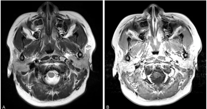
인두위턱 양성신경종양에 의한 반복실신
한림대학교 의과대학 강동성심병원 신경과,
a한림대학교 의과대학 동탄성심병원 신경과
안소현·최미송a·조수진a·김주용a·권기한a·강석윤a
Recurrent Syncope Due to a Benign Neurogenic Tumor in the Parapharyngeal Space
So Hyun Ahn, MD, Mi-Song Choi, MD
a, Soo-Jin Cho, MD, PhD
a, Joo Yong Kim, MD, PhD
a, Ki-Han Kwon, MD
a, Suk Yun Kang, MD, PhD
aDepartment of Neurology, Hallym University Kangdong Sacred Heart Hospital, Hallym University College of Medicine, Seoul;
a