J. Exp. Biomed. Sci. 2010, 16(3): 133~138
Isolation of Environmental Mycobacteria from Diverse Water Samples Using Cetylpyridinium Chloride
Yeonim Choi1, Hyunwoo Jin1, Gyusang Lee1, Jong Bae Kim1, In Keun Song2, Young Joon Kim2 and Hyeyoung Lee1,†
1Department of Biomedical Laboratory Science, College of Health Sciences, Yonsei University, Wonju-si, Gangwon-do 220-710, 2Division of Biotechnology, The Catholic University, 43-1 Yeokgok 2-dong,
Wonmi-gu, Buchoen, Gyeonggi-do 420-743, Korea
Despite of the increasing importance of environmental mycobacteria, detection and identification of mycobacteria from environmental sources including water have been fraught with technical difficulties. Although, several protocols to optimize isolation of mycobacteria from water sources have been reported, standard method has not yet been established. In this study, usefulness of cetylpyridinium chloride (CPC), a cationic quaternary ammonium compound, for the isolation of environmental mycobacteria from diverse water samples was evaluated. For this, water samples from diverse water sources such as effluent water, lake water, and underground water were collected, treated with diverse concentrations of CPC, and plated on the solid agar plates. Subsequently individual colonies grown on the plates were sequence analyzed for identification of each colony. In brief, the results from this study showed that the growth of mycobacteria was enhanced by use of CPC as a pre-treatment reagent to water samples by inhibiting growth of other non-mycobacteria in water. In fact, the effect of CPC to decontaminate non-mycobacteria for isolation of mycobacteria was better than 1~4% of NaOH, which is a routinely used decontaminating reagent widely employed for culturing mycobactera from sputum specimens. Therefore, the results from this study seems to support that the CPC pre-treatment may be useful for isolation of mycobacteria from diverse sources including clinical specimens which are often contaminated with other bacteria.
Key Words: Environmental mycobacteria; Decontamination reagents; Cetylpyridinium chloride; Environmental water samples
INTRODUCTION
Recently, there have been increasing evidences showing that nontubuerculous mycobacteria from environmental sources are implicated in a variety of human diseases.
mycobacteria are common saprophytes found in all natural ecosystems including water, soil, food, dust, and aerosols (Dailloux et al., 1999). Mycobacteria have been also demonstrated to be present in drinking water (Goslee et al.,
1976; Kubalek et al., 1995; Kubalek et al., 1996; Covert et al., 1999; Falkinham et al., 2001; Le Dantec et al., 2002;
Vaerewijck et al., 2005), drinking water distribution systems (Chang et al., 2002; Le Dantec et al., 2002a; Le Dantec et al., 2002b; Vaerewijck et al., 2005), hot water systems (Du Moulin et al., 1988), spas (Embil et al., 1997), and pools (Leoni et al., 1999; Livanainen et al., 1999). Mycobacteria are not killed by common disinfectants and can tolerate wide ranges of pHs and temperatures, these organisms can persist in water for long periods (Le Dantec et al., 2002).
Despite of the increasing importance of mycobacteria in clinical aspects, detection and identification of mycobacteria from environmental sources have been extremely hindered (Kazda, 1983; Du Moulin, 1986; Falkinham, 1996). This difficulty is often due to various growth rates and specific growth requirements of mycobacteria, contamination by
*Received: 18 June, 2010 / Revised: 16 September, 2010 Accepted: 27 September, 2010
†Corresponding author: Hyeyoung Lee, Department of Biomedical Laboratory Science College of Health Sciences, Yonsei University, Wonju-si, Gangwon-do 220-710, Korea.
Tel: 82-33-760-2740, Fax: 82-33-760-2561 e-mail: [email protected]
other bacteria that outgrow mycobacteria, and the nature of diverse sources of environmental samples. For this reason, choice of pretreatment methods to limit other bacteria than mycobacteria and fungal overgrowth and enhance growth of mycobacteria (Thomson et al., 2008) is very important.
In past, substances which can kill other bacteria and pathogenic fungi have shown to increase the mycobacterial culture positivity and reduce the contamination rate. Mineral acids (HCl) and bases (eg, NaOH), organic acids (eg, oxalic acid), and detergents have been used as decontaminating agents (Songer JG, 1981). However, the pretreatment method can also prevents the detection of certain species of mycobacteria and reduce the rate of positive samples and the number of colonies seen. Several authors have described the use of cetylpyridinium chloride (CPC) for decontami- nation of diverse water samples from environmental sources (Du Moulin, 1978; Schulze-Robbecke et al., 1991; Le Dantec et al., 2002b; Falkinham, 2004; Thomson et al., 2008). CPC is an antiseptic that kills bacteria and other microorganisms, and used in some types of mouthwashes, toothpastes, anti- sore throat sprays.
The present study was set to evaluate the usefulness of CPC as a method for pretreatment of water samples for isolation of mycobacteria.
MATERIALS AND METHODS Collection of water samples
A total of 10 water samples were collected from 3 different types of water sources; effluent water, lake water, and underground water. A total of 8 water samples from waste water treatment plants, 1 surface water from the lake Maeji, and 1 from underground water at Wonju-si, Gangwon province, Korea were taken according to the standard methods (American Public Health Association, 1995; Korea ministry of Environment. 2002). Samples were transported to the laboratory and were kept at 4℃ until analysis. All samples were analyzed within 24 h of collection.
Decontamination and filtration of water samples In order to evaluate the effect of CPC as a pretreatment method for water samples for detecting environmental
mycobacteria, CPC and NaOH were compared using lake and ground water samples. For this, water samples were analyzed by the method reported by previous researchers (Neumann et al., 1997; Covert, 1999; Falkinham et al., 2001). In brief, 200 ml of water samples were exposed to from 0.01% to 0.1% of CPC (Sigma Chemical Co., St.
Louis, U.S.A) and to 1% to 4% of NaOH for 30 min at RT.
All water samples were filtered through 0.45 μm nitro- cellulose membrane filter (Millipore Corp., Bedford, Mass.) by vaccum filtration after exposure to CPC and NaOH.
Water samples were decontaminated by CPC and NaOH.
In the second set of experiments, evaluate the effect of CPC for the isolation of mycobacteria from 10 water samples. Water samples were exposed to from 0.01% to 0.1% of CPC (Sigma Chemical Co., St. Louis, U.S.A.) at RT. And then, water samples were filtered through 45 μm nitrocellulose membrane filter (Millipore Corp., Bedford, Mass.) by vaccum filtration after exposure to CPC.
Processing of cultural detection of nontubuerculous mycobacteria
After filtration, the membranes were rinsed with PBS and transferred to Middlebrook 7H10 agar plates (Difco Laboratories, Detroit, Mich.) containing 500 mg of Amphotericin B (Sigma Chemical Co. St. Louis, U.S.A.) per liter. The plates were then sealed and incubated at 37℃
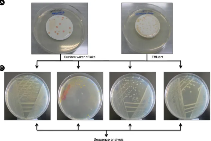
with 10% CO2 for 8 weeks. All plates were examined at weekly intervals for 8 weeks. After 8 weeks of incubation, all the colonies grown on the plate were enumerated, acid fast stained and individual colonies which looked distinct each other were then restreaked onto the new Middlebrook 7H10 agar plate (Fig. 1).
DNA extraction for PCR amplification
Subsequently, DNA was extracted from a single isolated colony by CTAB method as previously described (Lemarchand et al., 2005), and subjected for PCR ampli- fication of 16S rRNA gene. The quality and the quantity of the extracted DNA were determined by electrophoresis using a 1% agarose gel.
Identification of nontubuerculous mycobacteria by 16S rRNA gene sequencing
All Mycobacterium isolates that were cultured on the filter membranes were subjected to 16S rRNA gene sequencing. To design primer molecules, data base of 16S rRNA sequences of mycobacteria were compiled from GenBank database of National Center for Biotechnology Information web site (http://www.ncbi.nlm.nih.gov), and the sequence data for mycobacteria were aligned by using the Clustral method (http://www.cmbi.kun.nl/bioinf/tools/
clustalws.html). The PCR primers were as following: 16S - SF: 5'-AAYACATGCAAGTCGARCK-3' and 16S - R5H-A:
5'-ACRDAKTTRGCCGKKGCTT-3' (479 base pairs in length). The amplification reactions were performed with a Thermal cycler (GeneAmp PCR® System 2700). The PCR condition used was denaturation at 95℃ for 30 s, annealing
at 52℃ for 30 s, and extension at 72℃C for 30 s and a total of 35 cycles were used. Successful amplification was confirmed by using agarose gel electrophoresis. Sequencing of the PCR products was carried out at genotech (Daejeon, Korea). The sequences were analyzed with databases GenBank. Species identification was made on the basis of the sequence homology to the respective sequences in the GenBank database of National Center for Biotechnology Information web site (http://www. ncbi.nlm.nih.gov).
RESULTS AND DISCUSSION
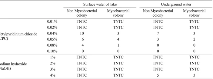
First of all, in order to compare the effect of CPC and NaOH treatment on mycobacterial isolation, water samples were taken from the lake and underground water. Water samples were then exposed to from 0.01% to 0.1% of CPC and from 1% to 4% of NaOH for 30 min at room A
B
Fig. 1. Isolation of non mycobacterial and mycobacterial colones from effluent and surface water of lake by treated 0.05% CPC.
(A) Microorganisms grown on the filtered membrane were incubated for 8 weeks at Middlebrook 7H10 agar plate, and then (B) single isolated individual colonies obtained from plate (A) were restreaked at new Middlebrook 7H10 agar plate for further identification of isolates. The filter membranes in (A) had been used for passing water samples treated with CPC. Subsequently DNAs were extracted from individual colonies and then their 16S rRNA gene were PCR-sequence analyzed for identification of species.
temperature, and subsequently plated on solid agar media.
As shown in the Table 1, when 1% to 4% of NaOH were used, too many microbial organisms grew on the plate (to numerous to count; TNTC), making impossible to identify any colonies. On the other hand, at the concentration of 0.04% or more of the CPC, countable numbers of individual colonies were grown on the plate, making possible to identify colony isolates. Next, for identification of individual colonies, each colonies grown on the plates were subjected for sequence analysis, and numbers of non-mycobacterial colonies and mycobacterial colonies were counted (Table 1).
Subsequently, in order to evaluate the usefulness of CPC with more diverse water samples, 10 water samples were taken from various water sources: 8 waste water treatment plants, 1 surface of the lake, and 1 underground water. Then, the water samples were treated with different concentration of CPC and cultured on the plates. As shown in the Table 2,
the counts of mycobacterial colonies isolated fro water samples ranged from 1 CFU/200 ml to too numerous to count. The highest level of mycobacterial growth was obtained with diverse water samples by using 0.04% CPC treatment. From the concentration of 0.04% CPC, the number of non-mycobacterial growth started to be control- lable, and mycobacterial colonies were countable. In fact, the optimized concentration of the CPC for isolation of Mycobacteria seems to reside between 0 04~0.05%. The identification of each colony to be mycobacteria or non- mycobacteria was made by sequence analysis of each individual colonies as previous experiment. Overall, 0.04%
CPC appeared to be the most effective both in reducing non-mycobacterial contaminants and in allowing good recovery of mycobacteria from water samples.
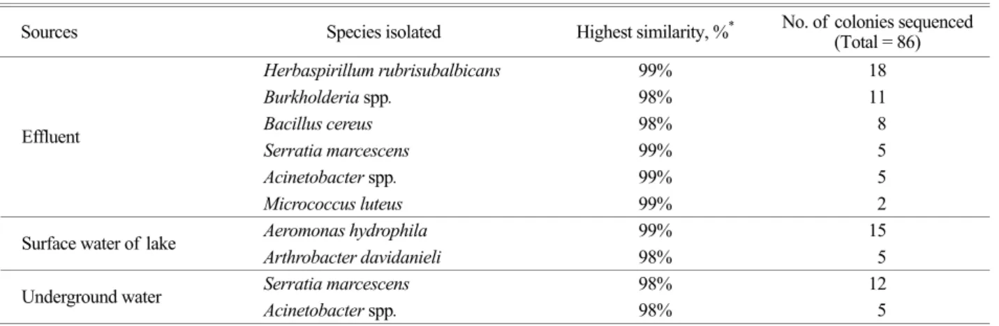
As a result of sequence analysis of a total of 134 individual colonies, 86 colonies (64%) were determined to Table 1. Effects of different concentrations of CPC and NaOH on microbial growth isolated from surface water of lake and underground
water
Surface water of lake Underground water Non Mycobacterial
colony Mycobacterial
colony Non Mycobacterial
colony Mycobacterial colony
0.01% TNTC TNTC TNTC TNTC
0.02% TNTC TNTC TNTC TNTC
0.04% 10 3 7 3
0.05% 6 4 3 2
0.08% 4 1 0 0
Cetylpyridinium chloride (CPC)
0.10% 0 0 0 0
1% TNTC TNTC TNTC TNTC
2% TNTC TNTC TNTC TNTC
3% TNTC TNTC TNTC TNTC
Sodium hydroxide (NaOH)
4% TNTC TNTC 5 3
*TNTC: Too Numerous To Count
Table 2. Effects of different concentrations of CPC on microbial growth isolated from diverse water samples Effluent (n=8) Surface water of lake (n=1) Underground water (n=1) Non Mycobacterial
colony Mycobacterial
colony Non Mycobacterial
colony Mycobacterial
colony Non Mycobacterial
colony Mycobacterial colony
0.01% TNTC TNTC TNTC TNTC TNTC TNTC
0.02% TNTC TNTC TNTC TNTC 12 4
0.04% 26 11 12 9 5 1
0.05% 18 9 8 8 0 1
0.08% 5 2 0 2 0 0
0.1% 0 1 0 0 0 0
*TNTC: Too Numerous To Count
be 8 non-mycobacterial species (Table 3), whereas 48 colonies (36%) to be 5 mycobacterial species (Table 4).
Despite measures to control contamination with CPC, Herbaspirillum spp. and Bacillus spp. overgrowth were found to be a major problem with water samples. On the other hand, Mycobacterium mageritense was the most frequently isolated mycobacterial species being isolated from all 3 kinds of water samples tested. Besides, M. gilvum, M. lentiflavum, M. peregrinum, and M. wolinskyi which are considered to be human opportunistics were also isolated.
There have been a number of studies using different methods to isolate mycobacteria from water samples. In this study, with the use of CPC at the optimized concentration between 0.04% and 0.05%, mycobacteria were successfully isolated from diverse water samples. This study confirms the findings by previous authors: that is, CPC inhibits growth of non-mycobactera, while enhances growth of mycobacteria. In addition to previous findings, however, this study further suggests the optimum concentration of the
CPC (0.04% and 0.05%) for the isolation of mycobacteria from diverse water samples. In summary, the present study showed that treatment with CPC is an effective procedure for retrieving pure cultures of mycobacteria from cultures initially positive with contamination. The correlation between the quality of water and the contamination level of water with mycobacteria seemed to need further investigation. In short, we believe that the experience from our study can be benefit to the future studies which aim to investigate presence of mycobacteria in diverse environmental sources including water samples.
Acknowledgements
This work was supported by the Eco-Technopia 21 Project (102-061-046) of Ministry of Environment, Republic of Korea and authors appreciate the support.
Table 3. Identification of non mycobacterial isolates based on 16S rRNA-gene sequence analysis
Sources Species isolated Highest similarity, %* No. of colonies sequenced (Total = 86)
Herbaspirillum rubrisubalbicans 99% 18
Burkholderia spp. 98% 11
Bacillus cereus 98% 8
Serratia marcescens 99% 5
Acinetobacter spp. 99% 5
Effluent
Micrococcus luteus 99% 2
Aeromonas hydrophila 99% 15
Surface water of lake
Arthrobacter davidanieli 98% 5
Serratia marcescens 98% 12
Underground water
Acinetobacter spp. 98% 5
*Identified by comparison against NCBI Blast (http://www. ncbi.nlm.nih.gov/blast/).
Table 4. Identification of mycobacterial isolates based on 16S rRNA-gene sequence analysis
Sources Species isolated Highest similarity, %* No. of colonies sequenced (Total = 48)
Mycobacterium gilvum 99% 3
Mycobacterium lentiflavum 98% 2
Mycobacterium mageritense 100% 8
Mycobacterium peregrinum 98% 5
Effluent
Mycobacterium wolinskyi 98% 5
Surface water of lake Mycobacterium mageritense 100% 20
Underground water Mycobacterium mageritense 98% 5
* Identified by comparison against NCBI Blast (http://www.ncbi.nlm.nih.gov/blast/).
REFERENCES
American Public Health Association. Standard methods for the examination of water and wastewater. 1995. p.120-150 19th ed. American Public Health Association, Washington, D.C., USA.
Chang CT, Wang LY, Liao CY, Huang SP. Identification of nontuberculous mycobacteria existing in tap water by PCR- restriction fragment length polymorphism. Appl Environ Microbiol. 2002. 68: 3159-3161.
Covert TC, Rodgers MR, Reyes AL, Jr Stelma GN. Occurrence of nontuberculous mycobacteria in environmental samples. Appl Environ Microbiol. 1999. 65: 2492-2496.
Dailloux MC, Weber LM, Hartemann P. Water and nontuberculous mycobacteria. Water Res. 1999. 33: 2219-2228.
Du Moulin GC, Stottmeier KD. Use of cetylpyridinium chloride in the decontamination of water for culture of mycobacteria.
Appl Environ Microbiol. 1978. 36: 771-773.
Du Moulin GC, Stottmeier KD. Waterborne mycobacteria: an increasing threat to health. ASM News. 1986. 52: 525-529.
Du Moulin GC, Stottmeier KD, Pelletier PA, Tsang AY, Hedley- Whyte J. Concentration of Mycobacterium avium by hospital hot water systems. JAMA. 1988. 260: 1599-1601.
Embil J, Warren P, Yakrus M, Stark R, Corne S, Forrest D, Hershfield E. Pulmonary illness associated with exposure to Mycobacterium avium complex in hot tub water. Hyper- sensitivity pneumonitis or infection? Chest. 1997. 111: 813 -816.
Falkinham JO 3rd. Epidemiology of infection by nontuberculous mycobacteria. Clin Microbiol Rev. 1996. 9: 177-215.
Falkinham JO 3rd, Norton CD, LeChevallier MW. Factors influencing numbers of Mycobacterium avium, Mycobacterium intracellulare, and other Mycobacteria in drinking water distribution systems. Appl Environ Microbiol. 2001. 67: 1225 -1231.
Falkinham JO 3rd, nontuberculous mycobacteria in the environ- ment. Clin Chest Med. 2002. 23: 529-551.
Falkinham J. Environmental sources of Mycobacterium avium linked to routes of exposure. Pathogenic mycobacteria in water: a guide to public health consequences, monitoring management. 2004. IWA Publishing, London, United Kindom.
Goslee S, Wolinsky E. Water as a source of potentially pathogenic mycobacteria. Am Rev Respir Dis. 1976. 113: 287-292.
Kazda JF. The principles of the ecology of mycobacteria, The biology of mycobacteria, 1983. vol. 2. cademic Press, London, United Kingdom.
Korea ministry of Environment. Bacterial contamination guidelines for drinking water in Korea. 2002.
Kubalek I, Komenda S. Seasonal variations in the occurrence of environmental mycobacteria in potable water. APMIS. 1995.
103: 327-330.
Kubalek I, Mysak J. The prevalence of environmental mycobacteria in drinking water supply systems in a demarcated region in Czech Republic, in the period 1984~1989. Eur J Epidemiol.
1996. 12: 471-474.
Le Dantec C, Duguet JP, Montiel A, Dumoutier N, Dubrou S, Vincent V. Chlorine disinfection of atypical mycobacteria isolated from a water distribution system. Appl Environ Microbiol. 2002. 68: 1025-1032.
Le Dantec C, Duguet JP, Montiel A, Dumoutier N, Dubrou S, Vincent V. Occurrence of mycobacteria in water treatment lines and in water distribution systems. Appl Environ Microbiol. 2002. 68: 5318-5325.
Leoni E, Legnani P, Mucci MT, Pirani R. Prevalence of mycobacteria in a swimming pool environment. J Appl Microbiol. 1999. 87: 683-688.
Lemarchand K, Berthiaume F, Maynard C, Harel J, Payment P, Bayardelle P, Masson L, Brousseau R. Optimization of microbial DNA extraction and purification from raw wastewater samples for downstream pathogen detection by microarrays. J Microbiol Methods. 2005. 63: 115-126.
Livanainen E, Northrup J, Arbeit RD, Ristola M, Katila ML, Von Reyn CF. Isolation of mycobacteria from indoor swimming pools in Finland. APMIS. 1999. 107: 193-200.
Schulze-Robbecke R, Weber A, Fischeder R. Comparison of decontamination methods for the isolation of mycobacteria from drinking water samples. J Microbiol Methods. 1991.
14: 177-183.
Songer JG. Methods for selective isolation of mycobacteria from the environment. Can J Microbiol. 1981. 27: 1-7.
Vaerewijck MJ, Huys G, Palomino JC, Swings J, Portaels F.
Mycobacteria in drinking water distribution systems: ecology and significance for human health. FEMS Microbiol Rev.
2005. 29: 911-934.