Introduction
The frequency of congenital coronary anomalies has been reported from 0.3% to 5.6% in anatomic and angio- graphic studies [1]. Hypoplastic coronary artery disease (HCAD) refers to congenital underdevelopment of one or more epicardial coronary arteries or their major branches with greatly decreased luminal diameter or length [1-3].
HCAD is associated with myocardial ischemia and related consequences based on the severity of hypoplasia. There-
fore, HCAD often described in the young population, which suffered from myocardial infarction [2,3] or sudden death [1].
On the other hand, the term, pseudohypoplasia might be defined as an anatomical hypoplasia based on the diameter of an artery compared with contralateral artery, but is not the real hypoplasia both anatomically and histopathologi- cally. During a routine dissection course, we encountered a pseudohypoplasia in right coronary artery (RCA) in an 82-year-old Korean female cadaver who had a common mesenteric trunk [4] and a fibromuscular dysplasia of left vertebral artery [5].
Case Report
During a routine dissection carried out at Jeju National University Medical School in 2011, we found a case of pseudohypoplasia in RCA. This variation was observed in
Pseudohypoplasia of Right Coronary Artery in a Korean Female Cadaver
Misun Kim
1, Sang Pil Yoon
21Medical Course, School of Medicine, Jeju National University
2Department of Anatomy, School of Medicine, Jeju National University
(Received 3 January 2013, revised 25 February 2013, accepted 13 March 2013, Published Online 30 March 2013)
Abstract : Stenotic or hypoplastic coronary arteries occupy vital features in the clinical situations. The pseudo- hypoplasia might be defined as a macroscopic hypoplasia based on the diameter of an artery compared with con- tralateral artery but is not hypoplasia both anatomically and histopathologically.
During a routine dissection course, a pseudohypoplasia in right coronary artery was recognized in an 82-year-old Korean female cadaver who had a common mesenteric trunk and a fibromuscular dysplasia in the left vertebral artery.
Although macroscopic hypoplasia was discovered, the right coronary artery had normal anatomical features and the left coronary artery showed atherosclerotic changes on the main trunk. The right coronary artery was misread to have hypoplasia due to atherosclerotic aneurysm on left coronary artery.
Since macroscopic hypoplasia in radiological and surgical situations may lead to confusion to interpret its patho- physiology just like this case, the better anatomical knowledge on the coronary artery can help to prevent mislead- ing approaches.
Keywords:Atherosclerosis, Coronary artery, Pseudohypoplasia, Variation
*This research was supported by the 2013 scientific promotion program funded by Jeju National University.
The author (s) agree to abide by the good publication practice guideline for medical journals.
The author (s) declare that there are no conflicts of interest.
Correspondence to : Dr. Sang Pil Yoon (Department of Anatomy, School of Medicine, Jeju National University, 102 Jejudaehakno, Jeju-si, Jeju-do 690- 756, Korea)
E-mail : [email protected]
대한체질인류학회지 제26권 제1호
Korean J Phys Anthropol Vol. 26, No. 1 (2013) pp. 51~54
http://dx.doi.org/10.11637/kjpa.2013.26.1.51 Case Report
an 82-year-old Korean woman cadaver, whose cause of death was ‘unknown’. The protocol for the current report did not include any specific issue that needed to be approved by the ethics committee of our institution and it conformed to the provisions of the Declaration of Helsinki in 1995.
Diameter reduction of ›50% in the coronary angiogram was considered as a significant stenosis and ⁄50% stenosis as mild [6]. However, the standard of hypoplastic vertebral artery was utilized for the hypoplastic coronary artery since there was no criterion for hypoplasia in an anatomical study [7]. According to the criteria for hypoplastic vertebral artery, hypoplastic coronary artery can be defined as a luminal diameter less than 66.2% of the contralateral side.
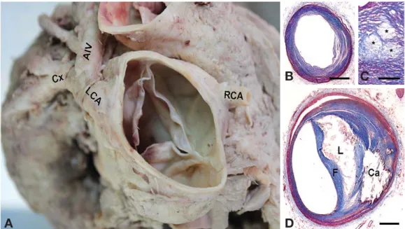
The left coronary artery (LCA) and RCA are respectively branched out of the left and right aortic sinus and respec- tively had external diameters of 6.5 mm and 3.9 mm. The LCA was separated into the anterior interventricular (5.0 mm) and the circumflex branches (4.8 mm). The first diag- onal branch (3.0 mm) arose from the anterior interventri- cular branch (3.0 mm away from the bifurcation) and cours- ed on the anterior wall of the left ventricle (Fig. 1A).
For histopathology, tissue sections were stained with Masson’s trichrome to differentiate between collagen and muscle fibers. The diameters of LCA (6.1 mm) and RCA
(3.4 mm) were estimated under microscopy. The RCA showed relatively normal histology (Fig. 1B). The main trunk of LCA had intimal fatty accumulation (Fig. 1C), atherosclerotic plaque and calcification (Fig. 1D), which result in narrowing the luminal over 50%.
Discussion
Various types of coronary arterial anomaly are frequently found in dissection laboratories of human cadaver and dur- ing radiological imaging. In this case we found an athero- sclerotic aneurysm on LCA and a hypoplasia of RCA, but the hypoplasia of RCA turned out to be a pseudohypopla- sia by further grounds.
RCA had an external diameter of 3.9 mm and 3.4 mm on macroscopic and microscopic measurement respectively, which correspond to the standard for hypoplasia (⁄4.30 mm and ⁄4.04 mm, respectively). Contrary to our expecta- tions of congenital hypoplasia, there were atherosclerotic involvements in LCA, but relatively normal histology in RCA. There is usually a difference between the diameter of LCA (4.44±1.79 vs 4.45±0.79 mm) and RCA (3.32±
0.79 vs 3.51±0.69 mm) in anatomic [8] and angiographic 52 Misun Kim, Sang Pil Yoon
Fig. 1. Macroscopic (A) and microscopic (B-D) features of the coronary artery. Right coronary artery (RCA) was considered as hypoplastic (A), but turned out to be normal (B). The main trunk of left coronary artery (LCA) had intimal fatty streak (asterisks in C), atherosclerotic plaque and calcification (Ca) (D). AIV, anterior interventricular branch; Cx, circumflex branch; L, lipid core; F, fibrous cap. Scale bar==1 mm for B and D==250 μm for C.
B C
A D
[9] studies. Based on the previous reports, our results did not meet the requirements for congenital coronary anom- alies. However it did satisfy the relatively common results from atherosclerotic dilatation in LCA based on the histo- pathology also.
In addition, we reported that a common mesenteric trunk [4] and a hypoplasia of left vertebral artery with intimal fibromuscular dysplasia [5] in this cadaver. Combined anomalies of different arteries shown in such the case can indicate the possibility of an abnormal genetic change. A few genes such as angiotensinogen, angiotensin I convert- ing enzyme (ACE), and apolipoprotein E are suggested as a genetic factor predisposing to peripheral arterial disease [10]. ACE genotype has been associated with an increased risk of coronary artery disease [11,12], cerebral infarction [13], and extracranial artery stenosis [14]. The cause of each anomaly that was differentially presumed in this cadaver may lead to confusion to interpret the genetic factors.
The anatomical variation concerning the simultaneous hypoplasia in vertebral and coronary arteries is of clinical importance, since concomitant arterial diseases may be associated with increased mortality and morbidity rates in patients undergoing procedures on various coronary artery diseases. Since vertebral artery disease with coronary artery disease is not uncommon [15] including this case, signs and symptoms of vertebrobasilar insufficiency should be carefully evaluated in patients with coronary artery disease including hypoplasia.
Taken together, macroscopic hypoplasia in radiological and surgical situations may lead to confusion to interpret its pathophysiology and its clinical outcomes. Better anato- mical knowledge on coronary artery can help to prevent misleading approaches to patients who suffering from coronary artery diseases or concomitant vertebrobasilar insufficiency.
References
1. De Giorgio F, Abbate A, Stigliano E, Capelli A, Arena V.
Hypoplastic coronary artery disease causing sudden death.
Report of two cases and review of the literature. Cardiovasc Pathol. 2010; 19:e107-11.
2. Kim MS, Han JK, Lee SE, Kim SY, Park KW, Koo BK, et al. Cases of right ventricular myocardial infarction in patients with an absent or hypoplastic right coronary artery. Korean Circ J. 2007; 37:84-6.
3. Sim DS, Jeong MH, Choi S, Yoon NS, Yoon HJ, Moon JY, et al. Myocardial infarction in a young man due to a hypoplastic coronary artery. Korean Circ J. 2009; 39:163- 7.
4. Yoo SJ, Ku MJ, Cho SS, Yoon SP. A case of the inferior mesenteric artery arising from the superior mesenteric artery in a Korean woman. J Korean Med Sci. 2011; 26:1382-5.
5. Hyun CL, Park HS, Shin HJ, Yoon SP. Hypoplasia of left vertebral artery with intimal fibromuscular dysplasia in a Korean woman. J Korean Med Sci. 2012; 27:811-3.
6. Thomas CS, Habib F, Varghese K, Abraham MT, Hayat NJ, Cherian G. Disease of proximal part of vertebral artery in patients with coronary artery disease. Angiology. 2003;
54:205-9.
7. Songur A, Gonul Y, Ozen OA, Kucuker H, Uzun I, Bas O, et al. Variations in the intracranial vertebrobasilar system.
Surg Radiol Anat. 2008; 30:257-64.
8. Fazliogullari Z, Karabulut AK, Unver Dogan N, Uysal II.
Coronary artery variations and median artery in Turkish cadaver hearts. Singapore Med J. 2010; 51:775-80.
9. Yun YK, Park KH, Choi YS, Kim K, Jun TG, Kim J, et al.
Computerized quantative analysis of coronary angiogram in patients without coronary pathology. Korean J Thorac Cardiovasc Surg. 1998; 31:488-93.
10. Fatini C, Sticchi E, Sofi F, Said AA, Pratesi G, Pulli R, et al. Multilocus analysis in candidate genes ACE, AGT, and AGTR1 and predisposition to peripheral arterial disease:
role of ACE D/-240T haplotype. J Vasc Surg. 2009; 50:
1399-404.
11. Bauters C, Amouyel P. Association between the ACE geno- type and coronary artery disease. Insights from studies on restenosis, vasomotion and thrombosis. Eur Heart J. 1998;
19 Suppl J:J24-9.
12. Acarturk E, Attila G, Bozkurt A, Akpinar O, Matyar S, Seydaoglu G. Insertion/deletion polymorphism of the ang- iotensin converting enzyme gene in coronary artery disease in southern Turkey. J Biochem Mol Biol. 2005; 38:486-90.
13. Um JY, Moon KS, Lee KM, Cho KH, Heo Y, Moon BS, et al. Polymorphism of angiotensin-converting enzyme, angio- tensinogen, and apolipoprotien E genes in Korean patients with cerebral infarction. J Mol Neurosci. 2003; 21:23-8.
14. Pfohl M, Fette M, Koch M, Barth CM, Rudiger W, Haring HU. Association between angiotensin I-converting enzyme genotypes, extracranial artery stenosis, and stroke. Athero- sclerosis. 1998; 140:161-6.
15. Chen WH, Ho DSWH, Ho SL, Cheung RTF, Cheng SWK.
Prevalence of extracranial carotid and vertebral artery disease in Chinese patients with coronary artery disease. Stroke.
1998; 29:631-4.
Pseudohypoplastic Coronary Artery 53
형성부전으로 오인된 오른심장동맥
김미선
1, 윤상필
21제주대학교 의학전문대학원 의학과,2제주대학교 의학전문대학원 해부학교실
간추림 : 심장동맥 협착이 임상적으로 중요한 지표로 사용되고 있지만, 형성부전은 선천적인 요소를 갖고 있어 의미가 있다. 혈관에서 거짓형성부전은 반대쪽 혈관 직경에 비해 작은 직경을 갖지만 어떤 형태학적 검사에서 도 형성부전의 증거를 보이지 않는 것으로 정의할 수 있다.
온창자간막동맥, 왼척추동맥 형성부전이 동반된 82세 여자 시신의 해부과정에서 오른심장동맥 거짓형성부전 이 관찰되었다.
맨눈으로 볼 때 형성부전으로 생각되었지만, 오른심장동맥은 정상적인 크기와 조직 구성을 보였다. 반면 왼심 장동맥 주 가지는 동맥경화로 인한 확장을 보였다. 결과적으로 이 시신에서 관찰된 오른심장동맥 형성부전은 왼심장동맥의 동맥경화성 동맥류에 기인한 거짓형성부전임을 알 수 있었다.
많은 임상 상황들이 맨눈에 의존하여 판단하게 되므로 심장동맥에 대한 보다 정확한 해부학 지식을 통해 그 릇된 접근을 예방할 수 있어야 할 것으로 생각한다.
찾아보기 낱말 : 거짓형성부전, 동맥경화, 심장동맥, 변이 54 Misun Kim, Sang Pil Yoon
교신저자 : 윤상필(제주대학교 의학전문대학원 해부학교실) 전자우편 : [email protected]