Brief Report
Vol. 31, No. 2, 2019 251
Received January 5, 2018, Revised April 2, 2018, Accepted for publication April 15, 2018
Corresponding author: Ji Won Byun, Department of Dermatology, Inha University School of Medicine, 27 Inhang-ro, Jung-gu, Incheon 22332, Korea. Tel:
82-32-890-2238, Fax: 82-32-890-2236, E-mail: [email protected] ORCID: https://orcid.org/0000-0003-0317-6725
This is an Open Access article distributed under the terms of the Creative Commons Attribution Non-Commercial License (http://creativecommons.org/
licenses/by-nc/4.0) which permits unrestricted non-commercial use, distribution, and reproduction in any medium, provided the original work is properly cited.
Copyright © The Korean Dermatological Association and The Korean Society for Investigative Dermatology and management. Dermatol Ther 2006;19:202-209.
4. Lucky AW, Biro FM, Huster GA, Leach AD, Morrison JA, Ratterman J. Acne vulgaris in premenarchal girls. An early sign of puberty associated with rising levels of dehydro- epiandrosterone. Arch Dermatol 1994;130:308-314.
5. Lucky AW. A review of infantile and pediatric acne. Derma- tology 1998;196:95-97.
6. Plewig G, Kligman AM. Acne and rosacea. 2nd ed. Berlin:
Springer-Verlag, 1993:674-675.
7. Sardana K, Sharma RC, Sarkar R. Seasonal variation in acne vulgaris--myth or reality. J Dermatol 2002;29:484-488.
8. Hancox JG, Sheridan SC, Feldman SR, Fleischer AB Jr. Sea- sonal variation of dermatologic disease in the USA: a study
of office visits from 1990 to 1998. Int J Dermatol 2004;43:
6-11.
9. Karciauskiene J, Valiukeviciene S, Gollnick H, Stang A. The prevalence and risk factors of adolescent acne among schoolchildren in Lithuania: a cross-sectional study. J Eur Acad Dermatol Venereol 2014;28:733-740.
10. Thiboutot D, Del Rosso JQ. Acne vulgaris and the epider- mal barrier: is acne vulgaris associated with inherent epider- mal abnormalities that cause impairment of barrier func- tions? Do any topical acne therapies alter the structural and/or functional integrity of the epidermal barrier? J Clin Aesthet Dermatol 2013;6:18-24.
https://doi.org/10.5021/ad.2019.31.2.251
Squamous Cell Carcinoma on the Fingers of Orthopedic Surgeon Induced by Occupational Radiation Exposure
Hee Seong Yoon, Ji Hye Heo, Si Hyub Lee, Jeonghyun Shin, Gwang Seong Choi, Ji Won Byun
Department of Dermatology, Inha University School of Medicine, Incheon, Korea
Dear Editor:
Recently, fluoroscopy is useful for diagnosis and treatment and its usage is increasing in the treatment of pain, ortho- pedic surgery and interventional radiology. The occupa- tional radiation exposure of physicians is also increasing and the hand is the most exposed area. Radiodermatitis of physicians caused by occupational radiation exposure has been reported1, but there is few report of the development of squamous cell carcinoma (SCC). We describe an ortho- pedic surgeon with multiple SCCs on the fingers occupa- tionally exposed to radiation.
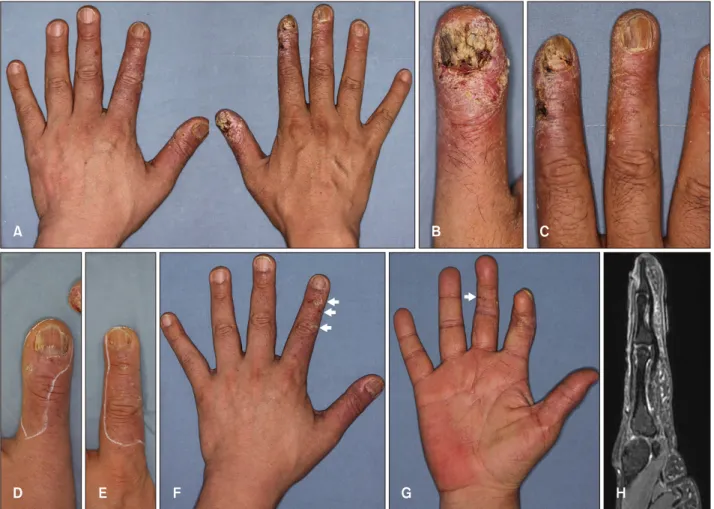
A 49-year-old man presented with painful crusted necrotic plaques and scale widely distributed in the both fingers 2
years ago (Fig. 1A∼E). He smoked 5∼6 packs of tobacco every day. He was an orthopedic surgeon and repeatedly exposed to radiation because of frequent use of fluo- roscopy. For 11 years, he performed an average of 700 cases of percutaneous vertebral augmentation and 70,000 cases of nerve block per year without radiation shielding gloves. The skin biopsy showed atypical squamous cells invading into dermis forming sheet and individual kerati- nization (Fig. 2). Magnetic resonance imaging of the right hand suspected bone invasion in the right index finger (Fig. 1H). Chest computed tomography revealed slightly enlarged lymph nodes in both axilla; however, they ap- peared benign. We diagnosed him with SCC and pre-
Brief Report
252 Ann Dermatol
Fig. 1. Painful crusted necrotic plaques and scale widely distributed in the right thumb, index, and 3rd finger, and left thumb and index finger. Each figure is as follows: (A) Both hands (B) right thumb (C) right index and 3rd finger (D) left thumb (E) left index finger. And photographs after 17 months of follow-up. Each figure is as follows: (F) Dorsal side of the left hand. There were three SCC lesions on the left index finger (arrows). (G) Ventral side of the right hand. There was a SCC lesion on the right third finger (arrow). (H) Right hand MRI image. On the T2 weighted image, SCC invasion is suspected in the medial cortex of the mid phalange and the marrow of the distal phalange of the right index finger. SCC: squamous cell carcinoma, MRI: magnetic resonance imaging.
scribed multidisciplinary cancer care. For the right thumb and index finger, the SCC lesions were widely excised.
The distal phalange of the right index finger, which had a high possibility of bone involvement, was partially sev- ered leaving a 6-mm of distal phalange, and the defect was repaired with a free flap. Mohs micrographic surgery (MMS) and a full thickness skin graft (FTSG) was per- formed on the left thumb. All fingers suspected of skin cancer in situ due to chronic radiation dermatitis were treated with ingenol mebutate. During 17 months of fol- low-up, he often wore protective gloves and continued to work. However, new SCC lesions of the right middle, left index finger appeared (Fig. 1F, G). MMS and FTSG was performed for those lesions. He is under observation with- out recurrence until 25 months later.
The effects of radiation exposure on the skin was de- scribed by the International Commission on Radiological
Protection in 20102. The annual equivalent dose is 500 mSv to the skin2. Direct beam contact with fluoroscopy causes radiation exposure of 40 mSv per minute, and no deep eye radiation exposure was observed at a distance of 91.4 cm from the fluoroscope3. The skin effects of radia- tion are divided into acute and chronic changes, depend- ing on the time period from the exposure4. Chronic changes include epidermal thinning, fibrosis, edema, der- mal thickening, dyspigmentation, telangiectasia, and der- mal necrosis4. The risk of cutaneous cancer increases after radiation exposure4.
Surgical methods are mainly used for the treatment of SCC.
Among the surgical methods, MMS has a 96% cure rate and is the gold standard5. If there is bone involvement, amputation should be performed5. Since cancer has been reported up to 50 years after exposure, continuous fol- low-up observation is needed1. There is concern in this case
Brief Report
Vol. 31, No. 2, 2019 253 Fig. 2. (A, B) Histopathologic find- ings of the nail bed of the right thumb (H&E: A, ×40; B, ×200). The skin biopsy showed atypical squa- mous cells invading into dermis forming sheet and individual kera- tinization.
that SCC will recur, so we will continue follow-up. This case demonstrates that physicians should be aware of oc- cupational radiation exposure.
ACKNOWLEDGMENT
We thank the patient for granting permission to publish this information. This work was supported by the Inha University Research Grant and grants (56990) from the National Research Foundation through the Korean govern- ment (NRF-2016R1C1B1014905).
CONFLICTS OF INTEREST
The authors have nothing to disclose.
ORCID
Hee Seong Yoon, https://orcid.org/0000-0001-8997-9697 Ji Hye Heo, https://orcid.org/0000-0002-0928-122X Si Hyub Lee, https://orcid.org/0000-0002-8246-8664 Jeonghyun Shin, https://orcid.org/0000-0002-4995-9533
Gwang Seong Choi, https://orcid.org/0000-0002-5766-0179 Ji Won Byun, https://orcid.org/0000-0003-0317-6725
REFERENCES
1. Shim DM, Kim YM, Oh SK, Lim CM, Kown BT. Radiation induced hand necrosis of an orthopaedic surgeon who had treated a patient with fluoroscopy-guided spine injection. J Korean Orthop Assoc 2014;49:250-254.
2. ICRP. Radiological protection in fluoroscopically guided procedures outside the imaging department. ICRP Publi- cation 117. Ann ICRP 2010;40.
3. Mehlman CT, DiPasquale TG. Radiation exposure to the orthopaedic surgical team during fluoroscopy: “how far away is far enough?”. J Orthop Trauma 1997;11:392-398.
4. Hegedus F, Mathew LM, Schwartz RA. Radiation dermatitis:
an overview. Int J Dermatol 2017;56:909-914.
5. Dika E, Fanti PA, Patrizi A, Misciali C, Vaccari S, Piraccini BM. Mohs surgery for squamous cell carcinoma of the nail unit: 10 years of experience. Dermatol Surg 2015;41:1015- 1019.