Opsonophagocytic Antibodies to Serotype Ia, Ib, and III Group B Streptococcus among Korean Infants and in Intravenous
Immunoglobulin Products
Group B streptococcus (GBS) infection is a leading cause of sepsis and meningitis among infants, and is associated with high rates of morbidity and mortality in many countries.
Protection against GBS typically involves antibody-mediated opsonization by phagocytes and complement components. The present study evaluated serotype-specific functional antibodies to GBS among Korean infants and in intravenous immunoglobulin (IVIG) products. An opsonophagocytic killing assay (OPA) was used to calculate the opsonization indices (OIs) of functional antibodies to serotypes Ia, Ib, and III in 19 IVIG products from 5 international manufacturers and among 98 Korean infants (age: 0–11 months). The GBS Ia, Ib, and III serotypes were selected because they are included in a trivalent GBS vaccine formulation that is being developed. The OI values for the IVIG products were 635–5,706 (serotype Ia), 488–1,421 (serotype Ib), and 962–3,315 (serotype III), and none of the IVIG lots exhibited undetectable OI values (< 4). The geometric mean OI values were similar for all 3 serotypes when we compared the Korean manufacturers. The seropositive rate among infants was significantly lower for serotype Ia (18.4%), compared to serotype Ib and serotype III (both, 38.8%). Infant age of ≥ 3 months was positively correlated with the seropositive rates for each serotype. Therefore, only a limited proportion of infants exhibited protective immunity against serotype Ia, Ib, and III GBS infections. IVIG products that exhibit high antibody titers may be a useful therapeutic or preventive measure for infants. Further studies are needed to evaluate additional serotypes and age groups.
Keywords: Streptococcus agalactiae; Antibodies; Opsonin Proteins; Infant;
Immunoglobulins; Intravenous Han Wool Kim,1 Ji Hyen Lee,1,2
Hye-Kyung Cho,3 Hyunju Lee,4 Ho Seong Seo,5 Soyoung Lee,1 and Kyung-Hyo Kim1,2
1Center for Vaccine Evaluation and Study, Medical Research Institute, Ewha Womans University School of Medicine, Seoul, Korea; 2Department of Pediatrics, Ewha Womans University School of Medicine, Seoul, Korea; 3Department of Pediatrics, Graduate School of Medicine, Gachon University, Incheon, Korea; 4Department of Pediatrics, Seoul National University College of Medicine, Seoul, Korea; 5Biotechnology Division, Korea Atomic Energy Research Institute, Jeongeup, Korea Received: 25 August 2016
Accepted: 27 January 2017 Address for Correspondence:
Kyung-Hyo Kim, MD, PhD
Department of Pediatrics, Ewha Womans University School of Medicine, 1071 Anyangcheon-ro, Yangcheon-gu, Seoul 07985, Korea
E-mail: [email protected]
Funding: This study was supported by a research grant from the Ewha Womans University School of Medicine (RP-grant 2014), and by research grants from the Ministry of Food and Drug Safety (14172MFDS245 and 16172MFDS267).
https://doi.org/10.3346/jkms.2017.32.5.737 • J Korean Med Sci 2017; 32: 737-743
INTRODUCTION
Group B streptococcus (GBS, Streptococcus agalactiae) is an important pathogen among infants (1). Although the introduc- tion of intrapartum antibiotic prophylaxis and maternal screen- ing has reduced the incidence of invasive GBS disease (e.g., sep- sis and meningitis) during recent years, the rates of morbidity and mortality from invasive GBS disease remain high in many countries (2). Preliminary epidemiological studies revealed a correlation between low concentrations of maternally derived antibodies against the capsular polysaccharide (CPS) of GBS and infants’ susceptibility to GBS infection (3-5). Therefore, an intrapartum vaccine was developed to protect infants from in- vasive diseases, which generated transplacental-acquired sero- type-specific capsular antibodies until the age of 3 months, as
> 95% of invasive GBS diseases occur during this period (6,7).
Similar to protection against other bacteria (e.g., Haemophi- lus influenzae type b, Streptococcus pneumoniae, and Neisseria
meningitides), protection against GBS generally involves anti- body-mediated opsonization by phagocytes and complement components (8). Thus, the opsonization assay has become an important tool for assessing the immunogenicity of pneumo- coccal vaccines (9), and a multiplexed opsonophagocytic kill- ing assay (OPA) was developed (10) to facilitate the widespread use of the pneumococcal opsonization assay. Burton and Nahm (The Bacterial Respiratory Pathogen Reference Laboratory, Na- tional Institutes of Health; University of Alabama at Birming- ham [UAB]) (10) performed minor modifications to the OPA protocol and detected opsonic antibodies against GBS CPS in rabbit antisera (the UAB GBS OPA assay). This killing-type assay measures antibodies’ specific abilities to actually kill bacteria by counting the surviving bacteria, although it has not been used to test human sera. In the present study, we used to UAB GBS OPA to evaluate the opsonization indices (OIs) of GBS Ia-, Ib-, and III-specific antibodies in the sera of Korean infants and in several commercial intravenous immunoglobulin (IVIG) prod- ORIGINAL ARTICLE
Infectious Diseases, Microbiology & Parasitology
ucts from different countries. The GBS Ia, Ib, and III serotypes were selected because they are included in a trivalent GBS vac- cine formulation that is being developed (CRM197-glycoconju- gate GBS vaccine) (11,12).
MATERIALS AND METHODS Bacterial strains
The GBS strains (serotype Ia: E-GBS 001, serotype Ib: E-GBS 002, and serotype III: E-GBS 003) were obtained from Dr. Hye-Kyung Cho (Gachon University, Gil Medical Center). The strains had been isolated from infants with invasive GBS diseases, and were stored at −80°C in 0.5 mL of Todd-Hewitt broth with 15% glyc- erol. GBS were identified based on the presence of gram-posi- tive cocci in pairs or short chains, beta hemolysis on blood agar plates, catalase-negative results, and formation of a substance (Christie-Atkins-Munch-Petersen [CAMP] factor) that enlarges the hemolysis area formed by β-hemolysin from Staphylococ- cus aureus. The GBS serotype was identified using a slide latex agglutination test (Denka Seiken, Tokyo, Japan).
Infants’ sera
This study evaluated sera from infants who underwent blood testing for health evaluations between 2009 and 2010. The sera were stored at −70°C until testing. Infants with known immune deficiencies or premature birth status (gestational age of < 37 weeks at birth) were excluded. Based on the available sera, we selected 103 sera from infants who were < 1-year old at the eval- uation. The infants were stratified according to age using 1-month intervals (0 months to 11 months). Among the 103 sera, 98 sera were found to be free from antibiotics (based on their inability to inhibit the growth of R36A pneumococcal bacteria), and were incubated at 56°C for 30 minutes to inactivate endogenous com- plement activity before our testing.
Commercial IVIG products
Nineteen lots of IVIG products were analyzed in this study. Ten lots (Korea-A) and 6 lots (Korea-B) were obtained from 2 Kore- an manufacturers, separate lots were also obtained from 2 Japa- nese manufacturers (Japan-A and Japan-B), and 1 lot was ob- tained from an American manufacturer (US). To purify the IVIG, Korea-A, Korea-B, and US products had been treated using cold ethanol solvent/detergent, while the Japan-A and Japan-B prod- ucts had been treated using polyethylene glycol. The Korea-A, Korea-B, and Japan-B products were formulated as liquids, while the Japan-A and US products were formulated as lyophilized powder. All IVIG products were formulated at a concentration of 50 mg/mL.
OPA for antibodies against GBS (the UAB GBS OPA assay) Duplicate samples of sera or IVIG (20 µL) were serially diluted
(1:3) in opsonization assay buffer B (OBB; Hanks’ balanced salt solution [with magnesium and calcium] with 0.1% gelatin and 5% defined fetal bovine serum [6000; Gibco, Waltham, MA, USA]) in 96-well round-bottom plates. Frozen working stocks of each of the target strains were thawed and washed twice with OBB and centrifuged (12,000 × g for 2 minutes), then the bacteria were prepared in OBB to an approximately concentration of 1
× 105 colony-forming units (CFUs)/mL. Ten microliters of the bacteria were added to each well, and the plates were incubat- ed at room temperature for 30 minutes with shaking. After the incubation, 10 μL of baby rabbit complement (BRC; Pel-Freez Biological, Rogers, AR, USA) were added to the wells, except for the control A wells, which received 10 μL of heat-inactivated BRC (heated at 56°C for 30 minutes). Forty microliters of differ- entiated HL60 cells (containing 4 × 10⁵ cells) were added to all wells, and the plates were incubated for 45 minutes in a 37°C/5%
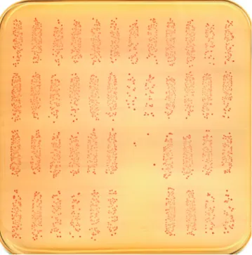
CO2 incubator with shaking. Afterwards, 5-μL aliquots of the re- action product from each well were spotted onto THYA-NR plates (Todd-Hewitt agar [1.5%] with yeast extract containing neutral red [30 μg/mL] and 1M Tris solution [0.02M]) and the plates were incubated overnight (37°C/5% CO2). After the overnight incubation, the plates were removed from the incubator and left at room temperature for 4–5 hours, which decreased the background color. The numbers of surviving colonies on the plates (Fig. 1) were enumerated using NICE colony counting
Fig. 1. Microcolonies of serotype III GBS on a THYA-NR plate (Todd-Hewitt agar with yeast extract containing neutral red and Tris solution) after overnight incubation (37°C/
5% CO2) and incubation for 4–5 hours at room temperature/room air. The numbers of surviving colonies were counted on the plates. The average diameter of the micro- colonies was approximately 0.3 mm, and the colonies were clearly red and spatially separated.
GBS = group B streptococcus.
Kim HW, et al. • Antibodies to Group B Streptococcus in Korean Infants and IVIG
software (National Institute of Standards and Technology’s In- tegrated Colony Enumerator; National Institute of Standards and Technology, Gaithersburg, MD, USA). OIs were calculated using linear interpolation. To facilitate the data analysis, an Ex- cel-based data processing program was used to transfer the colony counts to an “opsonization index” program (“opsotiter3”
from the UAB reference laboratory). The OI value was defined as the reciprocal of the interpolated dilution of serum that killed 50% of the bacteria. If an undiluted serum sample killed 50% of the bacteria, this produced an OI value of 4 in our system. A de- tailed protocol is posted at http://www.vaccine.uab.edu/.
Statistical analysis
The geometric mean indices (GMIs) and associated 95% confi- dence intervals (CIs) were calculated for each of the serotypes.
Differences in the GMIs among the IVIG products were analyzed using the Kruskall-Wallis test and the Mann-Whitney U test.
Box and whisker plots were used to report the median, mini- mum, and maximum values, as well as the 25th and 75th per- centiles for each group. Seropositivity was defined as the pres- ence of detectable OI (≥ 4). Pearson’s χ2 tests and Cochran’s Q test were used to compare seropositive rates. The χ2 test for trend was used to evaluate the relationship between the seropositive rates and age. The linear correlations between the OIs and in- fant age (≥ 3 months) for each serotype were evaluated using Spearman’s correlation coefficients. Serum samples with OIs of
< 4 were assigned a value of 2 for the present analyses. Differ- ences were considered statistically significant at a P value of
< 0.05. Statistical analyses were performed using SPSS software (version 23.0; SPSS Inc., Chicago, IL, USA).
Ethics statement
This study was approved by the Institutional Review Board of the Ewha Womans University Mokdong Hospital (EUMC 2016- 02-044). The board exempted submission of informed consent.
RESULTS
The OIs to serotype Ia, Ib, and III GBS among IVIG products
The box and whisker plots for OIs to serotype Ia, Ib, and III GBS among the 19 IVIG products are shown in Fig. 2. The OI values were 635–5,706 (serotype Ia), 488–1,421 (serotype Ib), and 962–
3,315 (serotype III). None of the IVIG lots exhibited undetectable OI values (< 4). We observed lot-to-lot variations between the Korean manufacturers for serotype Ia (Korea-A: 635–987; Ko- rea-B: 791–5,706) and serotype III (Korea-A: 1,223–1,823; Korea- B: 962–3,315), but not for serotype Ib (Korea-A: 488–568; Korea- B: 504–607). The GMI values were not significantly different be-
Table 1. OIs and seropositive rates for GBS (serotypes Ia, Ib, and III) in various IVIG products and in Korean infants
Variables GMIs (95% CIs) Seropositive rate, No. (%)
Serotype Ia Serotype Ib Serotype III Serotype Ia Serotype Ib Serotype III
IVIG (No.)
Korea-A (6) 778 (672–905) 525 (504–547) 1,614 (1,428–1,823) - - -
Korea-B (10) 1,195 (820–1,742) 546 (528–565) 1,948 (1,569–2,420) - - -
Korean products (16) 1,018 (786–1,319) 538 (524–553) 1,816 (1,567–2,103) - - -
Total (19) 1,185 (900–1,560) 607 (531–693) 1,981 (1,690–2,322) - - -
Infants’ sera, mon (No.)
0–2 (22) 2.7 (2.0–3.7) 6.2 (3.2–11.8) 6.4 (3.1–13.5) 4 (18.2) 9 (40.9) 8 (36.4)
3–5 (14) 2.3 (1.8–3.0) 3.3 (1.7–6.5) 3.8 (1.8–8.0) 1 (7.1) 2 (14.3) 3 (21.4)
6–8 (34) 2.7 (2.0–3.6) 7.3 (3.5–15.3) 7.7 (3.8–15.7) 5 (14.7) 11 (32.4) 12 (35.3)
9–11 (28) 3.5 (2.4–5.2) 14.7 (6.5–33.2) 22.0 (8.4–57.7) 8 (28.6) 16 (57.1) 15 (53.6)
Total (98) 2.7 (2.3–3.2) 6.1 (4.2–8.9) 6.9 (4.6–10.5) 18 (18.4)* 38 (38.8) 38 (38.8)
OIs = opsonization indices, GBS = group B streptococcus, CIs = confidence intervals, IVIG = intravenous immunoglobulin, GMIs = geometric mean indices.
*Significant difference in the seropositive rates based on Cochran’s Q test (P = 0.002).
Fig. 2. OIs for GBS according to serotype (Ia, Ib, and III) in various IVIG products (n = 19). The top bar is the highest value, the bottom bar is the lowest value, the top of the box indicates the third quartile, the bottom of the box indicates the first quartile, and the middle bar is the median value. The lower limit of detection for the opsoniza- tion index was 4. The numbers of lots are given in parentheses.
OIs = opsonization indices, GBS = group B streptococcus, IVIG = intravenous immu- noglobulin.
10,000
1,000
100
10 4 1
2 Fig 2.
4
Opsonization index
5 6
Serotype
Ia Ib III
Opsonization index
2 Fig 2.
4
Opsonization index
5 6
2 Fig 2.
4
Opsonization index
5 6
2 Fig 2.
4
Opsonization index
5 6
Korea-A (n = 6) Japan-A (n = 1) US (n = 1)
2 Fig 2.
4
Opsonization index
5 6
2 Fig 2.
4
Opsonization index
5 6
Korea-B (n = 10) Japan-B (n = 1)
Kim HW, et al. • Antibodies to Group B Streptococcus in Korean Infants and IVIG
tween the Korean manufacturers for all 3 serotypes (Table 1).
The OIs to serotype Ia, Ib, and III GBS among the infants’
sera
The OIs of the infants’ sera to GBS were 2–119 for serotype Ia, 2–2,486 for serotype Ib, and 2–1,557 for serotype III (Fig. 3). The seropositive rates were 18.4% (18/98) for serotype Ia, 38.8% (38/
98) for serotype Ib, and 38.8% (38/98) for serotype III (Table 1).
The seropositive rate for serotype Ia was significantly lower, com- pared to the rates for serotypes Ib or III. The seropositive rates were lowest for all 3 serotypes among infants who were 3–5 mon ths old. The seropositive rates for each serotype were higher among younger infants (0–2 months old). Infant age of ≥ 3 months was positively correlated with the seropositive rates for each sero- type. There were positive correlations between age and the OIs against serotypes Ia, Ib, and III (r = 0.49–0.57).
DISCUSSION
The present study revealed that several commercial IVIG prod- ucts had functional antibodies to serotype Ia, Ib, and III GBS, regardless of their manufacturer’s location. However, many in- fants did not have functional antibodies against the 3 GBS sero- types.
As IVIG is produced using pooled plasma from up to 100,000 donors (13), it contains a wide range of immunoglobulin G (IgG) to many microorganisms, which reflects the donors’ cumulative environmental exposures. Many previous studies have measured the titers of antibodies in commercial IVIG products to encap- sulated bacteria, such as H. influenzae type b (14), meningococ- ci (15), and pneumococci (14,16). However, to the best of our knowledge, no studies have measured the titers of functional
antibodies to GBS in IVIG products. The present study’s find- ings of functional antibodies to serotype Ia, Ib, and III GBS in commercial IVIG, regardless of the donor population, is to be expected, as GBS is a human commensal in the gastrointestinal and genital tract that does not typically cause infection or dis- ease (17). Among pregnant women, the rate of GBS carriage in the vaginal and rectal microbiota is similar in both developing and developed countries (18-20), although large variations in the colonization rates are observed in different populations (21).
In Korea, the reported carriage rates are 6.3%–11.5% among pregnant women (22-24). However, the titers of pathogen-spe- cific antibodies in IVIG may varying according to the plasma source, as they are influenced by the cumulative environmental exposure of the donor population. Therefore, continuous test- ing of antibody titers is needed to monitor different IVIG prod- ucts from different countries.
In contrast with our findings from the IVIG products, most infants in the present study (61.2%–81.6%) did not have func- tional antibodies to the 3 GBS serotypes. Furthermore, the sero- positive rate for serotype Ia was especially low, compared to the rates for serotypes Ib or III. Nevertheless, infant age of ≥ 3 months was positively correlated with the seropositive rates for each se- rotype. Moreover, there were positive correlations between age and OIs in the infants’ sera, which exhibited moderate variations according to serotypes. These results suggest that, even during infancy, specific functional immune responses to the 3 GBS se- rotypes can be achieved through a commensal in the gastroin- testinal or genital tract, without causing infection or disease at older ages.
The delayed immunoglobulin synthesis and low rate of trans- placental antibody transfer in neonates suggest that IVIG may be a useful adjunctive therapy for treating GBS infections. In addition, IVIG may reduce the morbidity and mortality that is associated with GBS infections in high-risk neonates (25). In this context, IVIG provides immune protection in experimental models of GBS infection. However, meta-analyses of IVIG for treating cases of suspected or proven neonatal sepsis have re- vealed conflicting results, although these studies did not specif- ically target GBS sepsis (26,27). Moreover, several studies have revealed that, compared to IVIG preparations from adults who were immunized with GBS (hyperimmune IVIG), standard IVIG preparations contain only modest levels of antibodies to GBS (28), exhibit dose- and preparation-dependent variations in their in vitro opsonophagocytosis, and are significantly less protec- tive in experimental infection models. Baker et al. (29) have re- viewed the studies regarding IVIG in human neonates and re- ported inconclusive findings, which were related to the small numbers of included infants, the lack of suitable controls, the use of a clinical definition for sepsis (vs. a strict microbiological definition), and single-center study designs. Thus, further stud- ies are needed to provide evidence regarding the efficacy of IVIG Fig. 3. OIs for GBS according to serotype (Ia, Ib, and III) and age among Korean in-
fants (n = 98). The lower limit of detection for the opsonization index was 4. The num- bers of individuals in each age group are given in parentheses.
OIs = opsonization indices, GBS = group B streptococcus.
10,000
1,000
100
10 4 1
3 Fig 3.
Opsonization index
Serotype
Ia Ib III
Opsonization index
3
Opsonization index
3
Opsonization index
3
Opsonization index
3
Opsonization index
0–2 months (n = 22) 6–8 months (n = 34)
3–5 months (n = 14) 9–11 months (n = 28)
Kim HW, et al. • Antibodies to Group B Streptococcus in Korean Infants and IVIG
for treating neonatal GBS sepsis.
The GBS Ia, Ib, and III serotypes selected in this study are in- cluded in the trivalent GBS vaccine formulation (11,12). Based on the serotype distribution in South Africa, a GBS vaccine that incorporates these 3 serotypes is estimated to help prevent 85%
and 98% of early-onset and late-onset invasive disease, respec- tively, among infants (30). Another study revealed that a pen- tavalent vaccine (serotypes Ia, Ib, II, III, and V) was estimated to prevent up to 98% of neonatal disease and 88% of pregnancy- associated disease in the US (1). Moreover, a conjugate vaccine incorporating the 5 GBS serotypes (Ia, Ib, II, III, and V) could prevent > 85% of GBS disease among < 3-month-old infants throughout the world (31). In Korea, the most prevalent sero- types are III, V, and Ia (in that order) (22-24), and different pop- ulations exhibit varying serotype distributions (30,31). There- fore, further studies are needed to consider the other serotypes (II and V) in their analyses.
In the present study, we used the UAB GBS OPA to measure the titers of serotype-specific antibodies to GBS. Similar to other encapsulated bacteria, protection against GBS is generally me- diated by serotype-specific antibodies, which can be evaluated using enzyme-linked immunosorbent assays (ELISA) (3,5). How- ever, ELISA typically only measures IgG, and recent evidence has revealed that IgM contributes to opsonization-related im- mune protection against pneumococci among children and adults (32,33). Moreover, ELISA does not measure the function- al capacity of the antibodies, and simply evaluates their capaci- ty to bind antigens that are coated on a plate (34). Thus, we be- lieve that the UAB GBS OPA function assay is useful for measur- ing serotype-specific antibodies to GBS in sera and IVIG. An OPA using adult polymorphonuclear leukocytes was developed in 1992 (35), and it expressed opsonophagocytic activity as the log10
reduction in the CFU/mL of GBS, compared to the control well (35). However, the UAB GBS OPA has several unique features:
the ability to use frozen aliquots of target bacteria, the use of cul- tured cells as phagocytes (HL60; effector:target ratio of 400) (36), and automated colony counting (37), based on the use of a che- mical that colors GBS colonies growing on the agar plate. More- over, the OI is calculated as the reciprocal of the interpolated dilution of serum that kills 50% of the bacteria. Thus, this OPA method is both quicker and simpler, compared to the previous OPA method. As the UAB GBS OPA assay provides an excess number of functional phagocytes (HL60 cells), the same assay conditions can be used for evaluating opsonic antibodies to pneu- mococci and the efficacy of pneumococcal vaccines (9,10).
The present study has several limitations. First, an OPA titer of 8 is an important threshold for providing protection against most pneumococcal serotypes among children (9), although no such threshold has been established for protecting against GBS. Nevertheless, GBS serotype-specific antibody titers have been suggested in recent studies (38-40), and those findings may
be useful for developing appropriate cut-off values. Second, we only included a small number of IVIG products from Japan and the US, which precluded any definitive comparisons with the Korean IVIG products. Third, we did not compare the titers of functional antibodies in the mothers and their infants, and fur- ther studies are needed to address this issue.
In conclusion, we established the clinical utility of the UAB GBS OPA. This assay revealed that current IVIG products have opsonic antibodies against the 3 GBS serotypes. However, most of the Korean infants in this study did not have opsonic antibo- dies. Therefore, further studies are needed to evaluate the clini- cal utility of IVIG in infants with limited immunity, as well as the seroprevalences of other serotypes. Moreover, studies are needed to target different age groups and IVIG products from a greater number of manufacturers.
ACKNOWLEDGMENT
The authors thank Moon H. Nahm and Robert L. Burton (De- partment of Medicine, University of Alabama at Birmingham, Birmingham, AL, USA) for developing the OPA to detect anti- bodies to GBS, which enabled us to conduct this study. The au- thors also thank the study participants, Green Cross Corp. (Yon- gin, Korea) and SK Chemicals (Seongnam, Korea) for providing the intravenous immunoglobulin (IVIG) products, and Soo Young Lim for assistance with the laboratory work.
DISCLOSURE
The authors have no potential conflicts of interest to disclose.
AUTHOR CONTRIBUTION
Conceptualization: Kim KH. Investigation: Kim HW, Lee JH, Cho HK, Lee H, Seo HS, Lee S. Resources: Kim HW, Lee JH, Cho HK, Lee S. Writing - original draft: Kim HW, Kim KH. Writing - re- view & editing: Cho HK, Lee H, Seo HS.
ORCID
Han Wool Kim http://orcid.org/0000-0003-3463-3060 Ji Hyen Lee http://orcid.org/0000-0002-2234-1055 Hye-Kyung Cho http://orcid.org/0000-0003-0990-1350 Hyunju Lee http://orcid.org/0000-0003-0107-0724 Ho Seong Seo http://orcid.org/0000-0002-7103-1344 Soyoung Lee http://orcid.org/0000-0001-9627-6607 Kyung-Hyo Kim http://orcid.org/0000-0002-0333-6808 REFERENCES
1. Phares CR, Lynfield R, Farley MM, Mohle-Boetani J, Harrison LH, Petit S,
Craig AS, Schaffner W, Zansky SM, Gershman K, et al. Epidemiology of invasive group B streptococcal disease in the United States, 1999-2005.
JAMA 2008; 299: 2056-65.
2. Le Doare K, Heath PT. An overview of global GBS epidemiology. Vaccine 2013; 31 Suppl 4: D7-12.
3. Baker CJ, Kasper DL. Correlation of maternal antibody deficiency with susceptibility to neonatal group B streptococcal infection. N Engl J Med 1976; 294: 753-6.
4. Boyer KM, Papierniak CK, Gadzala CA, Parvin JD, Gotoff SP. Transplacen- tal passage of IgG antibody to group B streptococcus serotype Ia. J Pedi- atr 1984; 104: 618-20.
5. Gotoff SP, Papierniak CK, Klegerman ME, Boyer KM. Quantitation of IgG antibody to the type-specific polysaccharide of group B streptococcus type 1b in pregnant women and infected infants. J Pediatr 1984; 105: 628- 30.
6. Baker CJ, Rench MA, McInnes P. Immunization of pregnant women with group B streptococcal type III capsular polysaccharide-tetanus toxoid con- jugate vaccine. Vaccine 2003; 21: 3468-72.
7. Edwards MS, Lane HJ, Hillier SL, Rench MA, Baker CJ. Persistence of func- tional antibodies to group B streptococcal capsular polysaccharides fol- lowing immunization with glycoconjugate vaccines. Vaccine 2012; 30:
4123-6.
8. Dangor Z, Kwatra G, Izu A, Lala SG, Madhi SA. Review on the association of Group B streptococcus capsular antibody and protection against inva- sive disease in infants. Expert Rev Vaccines 2015; 14: 135-49.
9. Romero-Steiner S, Frasch CE, Carlone G, Fleck RA, Goldblatt D, Nahm MH. Use of opsonophagocytosis for serological evaluation of pneumo- coccal vaccines. Clin Vaccine Immunol 2006; 13: 165-9.
10. Burton RL, Nahm MH. Development and validation of a fourfold multi- plexed opsonization assay (MOPA4) for pneumococcal antibodies. Clin Vaccine Immunol 2006; 13: 1004-9.
11. Heyderman RS, Madhi SA, French N, Cutland C, Ngwira B, Kayambo D, Mboizi R, Koen A, Jose L, Olugbosi M, et al. Group B streptococcus vacci- nation in pregnant women with or without HIV in Africa: a non-randomis- ed phase 2, open-label, multicentre trial. Lancet Infect Dis 2016; 16: 546- 55.
12. Madhi SA, Dangor Z, Heath PT, Schrag S, Izu A, Sobanjo-Ter Meulen A, Dull PM. Considerations for a phase-III trial to evaluate a group B strep- tococcus polysaccharide-protein conjugate vaccine in pregnant women for the prevention of early- and late-onset invasive disease in young-in- fants. Vaccine 2013; 31 Suppl 4: D52-7.
13. Radosevich M, Burnouf T. Intravenous immunoglobulin G: trends in pro- duction methods, quality control and quality assurance. Vox Sang 2010;
98: 12-28.
14. Mikolajczyk MG, Concepcion NF, Wang T, Frazier D, Golding B, Frasch CE, Scott DE. Characterization of antibodies to capsular polysaccharide antigens of Haemophilus influenzae type b and Streptococcus pneumoni- ae in human immune globulin intravenous preparations. Clin Diagn Lab Immunol 2004; 11: 1158-64.
15. Adam E, Church JA. Antibody levels to Bordetella pertussis and Neisseria meningitidis in immunodeficient patients receiving immunoglobulin re- placement therapy. J Clin Immunol 2015; 35: 213-7.
16. Takahashi Y, Ishiwada N, Hishiki H, Tanaka J, Akeda Y, Shimojo N, Oishi K, Kohno Y. IgG levels against 13-valent pneumococcal conjugate vaccine serotypes in non pneumococcal conjugate vaccine immunized healthy
Japanese and intravenous immunoglobulin preparations. J Infect Che- mother 2014; 20: 794-8.
17. Schuchat A. Group B streptococcus. Lancet 1999; 353: 51-6.
18. Kwatra G, Adrian PV, Shiri T, Buchmann EJ, Cutland CL, Madhi SA. Natu- ral acquired humoral immunity against serotype-specific group B strep- tococcus rectovaginal colonization acquisition in pregnant women. Clin Microbiol Infect 2015; 21: 568.e13-21.
19. Edwards MS, Munoz FM, Baker CJ. Antibodies to type III group B strepto- coccal polysaccharide in breast milk. Pediatr Infect Dis J 2004; 23: 961-3.
20. Baker CJ, Edwards MS, Kasper DL. Role of antibody to native type III poly- saccharide of group B streptococcus in infant infection. Pediatrics 1981;
68: 544-9.
21. Melin P, Efstratiou A. Group B streptococcal epidemiology and vaccine needs in developed countries. Vaccine 2013; 31 Suppl 4: D31-42.
22. Uh Y, Choi SJ, Jang IH, Lee KS, Cho HM, Kwon O, Yoon KJ. Colonization rate, serotypes, and distributions of macrolide-lincosamide-streptograminB resistant types of group B streptococci in pregnant women. Korean J Clin Microbiol 2009; 12: 174-9.
23. Oh CE, Jang HO, Kim NH, Lee J, Choi EH, Lee HJ. Molecular serotyping of group B streptococcus isolated from the pregnant women by polymerase chain reaction and sequence analysis. Korean J Pediatr Infect Dis 2009;
16: 47-53.
24. Hong JS, Choi CW, Park KU, Kim SN, Lee HJ, Lee HR, Choi EH, Park KH, Suh CS, Kim BI, et al. Genital group B streptococcus carrier rate and sero- type distribution in Korean pregnant women: implications for group B streptococcal disease in Korean neonates. J Perinat Med 2010; 38: 373-7.
25. Fischer GW. Immunoglobulin therapy for neonatal sepsis: an overview of animal and clinical studies. J Clin Immunol 1990; 10: 40S-46S.
26. Ohlsson A, Lacy J. Intravenous immunoglobulin for suspected or subse- quently proven infection in neonates. Cochrane Database Syst Rev 2010:
CD001239.
27. INIS Collaborative Group, Brocklehurst P, Farrell B, King A, Juszczak E, Darlow B, Haque K, Salt A, Stenson B, Tarnow-Mordi W. Treatment of neonatal sepsis with intravenous immune globulin. N Engl J Med 2011;
365: 1201-11.
28. Baker CJ, Noya FJ. Potential use of intravenous immune globulin for group B streptococcal infection. Rev Infect Dis 1990; 12 Suppl 4: S476-82.
29. Baker CJ, Rench MA, Noya FJ, Garcia-Prats JA. Role of intravenous immu- noglobulin in prevention of late-onset infection in low-birth-weight neo- nates. The Neonatal IVIG Study Group. Rev Infect Dis 1990; 12 Suppl 4:
S463-8.
30. Madzivhandila M, Adrian PV, Cutland CL, Kuwanda L, Schrag SJ, Madhi SA. Serotype distribution and invasive potential of group B streptococcus isolates causing disease in infants and colonizing maternal-newborn dy- ads. PLoS One 2011; 6: e17861.
31. Edmond KM, Kortsalioudaki C, Scott S, Schrag SJ, Zaidi AK, Cousens S, Heath PT. Group B streptococcal disease in infants aged younger than 3 months: systematic review and meta-analysis. Lancet 2012; 379: 547-56.
32. Cho HK, Park IH, Burton RL, Kim KH. Impact of IgM antibodies on cross- protection against pneumococcal serogroups 6 and 19 after immuniza- tion with 7-valent pneumococcal conjugate vaccine in children. J Korean Med Sci 2016; 31: 950-6.
33. Park S, Nahm MH. Older adults have a low capacity to opsonize pneumo- cocci due to low IgM antibody response to pneumococcal vaccinations.
Infect Immun 2011; 79: 314-20.
Kim HW, et al. • Antibodies to Group B Streptococcus in Korean Infants and IVIG
34. Lee H, Nahm MH, Burton R, Kim KH. Immune response in infants to the heptavalent pneumococcal conjugate vaccine against vaccine-related serotypes 6A and 19A. Clin Vaccine Immunol 2009; 16: 376-81.
35. Hall MA, Edwards MS, Baker CJ. Complement and antibody participation in opsonophagocytosis of type IV and V group B streptococci. Infect Im- mun 1992; 60: 5030-5.
36. Fleck RA, Romero-Steiner S, Nahm MH. Use of HL-60 cell line to measure opsonic capacity of pneumococcal antibodies. Clin Diagn Lab Immunol 2005; 12: 19-27.
37. Clarke ML, Burton RL, Hill AN, Litorja M, Nahm MH, Hwang J. Low-cost, high-throughput, automated counting of bacterial colonies. Cytometry A 2010; 77: 790-7.
38. Lin FY, Weisman LE, Azimi PH, Philips JB 3rd, Clark P, Regan J, Rhoads GG, Frasch CE, Gray BM, Troendle J, et al. Level of maternal IgG anti-group B streptococcus type III antibody correlated with protection of neonates against early-onset disease caused by this pathogen. J Infect Dis 2004; 190:
928-34.
39. Baker CJ, Carey VJ, Rench MA, Edwards MS, Hillier SL, Kasper DL, Platt R.
Maternal antibody at delivery protects neonates from early onset group B streptococcal disease. J Infect Dis 2014; 209: 781-8.
40. Dangor Z, Kwatra G, Izu A, Adrian P, Cutland CL, Velaphi S, Ballot D, Reu- benson G, Zell ER, Lala SG, et al. Correlates of protection of serotype-spe- cific capsular antibody and invasive Group B streptococcus disease in South African infants. Vaccine 2015; 33: 6793-9.