INTRODUCTION
Transarterial chemoembolization (TACE) is widely considered as the first-line therapy for patients with non-resectable hepato- cellular carcinoma (HCC) and those who are surgically contrain- dicated for other reasons (1, 2). Known complications of TACE include postembolization syndrome (3), transient or irreversible liver failure (4), liver infarction and abscess formation (5), isch- emic bile duct injury (6), and pulmonary oil embolism due to systemic shunting of excessive amounts of ethiodized oil during the procedure (7). Herein, we report a rare complication of TACE manifesting as an empyema secondary to the migration of Lipiodol content from the liver into the ipsilateral pleural cavity.
CASE REPORT
A 60-year-old male patient was referred to our hospital from
an outside hospital due to refractory pleural effusion and he- moptysis that had begun six weeks before his referral. The patient was a hepatitis B carrier who had undergone neoadjuvant TACE combined with left lobectomy at our hospital eight years ago due to a huge HCC in the left lobe of the liver. Regular surveillance for possible recurrence had been performed several times. The pa- tient remained free from recurrence for the next seven years.
However, six months before he was re-admitted on this occasion, CT revealed a recurrent mass in the right liver dome measuring at 2.9 cm in diameter, demonstrating typical radiological features of HCC (Fig. 1A). At the time, TACE was repeated using an emul- sion of doxorubicin and Lipiodol (ethiodized oil; Guerbet, Aul- nay-Sous-Bois, France) followed by gelatin sponge particles sus- pended in contrast media. They were administered via a 2.0-Fr microcatheter (Progreat, Terumo, Tokyo, Japan) after superse- lection of the feeding arteries of the mass (Fig. 1B). Chemoem- bolization was not performed in any extrahepatic arteries be-
J Korean Soc Radiol 2015;72(1):33-37 http://dx.doi.org/10.3348/jksr.2015.72.1.33
Received July 9, 2014; Accepted November 15, 2014 Corresponding author: Jinoo Kim, MD
Department of Radiology, Ajou University Hospital, Ajou University School of Medicine, 164 World cup-ro, Yeongtong-gu, Suwon 443-380, Korea.
Tel. 82-31-219-5829 Fax. 82-31-219-5862 E-mail: [email protected]
This is an Open Access article distributed under the terms of the Creative Commons Attribution Non-Commercial License (http://creativecommons.org/licenses/by-nc/3.0) which permits unrestricted non-commercial use, distri- bution, and reproduction in any medium, provided the original work is properly cited.
A 60-year-old male patient who previously underwent transarterial chemoemboliza- tion for recurrent hepatocellular carcinoma three months ago presented to the emer- gency department with pleural effusion and hemoptysis. On serial review of plain ra- diographs and chest CT, transdiaphragmatic migration of Lipiodol from the treated area of the liver into the ipsilateral pleural cavity was demonstrated. The patient con- sequently developed empyema in the right thorax. Therefore, percutaneous drainage was performed. Empyema and pleural effusion regressed after 10 days of medical treatment and drainage. After that, the patient was transferred back to the local clinic upon full symptomatic recovery. Herein, we describe a rare complication of transarte- rial chemoembolization for hepatocellular carcinoma manifesting as an empyema secondary to the migration of the ethiodized oil content from the liver into the ipsi- lateral pleural cavity.
Index terms
Transarterial Chemoembolization Lipiodol
Empyema
A Rare Case of Empyema Developed after Transarterial Chemoembolization for Hepatocellular Carcinoma
1 간암의 화학색전술 후 발생한 농흉 증례1Young Keun Sur, MD
1, Je Hwan Won, MD
1, Hee-Jung Wang, MD
2, Jinoo Kim, MD
1Departments of 1Radiology, 2Hepatobiliary Surgery and Liver Transplantation, Ajou University Hospital, Ajou University School of Medicine, Suwon, Korea
vative management. Plain radiography of his chest revealed loc- ulated pleural effusion above the right diaphragm. Chest CT was performed to disclose an area of infarcted liver around the Lipiodol-laden mass in the liver dome and focal herniation of Lipiodol through the right diaphragm (Fig. 2A). The patient’s blood cell counts and routine blood chemistry were unremark- able. The only serologic abnormality was that serum C-reactive protein level was elevated to 4.61 mg/dL. In general, the patient was clinically stable. Underlying co-morbidities associated with cause no extrahepatic collaterals was revealed on angiography.
Cone-beam CT scan acquired immediately after TACE demon- strated compact uptake of Lipiodol within the tumor surround- ed by oily portograms (Fig. 1C). Other than transient fever that spontaneously subsided after a few days, the remainder of the in-hospital period was uneventful for the patient. He was dis- charged without further problems. Three months after TACE, the patient visited a local clinic complaining of fever, cough, and blood-tinged sputum. Therefore, the patient underwent conser-
Fig. 2. Coronal images from sequential chest CT performed twelve (A), sixteen (B), and seventeen (C) weeks after TACE. Gradual migration of the Lipiodol content (arrows) from the liver into the pleural cavity is noted. The Lipiodol finally became enclosed within an empyema (C).
Note.—TACE = transarterial chemoembolization
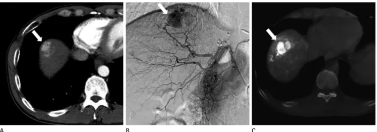
Fig. 1. Imaging characteristics of HCC demonstrated on CT before TACE, and on angiography and cone-beam CT during TACE.
A. Arterial phase of contrast-enhanced CT demonstrates a hyperattenuated mass (arrow) in the liver dome.
B. Digital subtraction angiography of the hepatic artery performed during TACE reveals a hypervascular HCC (arrow) in the liver dome.
C. Cone-beam CT acquired immediately after TACE demonstrates positive uptake of Lipiodol by the tumor (arrow) surrounded by oily portograms.
Note.—HCC = hepatocellular carcinoma, TACE = transarterial chemoembolization
B B
A A
C C
DISCUSSION
Various complications associated with TACE have been report- ed in the literature (8, 9). A frequent complication of TACE is postembolization syndrome (3), which encompasses symptoms such as abdominal pain, nausea, and vomiting. Such symptoms are usually self-limiting. They will subside with conservative man- agement. Irreversible liver failure is a more severe complication of TACE (4) that may occur in the presence of portal vein thrombo- sis or after extensive embolization of a large volume of the liver.
Other severe complications include ischemic bile duct injury (6), liver infarction, and intrahepatic abscess (5). As an extrahepatic complication of TACE, pulmonary oil embolism (7) has fre- quently been reported to occur secondary to shunting of the ethiodized oil in the hepatic arteries into the systemic circulation, particularly when a large dose of ethiodized oil is administered.
The current case of pleural empyema is, in a sense, a pulmonary complication of chemoembolization not related to pulmonary embolism. The plausible explanation for migration of intrahe- patic Lipiodol into the pleural cavity in our case could be infarc- tion around the HCC in the liver dome after chemoembolization that resulted in the rupture of the liver capsule. Even though che- moembolization was not performed via the right inferior phren- ic artery, we postulate that infarction of the diaphragm abutting the HCC may have occurred due to the shunting of the chemo- embolic agent through the peripheral anastomoses between the right hepatic artery and the right inferior phrenic artery. As a re- liver cirrhosis restricted the patient from undergoing surgical
treatment. As a result, the patient underwent intensive antibiotic therapy. Follow-up chest CT was performed sequentially four and five weeks later which showed progressive migration of the lump of Lipiodol until it had completely evacuated into the pleural cavity to become enclosed within an empyema (Fig. 2B, C). However, there was no residual Lipiodol in the liver. Only a region of low attenuation indicating where the Lipiodol accu- mulation once had been. No viable tumor was demonstrated ei- ther within the liver or in the pleural cavity on arterial phase of contrast-enhanced CT. A drainage catheter was inserted into the empyema via percutaneous approach through which turbid and yellowish material was drained. After further medical treatment and serial follow-up of chest radiographs, the patient’s symptoms regressed and the empyema was found to progressively resolved (Fig. 3). The drainage catheter was finally removed ten days after its insertion. Cytology of the pleural fluid was negative for malig- nancy. Bronchoscopy was performed to exclude other possible causes of hemoptysis. It only revealed old blood clots within the airway. Bronchial wash cytology did not reveal any evidence of malignancy. Positron emission tomography (PET)-CT disclosed no evidence of viable tumor in the chest or the liver. However, it did reveal distant metastasis to the right supraclavicular and dia- phragmatic lymph nodes. Following full symptomatic recovery, the patient was transferred back to the local clinic for palliative care. He has not returned since being discharged four months prior to the preparation of this report.
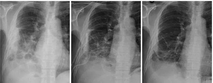
Fig. 3. Sequential plain radiographs of the chest demonstrating gradual resolution of pleural empyema following percutaneous drainage and antibiotic therapy.
conference. European Association for the Study of the Liver. J Hepatol 2001;35:421-430
3. Dhand S, Gupta R. Hepatic transcatheter arterial chemo- embolization complicated by postembolization syndrome.
Semin Intervent Radiol 2011;28:207-211
4. Hsin IF, Hsu CY, Huang HC, Huang YH, Lin HC, Lee RC, et al. Liver failure after transarterial chemoembolization for patients with hepatocellular carcinoma and ascites: inci- dence, risk factors, and prognostic prediction. J Clin Gas- troenterol 2011;45:556-562
5. Woo S, Chung JW, Hur S, Joo SM, Kim HC, Jae HJ, et al.
Liver abscess after transarterial chemoembolization in pa- tients with bilioenteric anastomosis: frequency and risk factors. AJR Am J Roentgenol 2013;200:1370-1377 6. Kim HK, Chung YH, Song BC, Yang SH, Yoon HK, Yu E, et
al. Ischemic bile duct injury as a serious complication after transarterial chemoembolization in patients with hepato- cellular carcinoma. J Clin Gastroenterol 2001;32:423-427 7. Chung JW, Park JH, Im JG, Han JK, Han MC. Pulmonary oil
embolism after transcatheter oily chemoembolization of hepatocellular carcinoma. Radiology 1993;187:689-693 8. Poggi G, Pozzi E, Riccardi A, Tonini S, Montagna B, Qua-
retti P, et al. Complications of image-guided transcatheter hepatic chemoembolization of primary and secondary tu- mours of the liver. Anticancer Res 2010;30:5159-5164 9. Xia J, Ren Z, Ye S, Sharma D, Lin Z, Gan Y, et al. Study of
severe and rare complications of transarterial chemoem- bolization (TACE) for liver cancer. Eur J Radiol 2006;59:
407-412
10. Negrini S, Zoppoli G, Andorno E, Picciotto A, Indiveri F. Io- dized oil pleural effusion in a patient previously treated with transarterial chemoembolization for hepatocellular carcinoma. Chest 2010;138:193-195
sult, the Lipiodol lump is thought to have herniated through the diaphragmatic defect into the ipsilateral pleural cavity. The re- sulting presence of chemoembolic material in the pleural space most probably triggered an inflammatory reaction that created an empyema.
With regards to treatment, surgical debridement and repair of the ruptured diaphragm was initially considered for our patient.
This would have revealed the nature of the Lipiodol lump in the pleural cavity and histological information, especially whether seeding of HCC had occurred. Unfortunately, histological con- firmation was not available in this case because a decision was reached to treat the patient medically and drain the empyema by percutaneous approach. Fortunately, such treatment sufficed in treating the empyema. No further complication developed in either the chest or the abdomen. There was no evidence to sug- gest viable tumor in the chest or liver on cytology of the pleural fluid and PET-CT.
Transdiaphragmatic herniation and consequent evacuation of ethiodized oil into the pleural cavity is an extremely rare compli- cation of TACE. To our knowledge, only one similar case has been reported previously in the literature (10). Although rare, the possible development of liver abscess with or without diaphragm rupture should be considered when a patient develops pleural ef- fusion or empyema after undergoing chemoembolization.
REFERENCES
1. Bruix J, Sherman M; Practice Guidelines Committee, Ameri- can Association for the Study of Liver Diseases. Manage- ment of hepatocellular carcinoma. Hepatology 2005;42:
1208-1236
2. Bruix J, Sherman M, Llovet JM, Beaugrand M, Lencioni R, Burroughs AK, et al. Clinical management of hepatocellu- lar carcinoma. Conclusions of the Barcelona-2000 EASL
간암의 화학색전술 후 발생한 농흉 증례1
서영균
1· 원제환
1· 왕희정
2· 김진우
160세 남성이 6주 전부터 시작된 흉막삼출과 객혈을 주소로 내원하였다. 환자는 약 3개월 전에 재발성 간암을 치료하기 위해 화학색전술을 받은 병력이 있었다. 연속적인 추적단순흉부촬영 및 전산화단층촬영에서 오른쪽 흉막강에 농흉의 발 생과 더불어, 간암부위에 주입한 리피오돌이 흉막강으로 유입되어 있음을 확인할 수 있었다. 저자들은 오른쪽 흉막강에 경 피적 배액관을 삽입하였고, 10일이 지난 후 환자의 흉막삼출액과 농흉은 소실되었다. 이 증례는 화학색전술을 한 간암 환 자에서 리피오돌의 유출에 의해 농흉이 발생할 수 있음을 보여준다.
아주대학교 의과대학 아주대학교병원 1영상의학과, 2외과