INTRODUCTION
Primary malignant melanoma of the esophagus (PMME) is one of the extremely rare tumor and the prognosis is very poor. This tumor has usually been reported as a pedunculat- ed, polypoid lesion in the middle and lower third of the eso- phagus. According to the reports, hematogenic and lympho- genic metastases are common for PMME cases. Surgical resec- tion has been the preferred method of treatment, despite the poor prognosis. Adjuvant chemotherapy has been employed in some cases, but the effectiveness has not been proved. We report a case of PMME which was treated by surgical resec- tion and post-operative chemotherapy.
CASE REPORT
A 60-yr-old male presented with a 3-month history of dys- phagia and upper abdominal discomfort. He was treated at a private clinic under the impression of gastritis without symp- tomatic improvement. He had no smoking or drinking histo- ry. There were no abnormalities noted on physical examina- tion or within the laboratory reports, and a pre-operative sim- ple chest radiography and electrocardiography showed no abnormal findings. Barium-contrast esophagogram showed irregular luminal narrowing with a 4 cm sized filling defect
at the distal esophagus. The proximal esophagus was dilated with markedly delayed dye passage (Fig. 1). Fiberoptic eso- phagoscopy showed a bell-shaped obstructing black mass with a 4 cm sized central ulcer located between 32 and 36 cm from the incisors. The pathology report of the biopsy speci- men diagnosed the mass as malignant melanoma. Chest com- puterized tomogram showed the mass to be 5 cm in size at the posterior wall of the distal esophagus with evidence of lymph node enlargement in the lesser curvature of the stom- ach (Fig. 2, 3). Pulmonary function tests, whole body bone scan with TC-99m, and a computerized brain tomogram were all unremarkable, and therefore the pre-operative clini- cal stage was classified as IVA (cT3N0M1a).
The patient underwent a total esophagectomy and cervical esophagogastric anastomosis via a right thoracotomy, laparo- tomy and cervical incision. Reconstruction was performed by pulling the gastric tube up through a retro-sternal route.
The cervical esophagogastric anastomosis was performed by a standard hand-sewn technique. There was no evidence of mediastinal lymph node enlargement and the post-operative course was uneventful but the patient suffered from post-oper- ative pain. The patient was discharged 24th post-operative day.
The resected mass was 5.5×4.2×1.3 cm in size, polypoid and pigmented from the distal esophagus with lymph node enlargement along the lesser curvature of the stomach (Fig.
4). Microscopic examination revealed junctional activities of Song Am Lee, Jae Joon Hwang,
Young Ho Choi*
Department of Thoracic and Cardiovascular Surgery, Konkuk University Hospital, Seoul; Department of Thoracic and Cardiovascular Surgery*, Korea University Medical College, Seoul, Korea
Address for correspondence Jae Joon Hwan, M.D.
Department of Thoracic and Cardiovascular Surgery, Konkuk University Hospital, 1 Hwayang-dong, Gwangjin-gu, Seoul 143-701, Korea Tel : +82.2-2030-7592, Fax : +82.2-2030-7592 E-mail : [email protected]
*This work was supported by a grant promoting research from Konkuk University.
149 J Korean Med Sci 2007; 22: 149-52
ISSN 1011-8934
Copyright � The Korean Academy of Medical Sciences
Surgical Treatment of Primary Malignant Melanoma of the Esophagus : A Case Report
Primary malignant melanoma of the esophagus (PMME) is an extremely rare tumor with only scattered cases reported. Although surgical resection has been consid- ered as the best possible option, the prognosis has been nonetheless poor. We report a case of PMME which was treated by surgical resection and additionally followed by chemotherapy. A 60-yr-old man underwent an esophagoscopy due to a 3-month history of dysphagia and upper abdominal discomfort. A pigmented poly- poid mass in the lower third of the esophagus was discovered, and a biopsy iden- tified the mass as a malignant melanoma. Consequently, a subtotal esophagecto- my and intrathoracic esophagogastrostomy was carried out. At follow-up four months after discharge, lymph node enlargements in the cervical area and celiac axis area were found. As a result, the patients was started on systemic chemotherapy treat- ment, which included Dacarbazine. The patient has been doing well and is now 35 months post-operative.
Key Words : Esophageal Neoplasms; Melanoma; Esophagectomy
Received : 20 July 2005 Accepted : 14 November 2005
150 S.A. Lee, J.J. Hwang, Y.H. Choi
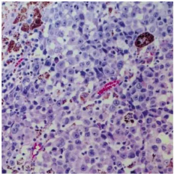
atypical melanocytes around base of the tumor and a defini- tive diagnosis of PMME was made (Fig. 5, 6). The tumor cells invaded the muscular layer. No regional lymph node was positive for metastasis. Three lymph nodes at the lesser curvature of the stomach were infiltrated by tumor cells. This case was then classified as esophageal cancer, stage IVA:T2- N0M1a, according to the TMN classification of AJCC.
Four months after discharge, at a follow-up, lymph node enlargements in the cervical area and celiac axis area were
found and systemic chemotherapy was started. The patient received Dacarbazine 800 mg/m2intravenously (IV) on day 1, Cisplatin 30 mg/m2IV on days 2-5, and Vinblastine 1.6 mg/
m2IV on days 1-5. The cycles were repeated every 4 weeks.
After the fifth cycle of chemotherapy, the size of the lymph nodes decreased significantly and at 35 months post-opera- tive, the patient is doing well without evidence of recurrence.
Fig. 1.Pre-operative esophagogram demonstrating a 4 cm sized mass with an irregular surface protruding into the lower esophagus.
Fig. 4.Gross appearance of the resected specimen showing the ulcerated, black, sessile polypoid tumor in the lower esophagus with lymph node enlargement along the lesser curvature of the stomach (arrow).
Fig. 3.Abdominal CT scan showing lymphadenopathy in the less- er curvature of the stomach (arrow).
Fig. 2.Contrast-enhanced chest CT scan showing a mass lesion from posterior wall of the lower esophagus (arrow).
Esophageal Melanoma 151
DISCUSSION
Since Baur made the first report of PMME in 1906, scat- tered cases have been reported in the literature and the inci- dence of PMME is reported to be less than 0.1% of all eso- phageal malignancies (1). Chalkiadakis and colleagues, in a review of 110 patients diagnosed with PMME found that men predominated the cases by 2:1, with an overall mean age of 60 yr (2). The tumors were usually located in the mid- dle and lower third of the esophagus (86%). The common symptoms were dysphagia (73%), retrosternal pain (24%), weight loss (16%), and regurgitation (12%).
The diagnosis of PMME is difficult and best deduced by a combination of several examinations including upper gas- trointestinal series, endoscopy, computed tomographic scan and biopsy with histopathologic assessment. Macroscopical- ly, the tumor usually presents as a polypoid mass, that is focal- ly ulcerated, but covered for the most part by intact squa- mous mucosa. According to the diagnostic criteria of prima- ry melanoma proposed by Allen and Spitz, which seems to be widely accepted, the tumor is considered as primary when (a) the mass has a characteristic structure of melanoma con- taining melanin, (b) the adjacent epithelium contains mela- nocytes, (c) the tumor is polypoid, and (d) it arises from the area of junctional changes in the squamous epithelium (3).
Thus, the proposed criteria for the origin of a tumor in the esophagus depends on the finding of junctional activity adja- cent to the tumor mass. Recently, immunohistochemical stain- ing utilizing antibodies specific to HMB-45 and/or S-100 cytoplasmic protein has been used to identify malignant melanoma of the esophagus (4, 5).
Hematogenic and lymphogenic metastasis are commonly associated with PMME and account for the poor prognosis.
Widespread metastases with a rapid deterioration in health, followed by an early death is the usual clinical course. The most common site of metastases reported is the liver, followed by the mediastinum, mediastinal lymph nodes, lung and brain (2).
The choice of therapy primarily depends on the functional status and the presence and extent of the metastatic disease at the time of diagnosis. Surgical resection with re-establish- ment of gastrointestinal continuity is the proposed methods of treatment, and looks like to provide the best result. The median survival rate after surgical resection was 13.4 months and longest reported survival was 12 yr (2, 6). Chemothera- py, radiation therapy, and immunotherapy have been report- ed, but with limited success (7, 8). Recently, several authors have reported a therapeutic effect of combination chemother- apy including Dacarbazine (9, 10). In this patient, post-opera- tive chemotherapy has thus far been effective in controlling recurrence. This case supports the combination of surgical resection and post-operative adjuvant chemotherapy, with Dacarbazine, and therefore should be considered as another treatment modality.
REFERENCES
1. Turnbull AD, Rosen P, Goodner JT, Beattie EJ. Primary malignant tumors of the esophagus other than typical epidermoid carcinoma.
Ann Thorac Surg 1973; 15: 463-73.
Fig. 5.Photomicrograph of the tumor showing junctional activities of atypical melanocytes around base of the tumor (H&E, ×100).
Fig. 6.Photomicrograph of the tumor revealing pigmented and non-pigmented malignant melanoma cells with vesicular nuclei, prominent nucleoli, and moderate amount of pale eosinophilic cytoplasm (H&E, ×200).
152 S.A. Lee, J.J. Hwang, Y.H. Choi
2. Chalkiadakis G, Wihlm JM, Morand G, Weill-Bousson M, Witz JP.
Primary malignant melanoma of the esophagus. Ann Thorac Surg 1985; 39: 472-5.
3. Allen AC, Spitz S. Malignant melanoma: A clinico-pathological anal- ysis of criteria for diagnosis and prognosis. Cancer 1953; 6: 1-45.
4. Stranks GJ, Mathai JT, Rowe-Jones DC. Primary malignant mela- noma of the esophagus: case report and review of surgical patholo- gy. Gut 1991; 32: 828-30.
5. DiCostanzo DP, Urmacher C. Primary malignant melanoma of the esophagus. Am J Surg Pathol 1987; 11: 46-52.
6. Hamdy FC, Smith JH, Kennedy A, Thorpe JA. Long survival after excision of a primary malignant melanoma of the oesophagus. Tho- rax 1991; 46: 397-8.
7. Caldwell CB, Bains MS, Burt M. Unusual malignant neoplasms of the esophagus. Oat cell carcinoma, melanoma, and sarcoma. J Tho- rac Cardiovasc Surg 1991; 101: 100-7.
8. Joob AW, Haines III Gk, Kies MS, Shields TW. Primary nalignant melanoma of the esophagus. Ann Thorac Surg 1995; 60: 217-22.
9. Suzuki Y, Aoyama N, Minamide J, Takata K, Ogata T. Amelanotic malignant melanoma of the esophagus: report of a patient with recur- rence successfully treated with chemoendocrine therapy. Int J Clin Oncol 2005; 10: 204-7.
10. Matsutani T, Onda M, Miyashita M, Hagiwara N, Akiya Y, Takubo K, Yamashita K, Sasajima K. Primary malignant melanoma of the esophagus treated by esophagectomy and systemic chemotherapy.
Dis Esophagus 2001; 14: 241-4.