INTRODUCTION
A shorter time to reperfusion is beneficial in treating pa- tients with fibrinolytic therapy for acute ST-segment eleva- tion myocardial infarction (STEMI) (1-4). In contrast, there is some controversy regarding the relationship between mor- tality and time to reperfusion with primary percutaneous coronary intervention (PCI), although the American College of Cardiology/American Heart Association (ACC/AHA) guidelines recommend that the door-to-balloon time (or medical contact-to-balloon) for PCI should be kept under 90 min (5). Some studies have shown that delays in symp- tom onset-to-balloon time (6-9) or door-to-balloon time adversely affects the prognosis in patients with STEMI (10, 11). However, in other studies, there was no significant dif- ference in clinical outcomes according to the time to reper- fusion (4, 12). In addition, because the initial treatment delay is an important quality indicator of the treatment for such patients, it is worthwhile to investigate how many cases are performed according to the guidelines. Therefore, the aim of this study was to evaluate the current status of delay in the time to reperfusion during primary PCI and its impact on patient mortality one month later using data from the
Korea Acute Myocardial Infarction Registry (KAMIR), the first nationwide multicenter registry of acute MI in Korea.
We also tried to define the subgroups that were mostly influ- enced by treatment delay.
MATERIALS AND METHODS Study design and subjects
A cohort of patients with STEMI who underwent prima- ry PCI was selected from the KAMIR, which is a nation- wide study for acute myocardial infarction in Korea. Between November 2005 and January 2007, 5,069 patients at 41 hos- pitals were registered.
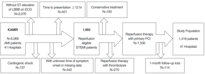
Patients were eligible for enrollment if they had STEMI, presented within 12 hr after symptom onset, and were treat- ed with primary PCI, and if they had completed a 30-day clinical follow-up at the analysis time point. The following patients were excluded sequentially: patients without ST elevation or left bundle branch block on the first electrocar- diogram (n=2,076), those with cardiogenic shock (n=137), those with unknown time of symptom onset or missing data
357
Young Bin Song1, Joo-Yong Hahn1, Hyeon-Cheol Gwon, Jun Hyung Kim, Sang Hoon Lee, Myung-Ho Jeong*, and KAMIR investigators
Department of Medicine, Samsung Medical Center, Sungkyunkwan University School of Medicine, Seoul;
Heart Center of Chonnam National University Hospital*, Chonnam National University Research Institute of Medical Sciences, Gwangju, Korea
1The first two authors contributed equally to this work.
Address for correspondence Hyeon-Cheol Gwon, M.D.
Cardiac and Vascular Center, Samsung Medical Center, Sungkyunkwan University School of Medicine, 50 Irwon-dong, Gangnam-gu, Seoul 135-710, Korea Tel : +82.2-3410-3418, Fax : +82.2-3410-0483 E-mail : [email protected] DOI: 10.3346/jkms.2008.23.3.357
The Impact of Initial Treatment Delay Using Primary Angioplasty on Mortality among Patients with Acute Myocardial Infarction: from the Korea Acute Myocardial Infarction Registry
The impact of treatment delays to reperfusion on patient mortality after primary percutaneous coronary intervention (PCI) for ST elevation myocardial infarction (STEMI) is controversial. We analyzed 5,069 patients included in the Korea Acute Myocardial Infarction Registry (KAMIR) between November 2005 and January 2007. We selected 1,416 patients who presented within 12 hr of symptom onset and who were treated with primary PCI. The overall mortality at one month was 4.4%. The medians of door-to-balloon time, symptom onset-to-balloon time, and symptom onset-to-door time were 90 (interquartile range, 65-136), 274 (185-442), and 163 min (90-285), respectively. One-month mortality was not increased signifi- cantly with any increasing delay in door-to-balloon time (4.3% for ≤≤90 min, 4.4%
for >90 min; p=0.94), symptom onset-to-balloon time (3.9% for ≤≤240 min, 4.8%
for >240 min; p=0.41), and symptom onset-to-door time (3.3% for ≤≤120 min, 5.0%
for >120 min; p=0.13). These time variables had no impact on one-month mortality in any subgroup. Thus, this first nationwide registry data in Korea showed a good result of primary PCI, and the patient prognosis may not depend on the initial treat- ment delay using the current protocols.
Key Words : Myocardial Infarction; Mortality; Angioplasty
Received : 15 March 2007 Accepted : 20 September 2007
(n=442), and those with symptom onset-to-door time ≥12 hr (n=421). Among the 5,069 patients, 1,993 patients had reperfusion eligible for STEMI and 1,800 (90.3%) received reperfusion therapy. Reperfusion therapy with primary PCI was performed in 1,530 patients (85.0% of 1,800) and with thrombolysis in 270 (15.0% of 1,800). Patients who did not undergo the one-month follow-up were excluded (n=114).
We finally included 1,416 patients with STEMI who were treated with primary PCI and who completed the one-month follow-up (Fig. 1).
Demographic and clinical characteristics recorded were gender, age, and medical history. The latter included any history of smoking, dyslipidemia, hypertension, diabetes mellitus, chronic renal insufficiency, stroke, ischemic heart disease, family history of coronary artery disease, previous PCI, previous coronary artery bypass graft surgery, or any past regular medication. The presentation characteristics included symptoms at admission, systolic and diastolic blood pressure, heart rate and rhythm, Killip classification, the results of the diagnostic electrocardiography (ECG), and ischemic location on ECG.
Definitions and outcomes measures
Time variables were defined as follows: door-to-balloon time was the time from arrival in the emergency department until initial balloon inflation; symptom onset-to-balloon time was the time from the onset of symptoms until the first balloon inflation; and symptom onset-to-door time was the time from the onset of symptoms until arrival in the emer- gency department. Patients were divided into two groups according to door-to-balloon time (≤90 min and >90 min), symptom onset-to-balloon time (≤240 min and >240 min), and symptom onset-to-door time (≤120 min and >120 min), respectively.
The main outcomes were mortality at one month and major cardiovascular adverse events (MACEs). MACEs included
death, reinfarction, and target vessel revascularization.
Statistical analysis
SPSS for Windows (version 12.0; SPSS Inc., Chicago, IL, U.S.A.) was used for all analyses. Continuous data are express- ed as the mean±SD or as the median and interquartile range (25th and 75th percentiles); categorical data were expressed as percentages. Statistical comparisons of baseline, angio- graphic, and outcome variables were performed for categori- cal variables using the Chi-squared test or Fisher’s exact test (if the expected value of the variable was <5 in at least one group). Student’s t test was applied to continuous variables.
p<0.05 was considered statistically significant. Multiple logistic regression analysis was performed to assess the rela- tion between predictor variables and one-month mortality.
RESULTS Baseline characteristics
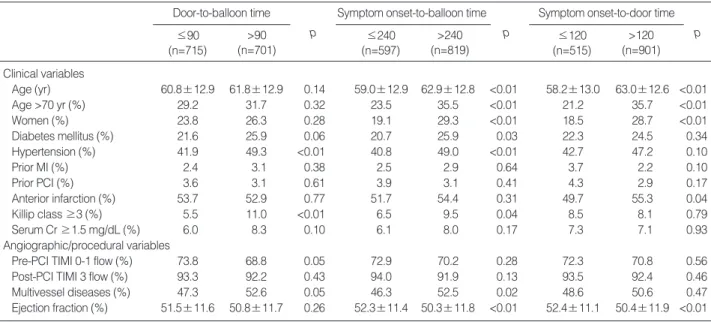
Medians of times to treatment were as follows: door-to- balloon time, 90 min (interquartile range, 65-136 min); symp- tom onset-to-balloon time, 274 min (185-442); and symp- tom onset-to-door time, 163 min (90-285). Of all patients, 36% presented to the hospital within 120 min of symptom onset. Door-to-balloon time was 90 min or less in 51% of patients and 42% of patients were reperfused within 240 min of symptom onset. A post-PCI thrombolysis in myocardial infarction (TIMI) grade 3 flow was achieved in 92.8% of the patients.
Demographic, clinical, and angiographic characteristics according to door-to-balloon time, symptom onset-to-bal- loon time, and symptom onset-to-door time are presented in Table 1. Patients with door-to-balloon times >90 min had hypertension and Killip class ≥3 more frequently than did
Fig. 1. Selection criteria.
Without ST-elevation of LBBB on ECG
N=2,076
Cardiogenic shock N=137 KAMIR N=5,069 AMI patients 41 Hospitals
With unknown time of symptom onset or missing data
N=442
Reperfusion therapy with thrombolysis
N=270
1-month follow-up loss N=114
Study Population 1,416 patients
41 Hospitals Time to presentation ≥12 hr
N=421
Conservative treatment N=193
1,993 Reperfusion
eligible STEMI patients
Reperfusion therapy with primary PCI
N=1,530
patients with door-to-balloon times ≤90 min. The preva- lences of older patients, women, having diabetes mellitus, hypertension, Killip class ≥3, multivessel disease, and lower left ventricular ejection fraction were higher in patients with
longer symptom onset-to-balloon times than in those with shorter symptom onset-to-balloon times. Patients with longer symptom onset-to-door times tended to be older, to be women, and to have anterior infarction more frequently and lower left ventricular ejection fraction than did patients with shorter symptom onset-to-door times.
Of the patients enrolled in the study, 262 patients (18.5%) were treated with glycoprotein IIb/IIIa inhibitors. Triple antiplatelet agents with aspirin, clopidogrel, and cilostazol were prescribed in 598 patients (42.1%). One-hundred and twenty-two patients (8.6%) with antiplatelet use before the index MI were not statistically different in mortality (5.7%
vs. 4.3%, p=0.44) and MACE (7.4% vs. 5.5%, p=0.39) com- pared with those without antiplatelet use. Sixty-three patients (4.5%) were treated with statin before the index MI.
Clinical outcomes by time variables
Sixty-two patients (4.4%) had died by one month after the procedure. Table 2 shows the association between treat- ment delay and clinical outcomes. Mortality at this point did not increase significantly with increasing delay in door-
Door-to-balloon time
≤90 p (n=715)
>90 (n=701)
Symptom onset-to-balloon time
≤240 p (n=597)
>240 (n=819)
Symptom onset-to-door time
≤120 p (n=515)
>120 (n=901) Clinical variables
Age (yr) 60.8±12.9 61.8±12.9 0.14 59.0±12.9 62.9±12.8 <0.01 58.2±13.0 63.0±12.6 <0.01
Age >70 yr (%) 29.2 31.7 0.32 23.5 35.5 <0.01 21.2 35.7 <0.01
Women (%) 23.8 26.3 0.28 19.1 29.3 <0.01 18.5 28.7 <0.01
Diabetes mellitus (%) 21.6 25.9 0.06 20.7 25.9 0.03 22.3 24.5 0.34
Hypertension (%) 41.9 49.3 <0.01 40.8 49.0 <0.01 42.7 47.2 0.10
Prior MI (%) 2.4 3.1 0.38 2.5 2.9 0.64 3.7 2.2 0.10
Prior PCI (%) 3.6 3.1 0.61 3.9 3.1 0.41 4.3 2.9 0.17
Anterior infarction (%) 53.7 52.9 0.77 51.7 54.4 0.31 49.7 55.3 0.04
Killip class ≥3 (%) 5.5 11.0 <0.01 6.5 9.5 0.04 8.5 8.1 0.79
Serum Cr ≥1.5 mg/dL (%) 6.0 8.3 0.10 6.1 8.0 0.17 7.3 7.1 0.93
Angiographic/procedural variables
Pre-PCI TIMI 0-1 flow (%) 73.8 68.8 0.05 72.9 70.2 0.28 72.3 70.8 0.56
Post-PCI TIMI 3 flow (%) 93.3 92.2 0.43 94.0 91.9 0.13 93.5 92.4 0.46
Multivessel diseases (%) 47.3 52.6 0.05 46.3 52.5 0.02 48.6 50.6 0.47
Ejection fraction (%) 51.5±11.6 50.8±11.7 0.26 52.3±11.4 50.3±11.8 <0.01 52.4±11.1 50.4±11.9 <0.01 Table 1. Baseline patient characteristics by time variables
MI, myocardial infarction; PCI, percutaneous coronary intervention; Cr, creatinine; TIMI, thrombolysis in myocardial infarction.
Door-to-balloon time (min)
≤90 p (n=715)
>90 (n=701)
Symptom onset-to-balloon time (min)
≤240 p (n=597)
>240 (n=819)
Symptom onset-to-door time (min)
≤120 p (n=515)
>120 (n=901)
Mortality (%) 4.3 4.4 0.94 3.9 4.8 0.41 3.3 5.0 0.13
MACEs (%) 5.5 5.8 0.75 4.5 6.5 0.12 4.5 6.3 0.15
Follow-up LVEF (%) 54.3±10.9 54.0±11.7 0.80 54.7±10.1 53.6±12.2 0.38 55.4±10.1 53.4±11.8 0.13 Table 2. One-month outcomes by time variables
MACEs, major adverse cardiovascular events; LVEF, left ventricular ejection fraction.
Fig. 2. Kaplan-meier estimates of cumulative survival stratified by door-to-balloon time.
Survival (%)
100
99
98
97
96
95
0 5 10 15 20 25 30
Days from MI onset
p=0.94
≤90 min
>90 min
to-balloon time (4.3% for ≤90 min vs. 4.4% for >90 min;
p=0.94), symptom onset-to-balloon time (3.9% for ≤240 min vs. 4.8% for >240 min; p=0.41), and symptom onset- to-door time (3.3% for ≤120 min vs. 5.0% for >120 min;
p=0.13) (Fig. 2). The rate of MACEs at one month was not significantly different according to door-to-balloon time, symptom onset-to-balloon time, or symptom onset-to-door
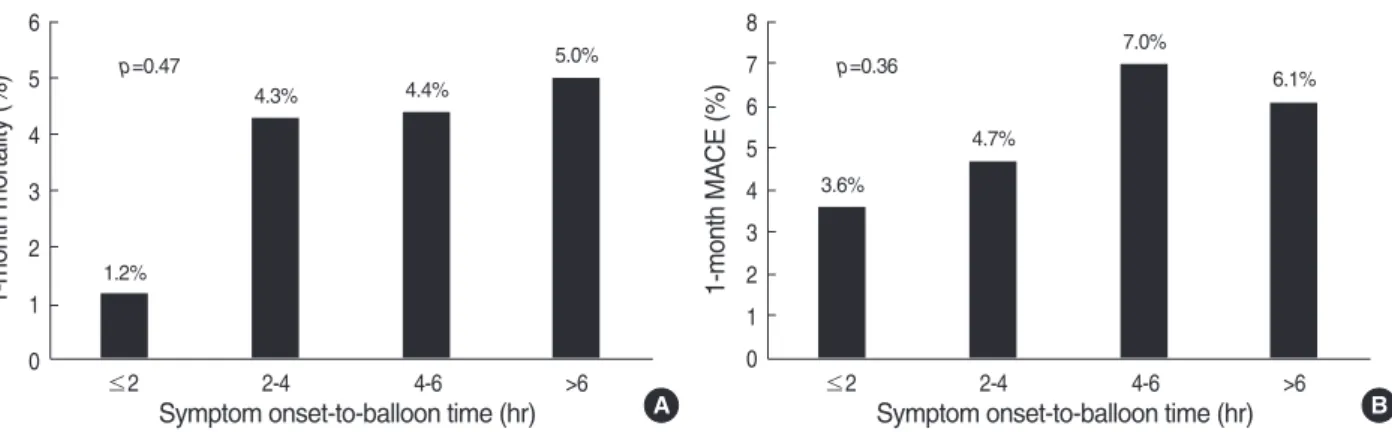
time. When patients were divided into 4 groups according to door-to-balloon time (≤90, 90-120, 120-180, ≥180 min) or symptom onset-to-balloon time (≤2, 2-4, 4-6, ≥6 hr), incremental delays in reperfusion appeared to have little effect on the one-month mortality and MACE rate (Fig. 3, 4). The left ventricular ejection fraction at the one-month follow-up was also similar between the early reperfused and late reper-
Fig. 3. The one-month mortality (A) and MACEs (B) stratified by door-to-balloon time.
1-month mortality (%)
6 5 4 3 2 1
0 ≤2 2-4 4-6 >6
Symptom onset-to-balloon time (hr)
1.2%
A
4.3% 4.4%
p=0.47 5.0%
1-month MACE (%)
8 7 6 5 4 3 2 1 0
≤2 2-4 4-6 >6
Symptom onset-to-balloon time (hr)
3.6%
B 4.7%
7.0%
p=0.36 6.1%
Fig. 4. The one-month mortality (A) and MACEs (B) stratified by symptom onset-to-balloon time.
1-month mortality (%)
6 5 4 3 2 1
0 ≤90 90-120 120-180 >180
Door-to-balloon time (min)
4.3%
A 5.7%
5.1%
2.3%
p=0.37 p=0.69
1-month MACE (%)
8 7 6 5 4 3 2 1 0
≤90 90-120 120-180 >180
Door-to-balloon time (min)
5.5%
B
6.8% 6.6%
4.1%
Mortality (n=62) p MACEs (n=80) p
Clinical variables
Age >70 yr 37/431 (8.6%) <0.01 41/431 (9.5%) <0.01
Women 27/354 (7.6%) <0.01 28/354 (7.9%) 0.03
Diabetes mellitus 16/331 (4.8%) 0.58 21/331 (6.3%) 0.49
Hypertension 30/637 (4.7%) 0.48 39/637 (6.1%) 0.42
Prior myocardial infarction 3/39 (7.7%) 0.31 6/39 (15.4%) <0.01
Prior PCI 2/48 (4.2%) 0.94 4/48 (8.3%) 0.41
Anterior infarction 35/750 (4.7%) 0.61 46/750 (6.1%) 0.44
Killip classification ≥3 20/115 (17.4%) <0.01 21/115 (18.3%) <0.01
Serum creatinine ≥1.5 mg/dL 14/101 (13.9%) <0.01 17/101 (16.8%) <0.01
Ejection fraction ≤40% 25/232 (10.8%) <0.01 29/232 (12.5%) <0.01
Angiographic/procedural variables
Pre-PCI TIMI 0-1 flow 48/994 (4.8%) 0.08 58/994 (5.8%) 0.43
Post-PCI TIMI 0-2 flow 15/100 (15.0%) <0.01 16/100 (16.0%) <0.01
Multivessel diseases 46/702 (6.6%) <0.01 63/702 (9.0%) <0.01
Table 3. Univariate predictors of one-month mortality and MACEs
MACEs, major adverse cardiovascular events; PCI, percutaneous coronary intervention; TIMI, thrombolysis in myocardial infarction.
fused patients regarding door-to-balloon time, symptom onset-to-balloon time, and symptom onset-to-door time.
Predictors of one-month clinical outcomes
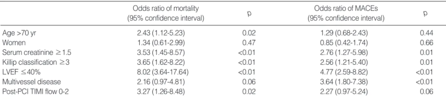
Applying univariate analysis, various clinical and angio- graphic or procedural variables were significantly associated with one-month mortality and MACEs (Table 3). In multi- variate analysis, old age (>70 yr), Killip class ≥3, serum creatinine ≥1.5 mg/dL, post-PCI TIMI flow grades of 0-2, and left ventricular dysfunction (ejection fraction ≤40%) were significantly associated with mortality. From the multi- variate analysis, Killip class ≥3, serum creatinine ≥1.5 mg/
dL, left ventricular dysfunction (ejection fraction ≤40%), and multivessel disease were independent predictors for MACEs (Table 4).
Clinical outcomes by time variables in subgroups
We performed subgroup analysis to identify those patients with outcomes most influenced by treatment delay. Based upon multivariate analysis for one-month mortality, we divid-
ed patients by age, serum creatinine, Killip classification, left ventricular ejection fraction, and post-PCI TIMI flow grade. Treatment delay was not significantly associated with increased short-term mortality in any subgroup. Moreover, among patients at high risk (age >70 yr, serum creatinine
≥1.5 mg/dL, Killip classification ≥3, left ventricular ejec- tion fraction ≤40%, post-PCI TIMI flow grades 0-2), the increase in door-to-balloon time, symptom onset-to-balloon time, or symptom onset-to-door time did not correlate with one-month mortality; nor were there any such associations among low-risk patients (Table 5).
DISCUSSION
Primary PCI was the preferred reperfusion strategy in this registry, and more than half of the patients underwent pri- mary PCI within the time window recommended in the guidelines. The one-month mortality was 4.4% and was not increased significantly with increasing delay in door-to- balloon time, symptom onset-to-balloon time, or symptom onset-to-door time. These time variables had no impact on
Odds ratio of mortality (95% confidence interval)
Odds ratio of MACEs (95% confidence interval)
p p
Age >70 yr 2.43 (1.12-5.23) 0.02 1.29 (0.68-2.43) 0.44
Women 1.34 (0.61-2.99) 0.47 0.85 (0.42-1.74) 0.66
Serum creatinine ≥1.5 3.53 (1.45-8.57) <0.01 2.76 (1.27-5.98) 0.01
Killip classification ≥3 3.65 (1.62-8.22) <0.01 2.56 (1.21-5.40) 0.01
LVEF ≤40% 8.02 (3.64-17.64) <0.01 4.77 (2.59-8.82) <0.01
Multivessel disease 2.16 (0.97-4.81) 0.06 3.64 (1.80-7.38) <0.01
Post-PCI TIMI flow 0-2 3.27 (1.26-8.48) 0.02 2.27 (0.97-5.24) 0.06
Table 4. Multivariate predictors of one-month mortality and MACEs
MACEs, major adverse cardiovascular events; LVEF, left ventricular ejection fraction; PCI, percutaneous coronary intervention; TIMI, thrombolysis in myocardial infarction.
Door-to-balloon time
≤90 min p (%)
>90 min (%)
Symptom onset-to-balloon time
≤240 min p (%)
>240 min (%)
Symptom onset-to-door time
≤120 min p (%)
>120 min (%)
Age >70 yr (n=431) 24/209 (11.5) 13/222 (5.9) 0.06 12/140 (8.6) 25/291 (8.6) 0.99 11/109 (10.1) 26/322 (8.1) 0.52 Age ≤70 yr (n=985) 7/506 (1.4) 18/479 (3.8) 0.06 11/457 (2.4) 14/528 (2.7) 0.81 6/406 (1.5) 19/579 (3.3) 0.08 Serum Cr ≥1.5 mg/dL (n=101) 9/43 (20.9) 5/58 (8.6) 0.08 5/36 (13.9) 9/65 (13.8) 0.99 2/37 (5.4) 12/64 (18.8) 0.06 Serum Cr <1.5 mg/dL (n=1,306) 21/668 (3.1) 26/638 (4.1) 0.37 18/557 (3.2) 29/749 (3.9) 0.54 15/473 (3.2) 32/833 (3.8) 0.53 Killip class 3-4 (n=115) 5/39 (12.8) 15/76 (19.7) 0.35 6/38 (15.8) 14/77 (18.2) 0.75 6/43 (14.0) 14/72 (19.4) 0.45 Killip class 1-2 (n=1,281) 26/666 (3.9) 16/615 (2.6) 0.19 17/549 (3.1) 25/732 (3.4) 0.75 11/463 (2.4) 31/818 (3.8) 0.17 LVEF ≤40% (n=232) 12/106 (11.3) 13/126 (10.3) 0.81 9/72 (12.5) 16/160 (10.0) 0.57 7/65 (10.8) 18/167 (10.8) 0.99 LVEF >40% (n=1,045) 6/543 (1.1) 4/502 (0.8) 0.61 0/454 (0) 10/591 (1.7) 0.06 0/392 (0) 10/653 (1.5) 0.06 Post-PCI TIMI <3 (n=100) 5/47 (10.6) 10/53 (18.9) 0.25 6/35 (17.1) 9/65 (13.8) 0.66 5/33 (15.2) 10/67 (14.9) 0.98 Post-PCI TIMI 3 (n=1,286) 26/657 (4.0) 18/629 (2.9) 0.28 15/549 (2.7) 29/737 (3.9) 0.24 10/472 (2.1) 34/814 (4.2) 0.06 High risk* (n=682) 29/318 (9.1) 23/364 (6.3) 0.17 19/228 (8.3) 33/454 (7.3) 0.62 14/199 (7.0) 38/483 (7.9) 0.71 Low risk (n=734) 2/397 (0.5) 8/337 (2.4) 0.06 4/369 (1.1) 6/365 (1.6) 0.51 3/316 (0.9) 7/418 (1.7) 0.401 Table 5. One-month mortality by time variables in patients subsets
*High risk: Age >70 yr; serum creatinine ≥1.5 mg/dL; Killip class ≥3; LVEF ≤40% or post-PCI TIMI <grade 3.
Cr, creatinine; LVEF, left ventricular ejection fraction; PCI, percutaneous coronary intervention; TIMI, thrombolysis in myocardial infarction.
one-month mortality in any subgroup.
Among 5,069 patients registered in the KAMIR between November 2005 and January 2007, 1,993 patients had STEMI and was eligible for reperfusion. Reperfusion thera- py was performed over 90% of these patients, and only a minority received conservative treatment. The rate of receiv- ing reperfusion therapy among these eligible patients with STEMI in the KAMIR seems to be higher than in previous data. From data of the National Registry of Myocardial Infarc- tion (NRMI) in the U.S.A., one-half of the patients with STEMI who were eligible for reperfusion received reperfu- sion therapy (13). Notably, primary PCI was the overwhelm- ingly preferred reperfusion strategy in this registry. Among those patients receiving reperfusion therapy, primary PCI was performed in 85%. This rate of primary PCI is much higher than results from other countries, which showed that primary PCI was performed in one-fourth to one-third (13, 14). In the KAMIR, the incidence of post-PCI TIMI grade 3 flow was also very high at 92.8%. This is higher than that reported in studies from the U.S.A. (15, 16). Moreover, the median door-to-balloon time was 90 min, which means that one-half of patients undergoing primary PCI received reper- fusion in the recommended time in this registry. Recently published studies in U.S.A. showed that fewer than one-half of patients with STEMI received reperfusion in the recom- mended door-to-balloon time, and the mean door-to-bal- loon time was 108.0 min (95% CI, 106.5-109.4 min) (17).
The growing interest in primary PCI and easy accessibility to the large-volume hospitals capable of performing prima- ry PCI, most of which participated in the KAMIR, may account for the higher performance of primary PCI in this registry than in those reports.
In our study, the overall one-month mortality was 4.4%.
This rate is similar to that reported in the NRMI in the U.S.A. (11). Our study found that old age, high Killip class, left ventricular dysfunction, elevated serum creatinine, and post-PCI TIMI flow grades of 0-2 were independent predic- tors of one-month mortality after primary PCI. These risk factors also predicted the mortality consistently with several other studies (18-20).
Despite the notion the efficacy of fibrinolytic therapy is largely dependent on time to reperfusion (1-4, 21, 22), sev- eral studies suggest that time to reperfusion may be less im- portant in PCI (10, 11). Myocardial salvage has been found to be related to time from symptom onset to fibrinolytic therapy but was not related to time from symptom onset to ballooning (12, 23). In some studies, delays in door-to-bal- loon time have an impact on late survival rates only in high- risk patients and in patients presenting early after the onset of symptoms (19). In our study, door-to-balloon time and symptom onset-to-balloon time also appeared to have little effect on one-month mortality and MACEs. Moreover, we could not identify any subgroups that were influenced by treatment delay.
There are several possible reasons for the poor relationship between early mortality and time variables of treatment delay in primary PCI, unlikely in fibrinolytic therapy. First, with thrombolytic therapy, the successful reperfusion rate decreas- es dramatically with delay to reperfusion (24, 25), whereas the procedural success rate of primary PCI remains high regardless of this (6, 12). Second, higher TIMI grade 3 flow was achieved even in the high-risk patients, regardless of time to reperfusion in patients with primary PCI (6, 12).
Third, death from myocardial rupture increases progressive- ly with increasing time to treatment with thrombolytic therapy (26) but is uncommon following primary PCI (27).
Fourth, the assessment of the time of symptom onset is often difficult because of patient’s reporting error. Patients fre- quently are unsure of the exact time of symptom onset and usually give an estimate. Finally, the patients with high risk factors are more prone to die before they arrive at hospital and are therefore selected out from this registry. It is likely that the exclusion of these patients decreased the effect of symptom onset-to-door time on mortality.
Because time to reperfusion did not have a major impact on clinical outcomes in our study, in patients presenting to local hospitals without interventional facilities, a transfer to interventional centers for primary PCI may be considered despite additional treatment delays. Several randomized tri- als suggest that this implication may be true (28-30). How- ever, we would like to emphasize that these data do not defend treatment delay without a justifiable cause. We consider that all efforts should be made to shorten time to reperfusion fol- lowing the ACC/AHA guidelines, which were based on six randomized controlled trials according to the Zwolle group meta-analysis (5).
Our study had several limitations. First, although the KAMIR is the nationwide multicenter trial in Korea, the number of patients, especially in this study cohort, was rela- tively small. Therefore, the statistical power to detect differ- ences in mortality between time variable groups was limited.
Second, we included only 1,416 patients from 5,069. More- over, 114 patients (7.5%) of 1,530 who had been treated with primary PCI did not undergo the one-month follow-up.
This may influence the one-month mortality and MACE rate. Third, as mentioned previously, only large hospitals that were capable of performing primary PCI participated in the KAMIR. Consequently, these subsets may not be representative of the entire cohort and this could have intro- duced selection bias. Finally, techniques of primary PCI vary with hospitals, which might have influenced the outcomes of primary PCI, and in turn, the results of our study.
In conclusion, this first nationwide registry of acute myocar- dial infarction in Korea showed a good result of primary PCI.
The one-month mortality was not associated with initial time variables to reperfusion, suggesting that patient prog- nosis may not depend on the initial treatment delay with the current practice of primary PCI. However, further stud-
ies are warranted to validate our observations.
REFERENCES
1. Indications for fibrinolytic therapy in suspected acute myocardial infarction: collaborative overview of early mortality and major mor- bidity results from all randomised trials of more than 1000 patients.
Fibrinolytic Therapy Trialists’ (FTT) Collaborative Group. Lancet 1994; 343: 311-22.
2. Newby LK, Rutsch WR, Califf RM, Simoons ML, Aylward PE, Armstrong PW, Woodlief LH, Lee KL, Topol EJ, Van de Werf F.
Time from symptom onset to treatment and outcomes after throm- bolytic therapy. GUSTO-1 Investigators. J Am Coll Cardiol 1996;
27: 1646-55.
3. Goldberg RJ, Mooradd M, Gurwitz JH, Rogers WJ, French WJ, Bar- ron HV, Gore JM. Impact of time to treatment with tissue plasmino- gen activator on morbidity and mortality following acute myocar- dial infarction (The second National Registry of Myocardial Infarc- tion). Am J Cardiol 1998; 82: 259-64.
4. Zijlstra F, Patel A, Jones M, Grines CL, Ellis S, Garcia E, Grinfeld L, Gibbons RJ, Ribeiro EE, Ribichini F, Granger C, Akhras F, Weaver WD, Simes RJ. Clinical characteristics and outcome of patients with early (<2 h), intermediate (2-4 h) and late (>4 h) presentation treated by primary coronary angioplasty or thrombolytic therapy for acute myocardial infarction. Eur Heart J 2002; 23: 550-7.
5. Antman EM, Anbe DT, Armstrong PW, Bates ER, Green LA, Hand M, Hochman JS, Krumholz HM, Kushner FG, Lamas GA, Mullany CJ, Ornato JP, Pearle DL, Sloan MA, Smith SC Jr, Alpert JS, Ander- son JL, Faxon DP, Fuster V, Gibbons RJ, Gregoratos G, Halperin JL, Hiratzka LF, Hunt SA, Jacobs AK. ACC/AHA guidelines for the management of patients with ST-elevation myocardial infarction: a report of the American College of Cardiology/American Heart Asso- ciation Task Force on Practice Guidelines (Committee to Revise the 1999 Guidelines for the Management of Patients with Acute Myocar- dial Infarction). Circulation 2004; 110: e82-292.
6. Brodie BR, Stuckey TD, Wall TC, Kissling G, Hansen CJ, Muncy DB, Weintraub RA, Kelly TA. Importance of time to reperfusion for 30-day and late survival and recovery of left ventricular function after primary angioplasty for acute myocardial infarction. J Am Coll Cardiol 1998; 32: 1312-9.
7. Antoniucci D, Valenti R, Migliorini A, Moschi G, Trapani M, Buon- amici P, Cerisano G, Bolognese L, Santoro GM. Relation of time to treatment and mortality in patients with acute myocardial infarction undergoing primary coronary angioplasty. Am J Cardiol 2002; 89:
1248-52.
8. De Luca G, Suryapranata H, Zijlstra F, van’t Hof AW, Hoorntje JC, Gosselink AT, Dambrink JH, de Boer MJ. Symptom-onset-to-bal- loon time and mortality in patients with acute myocardial infarction treated by primary angioplasty. J Am Coll Cardiol 2003; 42: 991-7.
9. Brodie BR, Stone GW, Cox DA, Stuckey TD, Turco M, Tcheng JE, Berger P, Mehran R, McLaughlin M, Costantini C, Lansky AJ, Grines CL. Impact of treatment delays on outcomes of primary percutaneous coronary intervention for acute myocardial infarction: analysis from
the CADILLAC trial. Am Heart J 2006; 151: 1231-8.
10. Berger PB, Ellis SG, Holmes DR Jr, Granger CB, Criger DA, Betriu A, Topol EJ, Califf RM. Relationship between delay in performing direct coronary angioplasty and early clinical outcome in patients with acute myocardial infarction: results from the global use of strategies to open occluded arteries in Acute Coronary Syndromes (GUSTO-IIb) trial. Circulation 1999; 100: 14-20.
11. Cannon CP, Gibson CM, Lambrew CT, Shoultz DA, Levy D, French WJ, Gore JM, Weaver WD, Rogers WJ, Tiefenbrunn AJ. Relation- ship of symptom-onset-to-balloon time and door-to-balloon time with mortality in patients undergoing angioplasty for acute myocardial infarction. JAMA 2000; 283: 2941-7.
12. Brodie BR, Stone GW, Morice MC, Cox DA, Garcia E, Mattos LA, Boura J, O’Neill WW, Stuckey TD, Milks S, Lansky AJ, Grines CL.
Importance of time to reperfusion on outcomes with primary coro- nary angioplasty for acute myocardial infarction (results from the Stent Primary Angioplasty in Myocardial Infarction Trial). Am J Cardiol 2001; 88: 1085-90.
13. Pinto DS, Kirtane AJ, Nallamothu BK, Murphy SA, Cohen DJ, La- ham RJ, Cutlip DE, Bates ER, Frederick PD, Miller DP, Carrozza JP, Jr, Antman EM, Cannon CP, Gibson CM. Hospital delays in reper- fusion for ST-elevation myocardial infarction: implications when selecting a reperfusion strategy. Circulation 2006; 114: 2019-25.
14. Stenestrand U, Lindback J, Wallentin L. Long-term outcome of pri- mary percutaneous coronary intervention vs prehospital and in-hos- pital thrombolysis for patients with ST-elevation myocardial infarc- tion. JAMA 2006; 296: 1749-56.
15. Bradley EH, Herrin J, Wang Y, McNamara RL, Radford MJ, Magid DJ, Canto JG, Blaney M, Krumholz HM. Door-to-drug and door- to-balloon times: where can we improve? Time to reperfusion ther- apy in patients with ST-segment elevation myocardial infarction (STEMI). Am Heart J 2006; 151: 1281-7.
16. Nallamothu BK, Wang Y, Magid DJ, McNamara RL, Herrin J, Bradley EH, Bates ER, Pollack CV Jr, Krumholz HM. Relation between hospital specialization with primary percutaneous coro- nary intervention and clinical outcomes in ST-segment elevation myocardial infarction: National Registry of Myocardial Infarction- 4 analysis. Circulation 2006; 113: 222-9.
17. McNamara RL, Herrin J, Bradley EH, Portnay EL, Curtis JP, Wang Y, Magid DJ, Blaney M, Krumholz HM. Hospital improvement in time to reperfusion in patients with acute myocardial infarction, 1999 to 2002. J Am Coll Cardiol 2006; 47: 45-51.
18. De Luca G, Suryapranata H, Ottervanger JP, Antman EM. Time delay to treatment and mortality in primary angioplasty for acute myocardial infarction: every minute of delay counts. Circulation 2004; 109: 1223-5.
19. Brodie BR, Hansen C, Stuckey TD, Richter S, Versteeg DS, Gupta N, Downey WE, Pulsipher M. Door-to-balloon time with primary percutaneous coronary intervention for acute myocardial infarction impacts late cardiac mortality in high-risk patients and patients pre- senting early after the onset of symptoms. J Am Coll Cardiol 2006;
47: 289-95.
20. McNamara RL, Wang Y, Herrin J, Curtis JP, Bradley EH, Magid DJ, Peterson ED, Blaney M, Frederick PD, Krumholz HM. Effect of
door-to-balloon time on mortality in patients with ST-segment ele- vation myocardial infarction. J Am Coll Cardiol 2006; 47: 2180-6.
21. GISSI-2: a factorial randomised trial of alteplase versus streptokinase and heparin versus no heparin among 12,490 patients with acute myocardial infarction. Gruppo Italiano per lo Studio della Soprav- vivenza nell’Infarto Miocardico. Lancet 1990; 336: 65-71.
22. Prehospital thrombolytic therapy in patients with suspected acute myocardial infarction. The European Myocardial Infarction Project Group. N Engl J Med 1993; 329: 383-9.
23. Schomig A, Ndrepepa G, Mehilli J, Schwaiger M, Schuhlen H, Ne- kolla S, Pache J, Martinoff S, Bollwein H, Kastrati A. Therapy-depen- dent influence of time-to-treatment interval on myocardial salvage in patients with acute myocardial infarction treated with coronary artery stenting or thrombolysis. Circulation 2003; 108: 1084-8.
24. Bode C, Smalling RW, Berg G, Burnett C, Lorch G, Kalbfleisch JM, Chernoff R, Christie LG, Feldman RL, Seals AA, Weaver WD. Ran- domized comparison of coronary thrombolysis achieved with dou- ble-bolus reteplase (recombinant plasminogen activator) and front- loaded, accelerated alteplase (recombinant tissue plasminogen acti- vator) in patients with acute myocardial infarction. The RAPID II Investigators. Circulation 1996; 94: 891-8.
25. Zeymer U, Tebbe U, Essen R, Haarmann W, Neuhaus KL. Influence of time to treatment on early infarct-related artery patency after dif- ferent thrombolytic regimens. ALKK-Study Group. Am Heart J 1999;
137: 34-8.
26. Honan MB, Harrell FE Jr, Reimer KA, Califf RM, Mark DB, Pryor
DB, Hlatky MA. Cardiac rupture, mortality and the timing of throm- bolytic therapy: a meta-analysis. J Am Coll Cardiol 1990; 16: 359- 67.
27. Moreno R, Lopez-Sendon J, Garcia E, Perez de Isla L, Lopez de Sa E, Ortega A, Moreno M, Rubio R, Soriano J, Abeytua M, Garcia- Fernandez MA. Primary angioplasty reduces the risk of left ventric- ular free wall rupture compared with thrombolysis in patients with acute myocardial infarction. J Am Coll Cardiol 2002; 39: 598-603.
28. Grines CL, Westerhausen DR Jr, Grines LL, Hanlon JT, Logemann TL, Niemela M, Weaver WD, Graham M, Boura J, O’Neill WW, Balestrini C. A randomized trial of transfer for primary angioplasty versus on-site thrombolysis in patients with high-risk myocardial infarction: the Air Primary Angioplasty in Myocardial Infarction study. J Am Coll Cardiol 2002; 39: 1713-9.
29. Andersen HR, Nielsen TT, Rasmussen K, Thuesen L, Kelbaek H, Thayssen P, Abildgaard U, Pedersen F, Madsen JK, Grande P, Vil- ladsen AB, Krusell LR, Haghfelt T, Lomholt P, Husted SE, Vigholt E, Kjaergard HK, Mortensen LS. A comparison of coronary angio- plasty with fibrinolytic therapy in acute myocardial infarction. N Engl J Med 2003; 349: 733-42.
30. Widimsky P, Budesinsky T, Vorac D, Groch L, Zelizko M, Ascher- mann M, Branny M, St’asek J, Formanek P. Long distance trans- port for primary angioplasty vs immediate thrombolysis in acute myocardial infarction. Final results of the randomized national mul- ticentre trial--PRAGUE-2. Eur Heart J 2003; 24: 94-104.