Korean J Gastroenterol Vol. 70 No. 2, 103-106 https://doi.org/10.4166/kjg.2017.70.2.103 pISSN 1598-9992 eISSN 2233-6869
CASE REPORT
Korean J Gastroenterol, Vol. 70 No. 2, August 2017 www.kjg.or.kr
국가 건강 검진에서 우연히 발견된 전이성 악성 흑색종의 내시경적 진단 1예
이정석, 김수진, 강대환, 김형욱, 최철웅, 박수범, 여창우, 김형진
양산부산대학교병원 소화기내과
The Diagnosis of Metastatic Malignant Melanoma Incidentally Found during a National Health Screening Endoscopy: A Case Report
Jeong Seok Lee, Su Jin Kim, Dae Hwan Kang, Hyung Wook Kim, Cheol Woong Choi, Su Bum Park, Chang Woo Yeo and Hyeong Jin Kim
Division of Gastroenterology, Department of Internal Medicine, Pusan National University Yangsan Hospital, Yangsan, Korea
Malignant melanoma is one of the most common malignant diseases of the gastrointestinal tract. It has been reported that the malig- nant melanoma metastasizes not only to the small intestine due to the abundant blood supply, but also to the stomach, colon, and esophagus. Gastrointestinal metastasis is usually suspected depending on the clinical symptoms, as well as based on radiological or endoscopic findings. Imunohistochemical stains, such as Melan-A/Melanoma antigen recognized by T cell-1 or human melanoma black-45, are useful for confirming the diagnosis of malignant melanoma. A 44-year-old male received an operation due to a malignant melanoma at the left thumb two years ago. On the national health screening endoscopy, a submucosal tumor with hyperemic change on the top was found. The final diagnosis was a metastatic malignant melanoma in the stomach, pancreas, and pelvic bone. We recom- mend that endoscopists should consider the potential malignancy of subepithelial tumor with mucosa change, despite the tumor size being less than 1 cm. (Korean J Gastroenterol 2017;70:103-106)
Key Words: Melanoma; Stomach; Metastasis; Endoscopy
Received June 19, 2017. Revised July 25, 2017. Accepted July 26, 2017.
CC This is an open access article distributed under the terms of the Creative Commons Attribution Non-Commercial License (http://creativecommons.org/licenses/
by-nc/4.0) which permits unrestricted non-commercial use, distribution, and reproduction in any medium, provided the original work is properly cited.
Copyright © 2017. Korean Society of Gastroenterology.
교신저자: 김수진, 50612, 양산시 물금읍 금오로 20, 양산부산대학교병원 소화기내과
Correspondence to: Su Jin Kim, Division of Gastroenterology, Department of Internal Medicine, Pusan National University Yangsan Hospital, 20 Geumo-ro, Mulgeum-eup, Yangsan 50612, Korea. Tel: +82-55-360-1535, Fax: +82-55-360-1536, E-mail: [email protected]
Financial support: None. Conflict of interest: None.
서 론
악성 흑색종은 멜라닌 색소를 생성하는 세포에서 기원하는 악성 종양으로, 신체의 모든 부위로 전이할 수 있다. 가장 많이 전이되는 부위는 피부, 피하조직, 림프절이며 그 외에 폐, 간, 부신, 뼈, 비장, 뇌 및 위장관 등도 가능하다.1 소화관의 악성 종양 중 전이성 종양은 흔하지 않으나 악성 흑색종은 소화관으 로 전이되는 흔한 악성 질환 중 하나이며, 흑색종으로 사망한 환자의 60%가 부검에서 소화관 전이를 보이고 있다.2소화관 중에는 혈류 공급이 풍부한 소장이 가장 전이의 빈도가 높고
그 외에 위, 대장, 식도 등에도 전이가 되는 것으로 보고되고 있다.3위 전이는 약 10-26%로 보고되고 있으며, 질병이 진행 되었음을 시사한다.4 소화관 전이는 임상 증상을 동반하는 경우 가 흔하며, 방사선 및 내시경 검사로 진단할 수 있다. 저자들은 특이 증상 없이 위 내시경에서 우연히 진단한 전이성 악성 흑색 종 1예를 경험하였기에 문헌 고찰과 함께 보고하는 바이다.
증 례
44세 남자 환자가 국가 암 검진 위 내시경에서 종양이 관찰
104
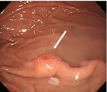
이정석 등. 전이성 악성 흑색종의 내시경적 진단The Korean Journal of Gastroenterology Fig. 1. Endoscopic finding. Subepithelial tumor-like lesion (8 mm)
with hypervascular change on the fundus (arrow).
A B C
Fig. 2. Pathologic finding. (A) Abnormal cell proliferation is observed. The nuclei of the cells are larger than other inflammatory cells (H&E stain, ×100). (B) Strongly positive immunohistochemical stains for HMB-45 protein (HMB-45 stain, ×100). (C) Strongly positive immunohistochemical stains for Melan-A (Melan-A stain, ×100). HMB-45, human melanoma black-45.
A B C
Fig. 3. PET-CT finding. (A) Increased FDG uptake at the right ilium (arrow). (B) Increased FDG uptake at the left hilar area in the mediastinum (arrow). (C) Focal increased FDG uptake at the pancreatic body (arrow). PET, photon emission tomography; CT, computed tomography; FDG, fluorodeoxyglucos.
되어 본원으로 의뢰되었다. 2년 전 좌측 엄지손가락의 악성 흑색종으로 좌측 엄지손가락 절단술과 좌측 액와 림프절 절제
술을 시행하고 인터페론으로 보조 항암 요법을 시행하던 중 인터페론의 심각한 합병증은 없었으나 환자 자의로 중단하고 본원에서 추적 관찰 중이었다. 내원 당시 계통 문진에서 발열, 오한은 없었고, 오심, 구토, 소화불량, 설사, 변비 등의 소화기 증상 역시 호소하지 않았다. 신체 검진에서 병색을 보이지 않 았으며, 결막은 창백하지 않았고, 공막의 황달 소견을 보이지 않았다. 간과 비장의 비대는 없었다. 혈압은 120/80 mmHg, 맥박 80회/분, 호흡수 18회/분, 체온 36,5oC였다.
검사실 소견으로 말초혈액 검사는 백혈구 7,270/mm3, 혈색 소 15.8 g/dL, 혈소판 356,000/mm3였고, 생화학 검사에서는 총 단백 8.1 g/dL, 알부민 4.7 g/dL, 총 빌리루빈 0.9 mg/dL, 나트륨 140 mEq/L, 칼륨 4.4 mEq/L, 아밀라아제 41 IU/L, 리 파아제 10 U/L였으며, aspartate aminotransferase/alanine aminotransferase 60/98 IU/L로 상승되어 있었다. 종양 표지 자 검사에서 alpha-fetoprotein (AFP)과 carcinoembryonic antigen (CEA) 모두 정상 범위였고, 흉부 X-선 검사에서 이 상 소견은 없었다. 위 내시경 검사에서 위저부에 과혈관상을 보이는 약 8 mm 정도의 단일 점막하 종양 형태의 병변이 관찰되어 조직 검사를 시행하였다. 체부와 전정부 및 십이지
Lee JS, et al. Endoscopic Diagnosis of Metastatic Malignant Melanoma
105
Vol. 70 No. 2, August 2017
장에서는 이상 소견이 관찰되지 않았다(Fig. 1). H&E 염색에 서 비정상적인 증식을 보이는 다양한 크기의 세포 내에 크고 진한 핵이 관찰되었다(Fig. 2A). 추가적인 면역조직화학 염색 에서 멜라닌 세포들은 human melanoma black-45에 강한 양성을 보였으며 Melan-A에 강한 양성을 보였다(Fig. 2B, C).
흉부 및 복부 전산화단층촬영에서 양측 폐엽 사이의 림프절 증식과 췌장 체부에 7 mm의 저음영 병변이 관찰되었다. 양전 자 방출 단층촬영-전산화단층촬영(photon emission tomog- raphy-computed tomography, [PET-CT])에서는 좌측 폐문 림 프절, 우측 장골 및 췌장체부의 섭취 증가가 관찰되었다(Fig. 3).
최종적으로 악성 흑색종의 다발성 전이로 진단하고 programmed death-1 (PD-1) 면역 치료인 pembrolizumab (KEYTRUDA® 200 mg; MSD Korea Ltd., Seoul, Korea)을 투여하였으며, 뼈 전이 부위에 방사선 치료를 시행하였다. Pembrolizumab을 2회 투여한 후 시행한 추적 복부 전산화단층촬영에서 췌장 전이 병변의 크기가 증가하였고, 양측 폐엽 내 전이 병변이 새로 발생하였다. 또한 대동맥 주위 림프절 및 대동정맥 림프 절, 양측 엉덩 림프절로 전이 소견을 보였다. 환자는 보존적 치료를 시행하면서 추적 관찰 중에 있다.
고 찰
악성 흑색종은 신경능에서 유래되어 조직의 색소를 생성하 는 멜라닌 세포의 악성 종양으로, 서구에서는 악성 종양의 약 1-3%를 차지하지만 국내에서는 비교적 드문 것으로 알려져 있다. 원발성 악성 흑색종은 피부에서 가장 흔하게 발생하며 전이는 비교적 조기에 나타난다. 인근 피부와 인접한 림프절 을 통한 전이와 혈행성 전이가 모두 일어날 수 있으며, 일반적 으로 림프계를 통해 액와부로, 혈행성 경로를 통해 소화관, 간, 폐, 뇌 및 뼈로 전이가 일어난다.5
소화관의 악성 종양 중 전이성 종양은 약 20% 정도이고, 악성 흑색종은 유방암, 폐암, 난소암과 더불어 소화기관으로 전이되는 흔한 악성 질환 중 하나이다.4,5 전이성 흑색종의 전 신 증상은 다른 악성 종양과 마찬가지로 피로감, 식욕 부진, 체중 감소 등이 있고, 소화관 전이 시에는 구역, 구토, 속쓰림, 소화불량 등의 증상을 호소하여 위 내시경 검사를 시행하여 진단하는 경우가 많다.
악성 흑색종의 위 전이는 내시경 검사에서 흑색소 침착이 관찰되는 경우 비교적 쉽게 진단할 수 있다. 육안적으로 색소 침착이 보이는 경우는 30-50% 정도이며 편평성 및 용종성 모 양의 병변을 나타내고, 대부분 다수의 결절 양상으로 보일 수 있으며, 궤양을 형성하는 경우는 ‘bull’s-eye’나 ‘target-like’
한 형태를 보일 수 있다.6,7 Nelson과 Lanza의 분류에 따르면 제1형은 정상 주름의 융기부에 다양한 크기의 흑색소 침착을
보이는 결절 형태로 궤양을 형성하며, 제2형은 중심부 궤양을 가진 용종성의 점막하 종양 형태를 보이고, 제3형은 다양한 크기의 궤양과 흑색소 침착을 가진 종괴로 나눌 수 있다.8 위 전이의 대부분은 대만부에서 발생하고, 병변 부위의 생검을 통해 멜라닌세포 연관 단 클론 항체인 Melan-A/Melanoma antigen recognized by T cell-1이나 premelanosome 당단 백 단클론 항체인 human melanoma black-45에서 양성을 보이면 확진할 수 있다.5
악성 흑색종의 위 전이에 대한 기존 보고들은 대부분 흑색 조 침착이 두드러지거나 결절 내 궤양이 관찰되었다. 기존의 보고들과는 달리 본 증례에서는 과혈관상 점막 변화를 가진 단일 점막하 종양 형태로 발견되었다. 검사 당시의 내시경 소 견만으로는 Nelson과 Lanza의 분류상 어느 아형에도 속하지 않지만 병변의 악성화가 진행한다면 제2형으로 진행되었을 것으로 생각된다.
악성 흑색종은 수술적 치료가 원칙이다. 특히, 소화관 전이 로 인한 심한 출혈, 장천공 등이 발생한 경우 조기에 수술적 치료를 통해 병변을 제거하는 것이 도움이 된다.9최근 PD-1 조절 면역치료제의 사용이 악성 흑색종의 생존율 향상을 보이 고 있으며, 새로운 면역 치료 약제들이 빠른 속도로 개발되고 있다.10,11
일반적으로 정상 점막으로 덮인 2 cm 미만의 점막하 종양 은 주기적인 내시경 추적 검사가 추천된다.12 본 증례에서는 8 mm 크기의 단일 점막하 종양 형태였지만 점막의 과혈관상 을 보여 조직 검사를 시행하였다. 점막의 과혈관상 변화를 보 이는 병변으로는 신경내분비 종양, 카포시육종, 혈관육종 혹 은 과혈관성 전이(hypervascular metastasis) 등이 있을 수 있으므로 반드시 조직 검사를 시행하여야 한다.13
저자들은 상부 위장관 내시경에서 우연히 발견된 표면에 과혈관상을 띈 1 cm 미만의 점막하 종양 형태의 전이성 악성 흑색종을 경험하였기에 이를 보고하는 바이다.
REFERENCES
1. Ryu JS, Oh HJ, Hu JW, et al. Two cases of endoscopically diag- nosed gastric metastatic malignant melanoma of unknown origin.
Korean J Gastrointest Endosc 2004;28:71-75.
2. Ihde JK, Coit DG. Melanoma metastatic to stomach, small bowel, or colon. Am J Surg 1991;162:208-211.
3. Caputy GG, Donohue JH, Goellner JR, Weaver AL. Metastatic mel- anoma of the gastrointestinal tract. Result of surgical management.
Arch Surg 1991;126:1353-1358.
4. Kadakia SC, Parker A, Canales L. Metastatic tumors to the upper gastrointestinal tract: endoscopic experience. Am J Gastroenterol 1992;87:1418-1423.
5. Schuchter LM, Green R, Fraker D. Primary and metastatic dis- eases in malignant melanoma of the gastrointestinal tract. Curr
106
이정석 등. 전이성 악성 흑색종의 내시경적 진단The Korean Journal of Gastroenterology Opin Oncol 2000;12:181-185.
6. Horowitz M, Nobrega MM. Primary anal melanoma associated with melanosis of the upper gastrointestinal tract. Endoscopy 1998;30:662-665.
7. McDermott VG, Low VH, Keogan MT, Lawrence JA, Paulson EK.
Malignant melanoma metastatic to the gastrointestinal tract.
AJR Am J Roentgenol 1996;166:809-813.
8. Nelson RS, Lanza F. Malignant melanoma metastatic to the up- per gastrointestinal tract: endoscopic and radiologic correla- tions, form and evolution of lesions, and value of directed biopsy in diagnosis. Gastrointest Endosc 1978;24:156-158.
9. Markovic SN, Erickson LA, Rao RD, et al. Malignant melanoma in the 21st century, part 2: staging, prognosis, and treatment.
Mayo Clin Proc 2007;82:490-513.
10. Hodi FS, O’Day SJ, McDermott DF, et al. Improved survival with Ipilimumab in patients with metastatic melanoma. N Engl J Med 2010;363:711-723.
11. Robert C, Schachter J, Long GV, et al. Pembrolizumab versus Ipilimumab in advanced melanoma. N Engl J Med 2015;372:
2521-2532.
12. Cho JW; Korean ESD Study Group. Current guidelines in the man- agement of upper gastrointestinal subepithelial tumors. Clin Endosc 2016;49:235-240.
13. Lee NK, Kim S, Kim GH, et al. Hypervascular subepithelial gastro- intestinal masses: CT-pathologic correlation. Radiographics 2010;30:1915-1934.