ORIGINAL ARTICLE
조기위암의 복강경하 원위부 위절제술 시 절제면 표시를 위한 내시경 클리핑의 최적 시기
박지영, 전태주, 신원창
인제대학교 의과대학 상계백병원 내과학교실
Optimal Timing of Endoscopic Clipping for Determining the Resection Line for Laparoscopy-assisted Distal Gastrectomy
Ji Young Park, Tae Joo Jeon and Won Chang Shin
Division of Gastroenterology, Department of Internal Medicine, Inje University Sanggye Paik Hospital, Inje University College of Medicine, Seoul, Korea
Background/Aims: Pre-operative endoscopic clipping for determining the resection line in patients with early gastric cancer has been used safely, and its efficacy has been demonstrated. However, the optimal timing of endoscopic clipping for determining the resection line in early gastric cancer patients undergoing laparoscopy-assisted distal gastrectomy has not been investigated.
Methods: A retrospective analysis of 92 patients with early gastric cancer who underwent gastric resection after endoscopic clipping at Inje University Sanggye Paik Hospital (Seoul, Korea) was performed. We analyzed the clinical and endoscopic features of patients, number of clips, time from clipping to surgery, and number of patients showing detachment of clips from the gastric wall before surgery. Patients were categorized according to the following two groups: group A included patients whose clips were applied within one day before surgery and group B included patients whose clips were applied more than one day before surgery.
Results: Of the 92 patients, 56 were included in group A and 36 were included in group B. In 11 patients (12.0%, five in group A and six in group B, p=0.329), the clips were detached from the gastric wall before surgery. The mean time from clipping to surgery did not differ significantly between the detached and non-detached groups (11 patients, mean 4.6±4.6 days vs. 81 patients, mean 3.0±4.0 days, p=0.227).
Conclusions: The timing of endoscopic clipping for localization of tumors in early gastric cancer patients undergoing gastrectomy is not important for determining the resection line. (Korean J Gastroenterol 2014;64:76-80)
Key Words: Stomach cancer; Laparoscopic surgery; Endoscopic clip; Gastrectomy
Received May 2, 2014. Revised May 19, 2014. Accepted June 2, 2014.
CC This is an open access article distributed under the terms of the Creative Commons Attribution Non-Commercial License (http://creativecommons.org/licenses/
by-nc/3.0) which permits unrestricted non-commercial use, distribution, and reproduction in any medium, provided the original work is properly cited.
교신저자: 신원창, 139-707, 서울시 노원구 동일로 1342, 인제대학교 상계백병원 소화기내과
Correspondence to: Won Chang Shin, Division of Gastroenterology, Department of Internal Medicine, Inje University Sanggye Paik Hospital, 1342 Dongil-ro, Nowon-gu, Seoul 139-707, Korea. Tel: +82-2-950-1341, Fax: +82-2-950-1955, E-mail: [email protected]
Financial support: None. Conflict of interest: None.
INTRODUCTION
The expanded use of diagnostic endoscopy has led to an increase in the detection rate of early gastric cancer (EGC).1 The therapeutic option for EGC is gastric resection with an ad- equate resection margin and perigastric lymph node
dissection.1-3 However, EGC cannot be detected by in- spection of the serosal surface in the operative field, and in general, it cannot be palpated manually because of its shal- low invasion depth.1,2,4,5
Endoscopic mucosal clipping was developed for hemo- stasis of gastrointestinal bleeding, but it is now widely used
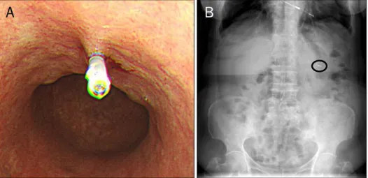
Fig. 1. Pre-operative endoscopic clipp- ing for early gastric cancer (HX-600- 090LⓇ; Olympus). Pre-operative appli- cation of a clip at the proximal side of the lesion (A) and plain abdominal x-rays obtained after clipping in the upright position (circle; B).
for other purposes; for example, as a marker for radiotherapy and for closing gastrointestinal perforations.2,6,7 Some stud- ies have demonstrated the value of endoscopic clipping of the proximal region outside the lesion for selection of the ap- propriate surgical procedure and determining the resection line for tumors located in the middle corpus or more distal portions of the stomach.2,8,9 This technique is safe, cost-ef- fective, and easily performed. In addition, the clips are not easily detached and remain firmly in place for several days.1,2 However, the optimal timing of pre-operative endoscopic clip- ping for determining the resection line has not been investigated. In the current study, we evaluated the optimal timing of endoscopic clipping for determining the resection line in patients with EGC undergoing laparoscopy-assisted distal gastrectomy (LADG).
MATERIALS AND METHODS
We performed a retrospective analysis of partial gastrec- tomies performed after endoscopic clipping for localization of EGC. A total of 92 patients with EGC who had undergone gastric resection after endoscopic clipping between January 2010 and December 2012 at Inje University Sanggye Paik Hospital (Seoul, Korea) were enrolled in the study. Pre-oper- ative endoscopic clipping was performed to determine the proximal resection line by identifying the exact site of the tu- mor in patients undergoing LADG. All data were reviewed retrospectively. The clinical and endoscopic features of all patients, number of clips, time from clipping to surgery, and number of patients whose clips were detached from the gas- tric wall before surgery were analyzed. Detachment of clips
was detected macroscopically from intra-operative and pathologic pictures of specimens. Patients were categorized according to the following two groups: group A included pa- tients whose clips were applied within one day before surgery and group B included those whose clips were applied more than one day before surgery.
1. Endoscopic clipping
Before the operation (mean, 3.1 days; range, 0-18 days), several endoscopic clips (HX-600-090L; Olympus, Tokyo, Japan) were placed at proximal sites outside the lesion to de- termine the resection line through a flexible endoscope using a roticulator clip-fixing device (HX-5QR-L; Olympus). The pro- cedure was performed separately by three endoscopists with extensive experience (>700 gastroscopies per year for a pe- riod of more than 10 years). The clips were 8 mm in length and 2 mm in diameter. After application of the clips, patients were evaluated by plain radiography of the chest and abdomen be- fore surgery to obtain general information on the location of the lesion and attachment of the clips to the gastric wall (Fig.
1).
2. Statistical analysis
The chi-square and Fischer’s exact tests were used for comparison of categorical data. All continuous data values were expressed as mean±SD. Differences in the mean val- ues were examined by Student’s t-test. The significance level was set at a p-value of less than 0.05. All statistical calcu- lations were performed using SAS ver. 8.1 (SAS Institute, Cary, NC, USA).
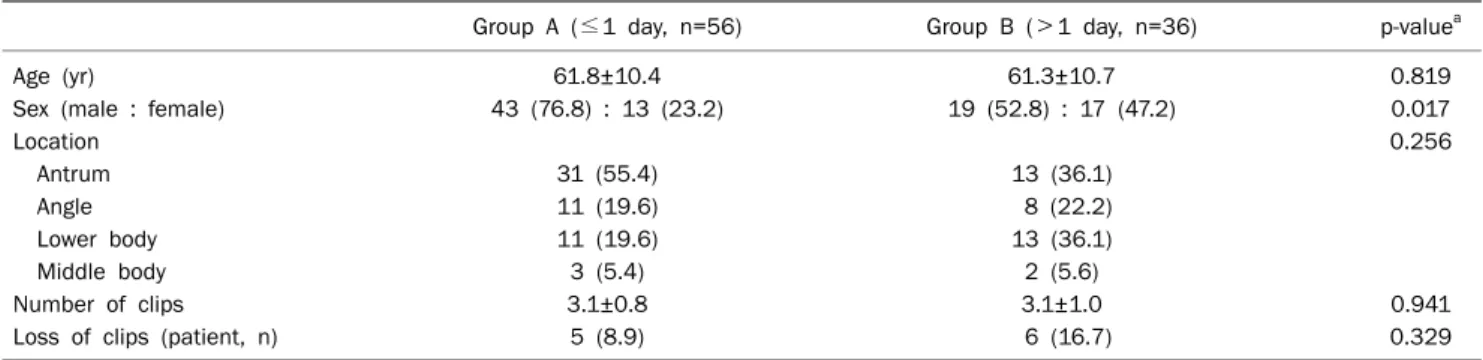
Table 1. Clinical and Endoscopic Features of the 92 Patients Who Underwent Endoscopic Clipping for Early Gastric Cancer
Group A (≤1 day, n=56) Group B (>1 day, n=36) p-valuea
Age (yr) 61.8±10.4 61.3±10.7 0.819
Sex (male : female) 43 (76.8) : 13 (23.2) 19 (52.8) : 17 (47.2) 0.017
Location 0.256
Antrum 31 (55.4) 13 (36.1)
Angle 11 (19.6) 8 (22.2)
Lower body 11 (19.6) 13 (36.1)
Middle body 3 (5.4) 2 (5.6)
Number of clips 3.1±0.8 3.1±1.0 0.941
Loss of clips (patient, n) 5 (8.9) 6 (16.7) 0.329
Values are presented as mean±SD or n (%).
ap<0.05.
Fig. 2. Resected specimen of early gastric cancer marked with a preoperative endoscopic clip (arrow).
Table 2. Clinical and Endoscopic Features of 11 Patients Who Showed Detachment of Clips Applied for Determining the Resection Line before Surgery
Detached (n=11, 12.0%) Non‐detached (n=81, 88.0%) p‐valuea
Age (yr) 65.4±9.0 61.1±10.6 0.202
Sex (male : female) 8 (72.7) : 3 (27.3) 54 (66.7) : 27 (33.3) 0.687
Location 0.187
Antrum 8 (72.7) 36 (44.4)
Angle 0 (0) 19 (23.5)
Lower body 2 (18.2) 22 (27.2)
Middle body 1 (9.1) 4 (4.9)
Number of clips 3.1±0.8 3.0±1.1 0.632
Time‐to‐surgery (day) 4.6±4.6 3.0±4.0 0.227
Values are presented as mean±SD or n (%).
ap<0.05.
RESULTS
The clinical and endoscopic characteristics of all patients are summarized in Table 1. All of the applied endoscopic clips were easily detected by intra-operative or pathologic pictures of specimens (Fig. 2). Proximal surgical margin identified by pathologic finding was free from carcinoma in all patients. Of the 92 patients, 56 were included in group A and 36 in group B. A total of 287 clips were used, with 2-4 clips per patient (mean, 3.1 clips/patient; range, 1-5 clips/patient). Detach- ment of clips from the gastric wall before surgery was ob- served in 11 patients (12.0%, five in group A, six in group B).
Age, tumor location, number of clips applied, and number of patients showing detachment of clips from the gastric wall before surgery did not differ significantly between the two groups (p>0.05).
The clinical and endoscopic features of the 11 patients showing detachment of clips applied before surgery for de- termining the resection line are shown in Table 2. One clip was lost in nine patients and two clips were lost in two
patients. The mean time from clipping to surgery did not differ significantly between the detached and non-detached groups (mean, 4.6±4.6 days [n=11] vs. mean, 3.0±4.0 days [n=81]; p=0.227).
DISCUSSION
Many studies have reported an association of EGC with a favorable prognosis; therefore, improving the quality of life of patients with EGC remains the primary therapeutic object- ive.1,4,10 LADG has many advantages over open distal gas- trectomy, including less pain, smaller wounds, fewer respira- tory complications, a shorter hospital stay, better quality of life postoperatively, and an earlier return to normal activ- ity.10,11 The success of LADG depends on precise determi- nation of the tumor site.1,5,12 Because accurate localization of the tumor is sometimes difficult, use of specific techniques is necessary in order to define an adequate gastric resection range, especially in patients undergoing laparoscopy-assisted gastrectomy.1,2 Several studies have shown that endoscopic clipping is a safe and reliable procedure for determining the resection line in tumors located in the stomach, and this tech- nique has been widely used for this purpose.1,2 However, the optimal timing of pre-operative endoscopic clipping for de- termining the resection line has not been investigated, and, in our institution, clipping has been routinely performed one day before surgery without definitive reference.
In the current study, the number of patients showing de- tachment of clips from the gastric wall before surgery did not differ significantly between groups A and B (five patients vs.
six patients, p=0.329). In addition, the mean time from clip- ping to surgery did not differ significantly between the de- tached and non-detached groups. Similarly, no significant differences in tumor location (p=0.187) and number of clips (p=0.632) were observed between the detached and non-de- tached groups. These results suggest that the timing of the clipping procedure was not important for localizing tumors in EGC patients.
None of the patients included in the study lost more than three clips, with nine patients losing one clip and two patients losing two clips. Based on this result, more than three clips should be applied for localization of lesions, although large-scale prospective randomized controlled studies are needed in order to prove this result. The exact period of ex- posure to clips tolerated by patients cannot be determined from our results. However, clips remained in place for >7 days in 16 cases, and the longest duration was 18 days in two cases. Further studies will be needed before an optimal or tol- erable duration can be suggested.
The main limitations of our study are inherent to its retro- spective study design. All data were collected retrospectively, and data were not adjusted for age and sex. In addition, the distances between the specimen, tumor margin, and depth of clipping were not determined after the gastrectomy.
Therefore, adequate placement of endoscopic clips could not be assessed. In addition, because specimens could not be obtained at the beginning of our study, clip detachment was assessed by plain radiography instead of analysis of specimens after gastrectomy. Therefore, the possible over- lapping of clips might have been missed in radiographic im- ages, and this could have affected our results. Despite these limitations, the current study is important, as it is the first study to evaluate the optimal timing of endoscopic clipping for determining the resection line for LADG in patients with EGC.
In conclusion, the results of the current study showed that the timing of clipping for localization of tumors in EGC pa- tients undergoing gastrectomy is not important. Conduct of further large-scale prospective randomized controlled stud- ies will be needed in order to confirm these findings.
REFERENCES
1. Hyung WJ, Lim JS, Cheong JH, et al. Intraoperative tumor local- ization using laparoscopic ultrasonography in laparoscopic-as- sisted gastrectomy. Surg Endosc 2005;19:1353-1357.
2. Ryu KW, Lee JH, Choi IJ, Bae JM. Preoperative endoscopic clip- ping: localizing technique of early gastric cancer. J Surg Oncol 2003;82:75-77.
3. Bozzetti F, Marubini E, Bonfanti G, Miceli R, Piano C, Gennari L.
Subtotal versus total gastrectomy for gastric cancer: five-year survival rates in a multicenter randomized Italian trial. Italian Gastrointestinal Tumor Study Group. Ann Surg 1999;230:
170-178.
4. Kikuchi S, Hirai K, Kuroyama S, et al. Role of endoscopic clipping for determining the resection line for tumors located in the mid- dle or upper corpus of the stomach: experience with 100 gas- trectomies for early gastric cancer. Anticancer Res 2004;24:
4163-4168.
5. Beretvas RI, Ponsky J. Endoscopic marking: an adjunct to laparo- scopic gastrointestinal surgery. Surg Endosc 2001;15:1202- 1203.
6. Cipolletta L, Bianco MA, Rotondano G, Marmo R, Piscopo R, Meucci C. Endoscopic clipping of perforation following pneu- matic dilation of esophagojejunal anastomotic strictures.
Endoscopy 2000;32:720-722.
7. Weyman RL, Rao SS. A novel clinical application for endoscopic mucosal clipping. Gastrointest Endosc 1999;49:522-524.
8. Kuwano H, Sadanaga N, Watanabe M, et al. Preoperative endo- scopic clipping for determining the resection line in early carci- noma of the esophagus. J Am Coll Surg 1995;180:97-99.
9. Okabayashi T, Gotoda T, Kondo H, et al. Usefulness of indigo car- mine chromoendoscopy and endoscopic clipping for accurate preoperative assessment of proximal gastric cancer. Endoscopy 2000;32:S62.
10. Lee SE, Kim YW, Lee JH, et al. Developing an institutional proto- col guideline for laparoscopy-assisted distal gastrectomy. Ann
Surg Oncol 2009;16:2231-2236.
11. Kim YW, Baik YH, Yun YH, et al. Improved quality of life outcomes after laparoscopy-assisted distal gastrectomy for early gastric cancer: results of a prospective randomized clinical trial. Ann Surg 2008;248:721-727.
12. Ohgami M, Otani Y, Kumai K, Kubota T, Kim YI, Kitajima M.
Curative laparoscopic surgery for early gastric cancer: five years experience. World J Surg 1999;23:187-192; discussion 192- 193.