대한밤시선의학회지 1997; 37 : 123-128
위선암의 나선식 CT 소견 l
이 동 호·고 영 태·윤 엽
위션암의 진단에 있어 CT는 중요한 역할을 한다 .CT를 이용하여 위선암 자체를 잘 볼 수 있을 뿐만 아니라 주위로의 파급 및 원위부 전이를 알 수 있다. 그러므로 위선암의 병기결정 에 CT가 중요하며, 또한 위에 생기는 다른 종양과의 감멸에도 도움이 된다. 최근에 와서 나 선식 CT를 이용하여 물을 먹인 후 검사를 하면 더 좋은 영상을 얻을 수 있어 CT의 역할이 더욱 중요하게 되였다. 이 임상화보에서 저자들은 위선암의 나선식 CT상의 특성과 병기결 정에서의 역할에 대하여 알아보았다.
위선암의 진단에는 과거부터 현재까지 상부위장관조영술 및 위내시경검사가 주로 이용되어 왔으며, 근래에 와서 초음파검 사, CT, MRI, 위내시경적 초음파검사 등이 이용된다. 그중에 서 CT는 종양의 침윤정도 뿐 아니라, 럼프절전이 빛 윈위부전 이 유무를비교적 객관적으로알수있어 위암병기판정에 꼭필 요한 검사로 간주되고 있다. 위의 CT검사는 고식적 방법으로 물을 먹이고 복와위에서 시행하였으나, 1990년대에 와서 나선 식 CT가 가능하게 되어, 이중시기 혹은 삼중시기의 영상이 가 능하게 되었다(1). 이 임상화보에서 저자들은 위선암의 나선식 CT소견 및 특징을 기술하고, 위암병기 결정에 있어서 나선식 CT의 유용성을 알아보고자 한다.
위선암의 나선식 CT
촬엉방법
위의 CT검사는 우선 위내강을 물로 가능한한 많이 팽창시켜 야 하고, 조영제 주사후적땅한시기에 촬영하여야좋은영상을 얻을 수 있다(1 ). 물은 약 600-800mL를 검사 직전에 먹이고,
병변이 위체부나 위동부에 있으면 복와위로, 병변이 분문 및 위 저부에 있으면 앙와위로 촬영한다. 조영증강은 비이온성 조영 제를 초당 3mL로 전체 120mL를 주사하며, 동맥강조영상은 조 영제주업 직후부터 35초후에, 지연기영상은 70-80초 후에 얻 는다. 간의 나선식 CT영상은 보통 25 ← 30초에 동맥강조영싱을 얻는데 반하여 약간 지연영상을 얻는 이유는 위선암은 결함조 직형성( desmoplasia) 과 섬유화가 많아 지연 조영증강되기 때 문에 좋은 영상을 얻기위해서는 5-10초정도 늦게 촬영해야 하 기 때문이다. 스캔방법은 절편두께 5mm, 테이블이동속도 초당
7mm, 재구성 간격 5mm로 한다.
l 경희대핵교 의파대학 진단방사선과학교실
이 논문은 1997년 2원 19일 접수하여 1997년 4 원 29일에 채택되었음‘
위선암의 형태 및 조직소견과 CT영상간의 관계
진행위암의 형태학적 분류에는 Borrmann의 분류가 널리 이 용된다 (Fig. 1). Borrmann행에 의한 빈도를 보면 제 l 형이 2
%, 제 2 형이 27%, 제 3 형이 43%, 제 4 형이 21%, 제 5 형이 7
%로 제 3 형이 가장 많다 (2). 고식적 CT에서는 Borrmann 제 1 형과 2 형에서 조영증강이 잘 안되고, 제 3 형과 4 행에서 조영 증강이 잘 되었으나 (3) , 나선식 CT를 시행하면 물론
Borrmann 제3형과 4 행은 대부분 강한 조영증강을 보이지만 (Fig. 1C and D), 저]1 행과 2 행에서도 많은 예에서 조영증강 을 보인다 (Fig. 1A and B). 이 결과는 혈관조영술상 위선암이 65-92%에서 조영증강되는 것과 일치하며 (4) , 나선식 CT의 장점이라 할 수 있다. 위선암의 발생위치를 보면 위동부에 호말 하지만, 근래에 와서 위체부 및 위저부의 발생빈도가 높아지고 있다 (Fig. 2). 조직학적으로 위암의 95%가 선암이며, 널리 이 용되는 분류는 WHO에 의한 것으로 분화도에 따라 유두상선 암, 관상선암, 인환세포암, 점액선암으로 나넌다. 조직학척 분류 에 의한 조영증강정도를 보면 인환세포암이 조영증강이 잘되며
(Fig. 1D) (3), 점액선암은 점액성분을 많이 갖고 있기 때문에 조영증강이 안되고 (Fig. 3A) (3), 간혹 석회화를 동반한다 (Fig.
3B) (5).
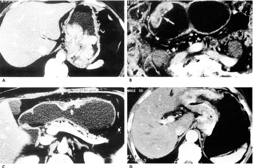
위선암의병기
위선암의 병기는 TNM 분류에 의한다.T병기를 보면 T1은 조기위암이고, T2는 암이 위장막내에 국한된 것으로 CT상 위 벽주위 지방층이 깨끗하게 보존되어 있으며 (Fig.
4A)
, T3는 암 이 위장막을 뚫고 밖으로 퍼져나간 것으로 CT상 위벽주위 지 방층에 지저분한 섬유선상의 침윤이 보이고 (Fig. 4B), T4는 주 위장기를 침범한 것으로 간, 횡행결장, 춰1 장으로의 파급이 관찰 된다 (Fig. 4C). 이들중 임상적으로는 T3와 T4의 감별이 가장 중요한데 나선식 CT를 이용하여도 정확한 감별이 어려운 경우123 -
이동호 오1: 우|선암의 나선석 CT
! ’P"~~ .• ‘ - -‘’‘• ’ ,
--- ...
• ‘
’ “ .
A B
C D
Fig. 1. Borrmann’s classification of advanced gastric carcinoma(AGC).
A. AGC, Borrmann type 1.
Spiral CT scan shows an endoluminal bulky mass at fundus and high body of the stomach. The tumor mass is highly enhanced and the outer margin is lobulated
B. AGC, Borrmann type 2
Spiral CT scan shows tumor mass at anterior wall of antrum. The mass has central and shallow ulceration (arrow), and the outer margin of tumor is well-demarcated. The tumor is moderately enhanced.
c.
AGC, Borrmann type 3Spiral CT scan shows infiltrative tumor mass at anterior wall of low body and proximal antrum. The mass has central ulceration (arrow), and the enhancement of the tumor is good and heterogeneous.
D. AGC, Borrmann type 4.
Spiral CT scan shows diffuse and marked wall thickening from high body to antrum, and the lumen is narrow. The tumor is heterogeneously and highly enhanced. Signet ring cell adenocarcinoma was confirmed at surgery
’i ‘ A
Fig. 2. Advanced gastric carcinoma at cardia.
Spiral CT scan shows irregular wall thickening at gastric fundus and cardia. The tumor is highly enhanced.
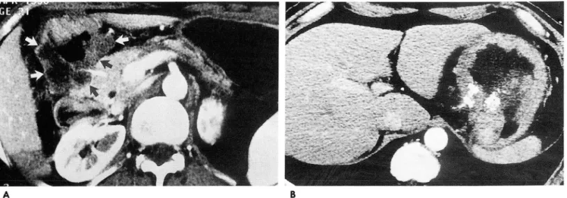
가 있다. 그 이유는 암환자에서는 지방층이 적으므로 정상적으 로 훼장과 위벽이 붙어보이는 경우가 있으며, 위와 춰1 장사이의 경계면이 CT 스캔면과 부분용적효과에 의하여 정확도가 떨어 지고, 지방층은 이차적인 춰l 장염이나 염증파급에 의하여서도 소실되기 때문이다.N 병기는 럼프절에 각각 변호를 붙여 3군 으로 분류하는데 제 2 군과 제 3 군사이의 구별이 중요하다. 그 이유는 제 3 군인 경우 수술이 어려울 때가 있고 예후가 나쁘기 때문이다. 럼프절 전이의 CT진단도 부정확한데 그 원인은 간 위간막인대나 위대만에 붙어있는 럼프절 비대는 종양과 구별이 안될 수 있으며 (Fig.5), 커진 럼프절의 경우 반응성 럼프절증 식인지 암이 퍼진것인지 구별이 안되며, 크기가 작아도 암이 퍼 진 럼프절이 있으며, 또한 정상크기와 커진 럼프절 사이에 수치 상의 기준점이 확립되어 있지않기 때문이다. 그러나 커진 럼프
• 124 •
대한망시선의학회지 1997;37: 123-128
절이 원형이며 내부에 괴사를 형성하고 있으면 전이된 럼프절 까지 내려가면 럼프종의 가능성이 높으나, 간흑 위션암에서도 로 취급한다 (Fig.6) 후복막강에 커진 럼프절이 신문보다 하방 신문하방의 럼프절 비대를 동반할 수 있다 (Fig. 7). M 병기는
A B
Fig. 3. Advanced gastric carcinoma, mucinous type.
A. Spiral CT scan shows a lobulated mass at pyloric antrum. The tumor mass contains portions of non-enhancing and low attenuation (arrows). At surgery, the tumor had extensive areas of mucin pools. Mucinous adenocarcinoma was confirmed at pathologic specimen.
B. Spiral CT scan shows wall thickening at high body. Punctate and conglomerated calcifications are seen within thickened wall
A
c
τ μ
B
Fig. 4. T stage of advanced gastric carcinoma‘
A. Spiral CT scan reveals highly enhancing and thickened wall (arrows) at anterior portion of proximal antrum. The outer margin of tumor is clearly defined and perigastric fat is preserved. Surgical stage of this patient was T2.
B. Spiral CT scan reveals poorly enhancing mass at py- loric antrum‘ Fibrostreaky infiltrations and spiculations (curved arrows) are seen at perigastric fat tissue. Surgical stage of this patient was T3
C. Spiral CT scan reveals irregular wall thickening at pos- terior wall of low body. The pancreas is attached and infiltrated by gastric tumor. Surgical stage of this patient was T4.
혈행성전이와 복막전이의 유무에 따라 결정된다. 위선암은 문 맥을 통하여 간으로의 혈행성전이를 제일 많이한다 (Fig.8A).
위암이 장막을 뚫고 복막으로 퍼지면 S자결장, 맹장, 장간막등 으로 전이되고 복수가 차게된다 (Fig.8B). 특히 인환세포암은 난소로 복막전이되어 Krukenberg 종o}을 형성한다 (Fig.8C).
니선식 CT에 의한 위암의 병기는 약 fìO-70%의 정확도를 보 인다 (6, 7).
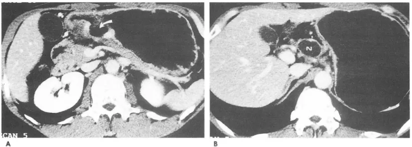
조기위암
나선식 CT상 국소적으로 미믿댁f 위벽비후를 보이며, 정막하 층인 저감쇠층이 보존되어 있으면 근육층을 침범하지 않은 조
Fig. S. N stage of advanced gastric carcinoma.
Spiral CT scan reveals wall thickening at antrum. A subpyloric Iymph node (N) is enlarged, but it is not separated from tumor mass by CT image. CT stage of this patient was NO, but stirgical stage was Nl.
A
Fig. 6. N stage of advanced gastric carcinoma
이동호 오1: 우|선암의 나선식 CT 소견
기위암으로 진단할 수 있다(Fig. 9) 조기위암의 조영증강은 일 반적으로 진행위암보다 약하게 되는데, 간흑 동맥강조영상에서 비후된 위점막의 강한 조영증강을 보이는 경우도 있다. 조기위 암의 고식적 CT상 발견율은 역동적 검사를 하여도
53 -65%
정도 밖에 안되며, 최근에 나선식 CT를 시행하면 63-75%의 발진율을 보인다 (fì, 8). 그러나 조기위암에서 나선식 CT의 역 할은 위암 자체를 발견하는 것보다는 다른 장기 및 복막, 럼프 절에 전이가 없음을 확인하는데 있다. 간흑 조기위암에서도 럼 프절전이가 생기기 때문이다. 조기위암의 유형에 의한 CT상
발견율은제
I
형과 IIa형의 발견이 용이하고 (Fig.9A) ,제 Dc 행과 Db형의 발견이 어렵다 (Fig.9B).Fig. 7. Advanced gastric carcinoma with multiple lymph nodes metastasis
Spiral CT scan reveals poorly enhancing tumor mass (ar- row) at anterior wall of body. Multiple lymphadeno- pathies (N) are seen at mesenteric, peripancreatic, retro- caval, paraaortic, and interaorticocaval areas, extending below renal hilum. Gastric adenocarcinoma was con- firmed by gastroscopic biopsy
B
A. Spiral CT scan reveals tumor mass with large ulceration (curved arrow) at posterior wall of proximal antrum
B. Spiral CT scan 4cm cranial to A. reveals totally necrotic lymphadenopathy (N) at hepatic artery area. Surgical stage of this patient was N2
- 126 -
대한방시선의학회지 1997; 37: 123-128
A
C
A
Fig. 9. Early gastric carcinoma(EGC) A. EGC type 1
B
Fig. 8. M stage of advanced gastric carcinoma A. Hepatic metastasis.
Spiral CT scan shows poorly enhancing tumor at pyloric antrum. Liver has multiple low attenuation masses due to hematogeneous metastasis.
B. Cancer peritonitis
Spiral CT scan shows highly enhancing tumor (curved ar row) at anterior wall of antrum. Moderate amount of as cites is seen at perihepatic space, perisplenic space, and lesser sac.
C. Krukenberg tumor‘
Spiral CT scan shows heterogeneous mass (arrows) within pelvic cavity‘ Metastatic adenocarcinoma at left ovary was confirmed at surgery. Three years ago, this patient received surgery due to advanced gastric carcinoma
8
Spiral CT scan shows focal protruding mass (arrow) at anterior wall of antrum‘ EGC type 1 was confirmed at surgery B. EGC type J] b+ J] c.
Spiral CT scan shows focal and mild wall thickening with enhancement (arrow) at anterior wall of antrum. Low attenu ation stripe representing submucosal layer is preserved (arrow heads). EGC type II b+ J] c was confirmed at surgery.
- 127 -
Fig. 10. Exophytic adenocarcinoma of the stomach‘ Spiral CT scan shows lobulated mass with massive necrosis protruding from gastric body. Gastric walls ad- jacent to the exogastric mass are thickened (curved arrows). Gastric adenocarcinoma was confirmed by gastroscopic biopsy.
감별진단
위선암의 독특한 형태로 돌출행이 있는데 이 경우는 위에서 생긴 평활근육종과의 감별이 어렵다 (Fig. 10). 이때의 감별점 은 암침윤에 의한 종양주위 위벽의 비후가 보이거나, 위유문폐 쇄 소견이 있을때 돌출형 위션암으로 진단이 가능하다 (9). 위 선암과 감별해야 할 질환으로 럼프종, MALToma, 평활근종 빛 평활근육종등이 있다(1이. 위선암과 구멸되는 럼프종의 특 정은 위벽이 전반적으로 심하게 비후되고, 외연이 매끄럽고 종 양의 조영증강이 낮고 균일한 양상을 보이며, 주위의 럼프절 종 대를 많이 동반한다. MALToma도 럼프종과 비슷한 소견을 보 이나 저등급 MALToma에서는 조기위암과의 감별이 어렵다 ( 0). 평활근종은 외연이 매끈하고, 위내강으로 자라며, 조영증
이동호 오1 : 우|선암의 니선석 CT
강이 미미하고, 종괴를 덮고 있는 정상 위점막을 볼 수 있다. 평 활근육종은 종괴가 크고, 주로 위외연 밖으로 돌출하여 자라고,
내부에 괴사를 잘 동반하는 특정이 있다. 이와 같은 나선식 CT 의 특정을 잘 이용하면 위선암과 나머지 종OJ:들간의 감별진단 이 가능하다.
*f -;") 드크 L!... E~ ι--'
l. Winter TC, Ager JD, Nghiem HV, Hill RS, Harrison SD, Freeny pc. Upper gastrointestinal tract and abdomen ’ water as an orally administered contrast agent for helical CT. Radiology 1996; 201 : 365-370
2. Maruyama M, Baba Y. Gastric carcinoma. Radio/ Clin North Am 1994;32: 1233-1252
3 신홍섭, 이동호, 김윤화, 고영태, 임주원, 윤 엽 위암의 역 동적 전산화 단층촬영에서의 조영증강 양상 병리 소견과의 비교연구 대한방사선의학회지 1996; 35: 81-86
4. Minami M, Kawauchi N, Itai Y, Niki T, Sasaki Y. Gastric tumors : radiologic-pathologic correlation and accuracy of T staging with dynamic CT. Radiology 1992; 185: 173-178
5. Hwang HY, Choi BL Han JK, et al. Calcified gastric carcinoma CT findings. Gastrointest Radiol 1992; 17:311-315
6. Lee DH, Ko YT, Park SJ. Comparison of hydro-US and spiral CT in the staging of gastric cancer (abstr). Radi%gy 1996; 201 (P): 251
7 이동호, 고영태 위질환에서 나선식 CT를 이용한 횡단 CT 영 상과 3D 업체영상의 소견 및 역할 대한방사선의학회지
1996; 35: 731-738
8 천정은, 최병인, 한준구, 김성현, 한만청 위선암 침윤도 결 정시 나선식 CT의 유용성 대한방사선의학희 추계학술대회 초록집 1995;311-312
9. Lee DH, Choi BL Lee MG, et al. Exophytic adenocarcinoma of the stomach: CT findings. AJR 1994; 163: 77-80
10 이동호1 이주희 I 고영태 위에 생긴 MALToma의 방사선학적 소견과 병리조직 소견과의 비교분석 대한방사선의학회지
1997; 36: 257-263
J Korean Radiol Soc 1997; 37 : 123-128
Spiral CT of the Gastric Adenocarcinoma
1Dong Ho Lee, M.D., Young Tae Ko, M.D., Yup Yoon, M.D. 1 Department of Diagnostic Radiology, Kyung Hee University Hospital
CT has an important role in the evaluation of gastric adenocarcinoma. It clearly demonstrates the primary tumor itself and reveals the spread of cancer to ad jacent or distant structures. It is there- fore useful in the staging of gastric carcinoma, and has proved valuable in the differential diagnosis of this and other gastric tumors. Recent ad vances in technology such as spiral CT with water inges tion, improve the value of CT. This report describes the characteristic findings of gastric adenocarcinomas by spiral CT, and elucidates its role in the staging of gastric carcinoma.
Index Words: Stomach, neoplasms Stomach, CT
Computed tomography(CT), helical
Address reprint requests to: Dong Ho Lee, M.D., Department of Diagnostic Radiology, Kyung Hee University Hospital
~ L Hoeki-dong, Dongdaemun-ku SeouL 130-702, Korea. Tel. 82-2-958-8615 Fax. 82-2-968-0787 - 128 -