대한소화기학회지 2006;48:355-359
접수: 2005년 9월 16일, 승인: 2006년 5월 25일
연락처: 박원규, 705-717, 대구광역시 남구 대명동 317-1 영남대학교 의과대학 영상의학과학교실 Tel: (053) 620-3048, Fax: (053) 653-5484 E-mail: [email protected]
Correspondence to: Won Kyu Park, M.D.
Department of Diagnostic Radiology, Yeungnam University College of Medicine, 317-1, Daemyeong-dong, Nam-gu, Daegu 705-717, Korea
Tel: +82-53-620-3048, Fax: +82-53-653-5484 E-mail: [email protected]
췌장에서 발생한 파골세포양 거대세포를 가지는 미분화 암종
영남대학교 의과대학 영상의학과학교실, 병리과학교실*, 외과학교실†
장한원․박원규․장재천․김재운․배영경*․최준혁*․윤성수
†․이동식
†Undiffentiated Carcinoma with Osteoclast-like Giant Cells of the Pancreas
Han Won Jang, M.D., Won Kyu Park, M.D., Jay Chun Chang, M.D., Jae Woon Kim, M.D., Young Kyung Bae, M.D.*, Jun Hyuk Choi, M.D.*,
Sung Su Yun, M.D.†, and Dong Shik Lee, M.D.†
Departments of Radiology, Pathology*, and Surgery†, Yeungnam University College of Medicine, Daegu, Korea
Undifferentiated carcinoma with osteoclast-like giant cells is a rare neoplasm of exocrine pancreas. Till recently, some cases have been reported, however histogenesis of the tumors are controversial and their characteristic findings have not been described yet. Thirty five-year-old men and 75-year-old men were presented with upper abdominal pain and a palpable mass. On computed tomography, one case showed a well enhancing solid tumor with low density and the other was showed a mainly cystic tumor with peripheral enhancement in the body and tail of the pancreas. One case accompanied multiple metastatic liver masses with subhepatic lymph node enlarge- ment. Tumor staining was seen on angiography. Biopsy and pancreatectomy were performed. Pathological findings revealed tumors composed of neoplastic spindle shaped or pleomorphic large cells with scattered non-neoplastic osteoclast-like giant cells. In one case, there were small foci of adenocarcinoma components in the periphery of the tumor. On immunohistochemical stain, neoplastic cells showed focal positivity for epithelial membrane antigen and vimentin. Tumors were diagnosed as undifferented carcinoma with osteoclast-like giant cells. We report these rare cases with a review of literature. (Korean J Gastroenterol 2006;48:355-359)
Key Words: Osteoclast-like giant cells; Undifferentiated carcinoma; Pancreas
서 론
췌장에 생긴 파골세포양 거대세포를 가지는 미분화 암종 (undifferentiated carcinoma with osteoclast-like giant cells)은 외분비 췌장암의 1% 이하로 매우 드물다.1 파골세포양 거대 세포를 가지는 미분화 암종의 기원에 대해 논란은 많았지만 WHO 분류기준(2000)에 따르면 관샘암종의 조직학적 아형 으로 분류한다.2 저자들은 수술로 확진된 1예와 초음파 유
도하 생검으로 확진된 1예를 방사선, 병리 소견을 중심으로 문헌 고찰과 함께 보고한다.
증 례
1. 증례 1
75세 남자 환자가 3개월 전부터 상복부에 종괴가 촉지되
356 대한소화기학회지: 제48권 제5호, 2006
A
B C
D E
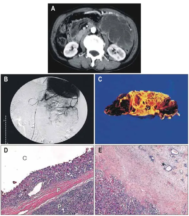
Fig. 1. Radiologic and pathologic findings of case 1. (A) Abdominal CT scan in arterial phase. Non-enhanced low density mass sparing the peripheral portion is located from body to tail. This indicates that mass consistes of necrosis or cystic changes. (B) Celiac angiographic finding. The tumor supplied by a branch of the splenic artery is noted. This tumor is also supplied by a branch of SMA (not shown). (C) Gross finding of resected pancreas specimen. Pancreas tissue is replaced by a cystic mass composed of variable sized cysts, some of which contain dirty hemorrhagic materials. (D) Histologic feature of resected pancreas specimen. The cystic mass is delineated by fibrous capsule (F) from adjacent normal pancreas (P). The cystic space (C) is surrounded by the mixed cellular components predominant of multinuclear giant cells (×40). (E) In the peripheral portion of the tumor, there is a focal area of conventional adenocarcinoma (*) (×40).
SMA, superior mesenteric artery.
었으며 최근 통증이 심해져 내원하였다. 본원에서 시행한 진 찰 소견에서 심와부 압통과 좌상복부 종괴가 촉진되었다. 검 사실 소견은 백혈구 5,900/uL, 혈색소 12.1 g/dL, 혈소판 293,000/uL, ESR 55 mm/hr였고, 생화학 검사 소견은 AST/
ALT 32/25 IU/L, 총 빌리루빈/직접 빌리루빈 1.2/0.5 mg/dL,
γ-GT/알칼리 포스파타제/LDH 737/1346/322 IU/L, Na/K/ Cl 142/5.3/102 mEq/L, 총 단백/알부민 7.2/3.4 g/dL이며, α- FP 5 ng/mL, CA19-9 16.6 u/mL, HBsAg/sAb/cAb -/-/-이었다.
복부 전산화단층촬영에서 췌장 체부와 미부에 걸쳐 약 18
×15 cm의 경계가 좋은 다방 낭성 종괴가 관찰되었다. 조영
장한원 외 7인. 췌장에서 발생한 파골세포양 거대세포를 가지는 미분화 암종 357
A B
C D
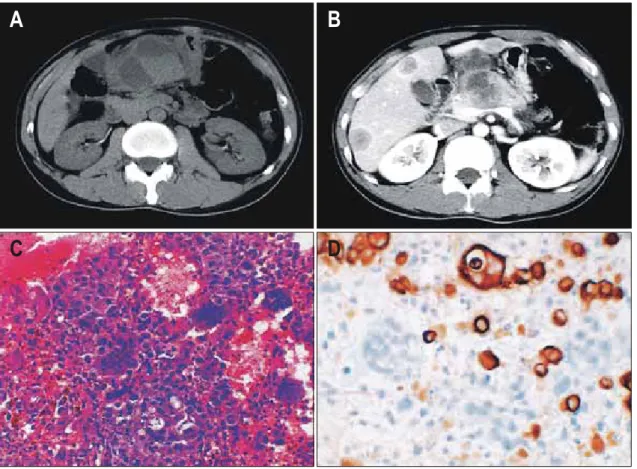
Fig. 2. Radiologic and pathologic findings of case 2. (A) In precontrast CT scan, pancreatic body mass is isodense with internal low density portion and subhepatic node has a low density with internal high density portion. (B) In arterial phase, this mass is enhanced except the low density portion. (C) Histologic features of biopsied subhepatic node. Large atypical mononuclear cells and multinuclear giant cells are admixed in the hemorrhagic background. There are also scattered lymphocytes and mononuclear inflammatory cells (×200). (D) Immunohistochemistry of the biopsied subhepatic node. Immunohistochemical stwining for cytokeratin 7 highlights mononuclear cancer cells (×200).
전 전산화단층촬영에서 낭종 일부는 고음영을 보여 종괴 내 부에 혈종의 존재를 의심하였다. 조영 후에는 낭성 부위를 제외한 종괴 주변부에 조영증강을 보였다(Fig. 1A). 복강 및 상장간막동맥 혈관조영에서 비장동맥과 상장간막동맥의 일 부에서 공급받는 종양염색이 관찰되었다(Fig. 1B). 종괴 주 위 혈관 침범으로 인해 근치 수술은 불가능하였으나 증상 완화를 위한 고식 수술로 원위부췌장절제술을 시행하였다.
적출된 종괴에서 육안 혈종을 포함하고 있는 다방 낭성 종 양이 관찰되었다(Fig. 1C). 현미경 소견에서 역형 방추상세 포와 다형 거대세포, 미만성으로 배열된 비종양 파골세포양 거대세포가 관찰되었고 종괴 주변부에서 국소적으로 샘암 종 부분이 관찰되었다(Fig. 1D, E). 면역조직화학염색에서 상피세포표지자인 epithelial membrane antigen (EMA)과 간질 세포표지자인 vimentin에 양성을 나타냈다.
2. 증례 2
35세 남자로 상복부 통증으로 내원하였다. 환자는 내원 한 달 전부터 심와부 동통이 지속되었으며, 한 달 동안 10 kg의
체중감소가 있었다. 다른 병원을 방문하여 상부위장관 내시 경 검사를 시행하여 위궤양으로 진단받고 약을 복용하였으 나, 증상 호전이 없어 복부 전산화단층촬영을 시행하였으 며, 전이로 의심되는 다발성 간 종괴가 발견되어 전원되었 다.
과거력상 특이 소견은 없었으며, 검사실 소견은 백혈구 12,800/uL, 혈색소 11.1 g/dL, 혈소판 367,000/uL, ESR 96 mm/hr이었고, 생화학 검사 소견은 AST/ALT 56/158 IU/L, 총 빌리루빈/직접 빌리루빈 1.4/0.6 mg/dL, γ-GT/알칼리 포스파 타제/LDH 737/1346/322 IU/L, Na/K/Cl 140/4.7/103 mEq/L, 총 단백/알부민 6.59/3.65 g/dL이며, α-FP 3 ng/mL, CA19-9 21.8 u/mL, HBsAg/sAb/cAb -/-/-이었다. 복부 전산화단층촬영 에서는 췌장 체부에 약 6×6 cm 크기의 낭성 종괴가 관찰되 었으며 조영 전 종괴 내부에 저음영과 고음영을 가지는 부 분이 있었고 조영 후에는 고형부위가 비교적 강한 조영증강 을 보였다. 췌장 주위에 림프절과 간 전이가 동반되어 있었 다(Fig. 2A, B). 췌장 체부 종괴와 간종괴 모두에서 세침흡인 생검을 통한 조직 검사를 시행하였고 췌장에서 발생한 파골
358 The Korean Journal of Gastroenterology: Vol. 48, No. 5, 2006
세포양 거대세포를 가지는 미분화 암종이 간으로 전이된 것 으로 진단하였다(Fig. 2C, D).
고 찰
관샘암종의 조직 아형 가운데 하나인 파골세포양 거대세 포를 가지는 미분화 암종은 상당히 드문 췌장 종양로서 Rosai3가 1968년 처음 언급한 이래로 문헌에 보고된 증례 수 는 극히 적은 편이다. 파골세포양 거대세포를 가지는 미분 화 암종은 두 가지 세포군으로 구성된다. 하나는 방추형 또 는 난형의 다형성이 심한 종양 단핵세포로, 흔히 세포 증식 능이 높고 비정형 세포분열상을 보인다. 종양에서 관찰되는 또 다른 세포군은 비종양 세포로서 보통 20개 이상의 일정 한 모양을 가지는 작은 핵들로 구성된 파골세포양 거대세포 와 단형 조직구양 단핵세포가 있다.2,4,5 형태학적으로 이 종 양은 매우 이질적이며, 종양의 여러 부위에서 철저한 조직 검사를 시행하면 통상적인 관샘암종과 다형성 거대세포암 종 부위를 종종 관찰할 수 있다.4-7 종양 일부에서는 점액낭 종양과 동반하여 발생하기도 한다.8-10 파골 세포양 거대세포 를 가지는 미분화 암종이 전형적인 관샘암종이나 점액낭성 종양과 함께 발견된다는 점, 종양 세포가 K-ras 돌연변이를 나타내고, cytokeratin과 vimentin 면역조직화학염색에 양성 인 점 등은 이 종양이 관세포 기원임을 시사한다.1,4,5 비종양 세포인 파골세포양 거대세포의 기원에 대해서는 논란이 많 지만 온순한 핵 모양(bland nuclear feature), 상피세포 표지자 에 음성인 점, 그리고 조직구표지자인 CD-68과 리소자임에 양성인 점 등은 세포 기원이 단핵 포식세포계라는 것을 강 력히 시사한다.1,4,5
남녀에서 발생빈도는 비슷하고 진단 당시 나이는 평균 62 세이지만, 32-93세까지 넓은 연령 분포를 보인다.1,11 주 증상 은 복통, 만져지는 종괴, 체중감소, 피로, 식욕부진, 황달 등 이었다. 종괴는 췌장 두부에서 발생한 경우가 가장 많았으 며 체부와 미부의 빈도는 비슷하다. 팽대부 주위에서 발생 한 경우도 있다. 주로 폐, 간, 림프절로의 전이가 많았으며 평균 생존율은 20개월이 안 되어 예후는 좋지 않은 편이다.
통상적인 관샘암종이나 다형성 거대세포암종보다는 천천히 자라며 전이하는 빈도가 낮다.12,13 이번 증례에서는 각각 35 세, 75세였으며 심와부 동통과 만져지는 종괴로 내원하였고 한 예에서는 발견 당시 국소 림프절 및 간 전이가 있었다.
파골세포양 거대세포를 가지는 미분화 암종의 전산화단 층촬영 소견에 대한 연구는 충분치 않으나 괴사에 해당하는 저음영을 가지는 큰 종괴로 동맥기에 강한 조영증강을 보이 는 것으로 기술되어 왔다.14,15 혈관조영 소견은 골에 생긴 거대세포암종과 같은 과혈관성이며 이는 조직학적으로 해 면 혹은 굴모양 혈액강(sinusoidal blood space)를 가지기 때
문이다.15 저자가 경험한 한 예에서 이중조영 CT상 동맥기 에 조영증강을 보이고 괴사에 해당하는 저음영 부위가 보였 으며 혈관조영상 비장동맥과 상장간동맥에서 공급 받는 종 괴염색이 보였다. 그리고 또 다른 예에서는 광범위한 괴사 부위로 나타났으며 동맥기에 변연부만 조영증강되는 양상 을 보였다. 감별해야 할 췌장 병변으로 비종양 병변인 췌장 가성낭종과 종양 병변으로 장액 혹은 점액 낭종, 고형 가성 유두상 종양, 관내 유두상 점액성 종양, 선방세포 낭종암종, 신경내분비암종 등이 있다.4,15 췌장 가성낭종은 내부로 격막 을 가지지 않으므로 감별이 가능하고, 장액낭종은 수많은 작은 다방성의 미세낭포로 이루어져 있어 감별하기 용이하 지만 선방세포낭종암종과 신경내분비암종은 감별하기 곤란 한 경우가 있다. 낭성 및 고형 종괴로 관찰되면서 내부 혈종 을 동반한 경우에는 고형 가성유두상 종양과 감별을 필요로 하나 이 종양의 경우 젊은 여자에서 흔히 발생한다는 점이 감별에 도움이 된다.
조직학적으로 파골세포양 거대세포를 가지는 미분화 암 종은 관샘암종 부위를 포함하는 종양 미분화방추형 혹은 난 형세포와 비종양 파골세포양 거대세포를 보이므로 췌장 가 성낭종이나 다른 종양과 쉽게 감별할 수 있다.4
파골세포양 거대세포를 가지는 미분화 암종에 대한 조직 기원은 밝혀졌으나 이에 대한 방사선 소견은 확립되어 있지 않다. 하지만 괴사와 출혈을 동반하는 낭성 및 고형 종괴가 보이는 경우 파골세포양 거대세포를 가지는 미분화 암종을 감별 진단에 포함시켜야 할 것으로 생각한다.
참고문헌
1. Molberg KH, Heffess C, Delgado R, Albores-Saacdra J.
Undifferentiated carcinoma with osteoclast-like giant cells of the pancreas and periampullary region. Cancer 1998;82:
1279-1287.
2. Klöppel G, Adler G, Hruban RH, Kern SE, Longnecker DS, Partanen TJ. Ductal adenocarcinoma of the pancreas. In:
Hamilton SR, Aaltonen LA, eds. Pathology and genetics of tumours of the digestive system. 1st ed. Lyon: IARC Press, 2000:221-230.
3. Rosai J. Carcinoma of the pancreas simulating giant cell tumor of the bone. Cancer 1968;22:333-344.
4. Oehler U, Jürs M, Klöppel G, Helpap B. Osteoclast-like giant cell tumour of the pancreas presenting as a pseudocyst-like lesion. Virchows Arch 1997;431:215-218.
5. Hansen T, Burg J, Kirkpatrick CJ, Kriegsmann J. Osteoc- last-like giant cell tumor of the pancreas with ductal ade- nocarcinoma: case report with novel data on histogenesis.
Pancreas 2002;25:317-320.
Jang HW, et al. Undiffentiated Carcinoma with Osteoclast-like Giant Cells of the Pancreas 359
6. Nojima T, Nukamura F, Ishikura M, Inoue K, Nagashima K, Kato H. Pleomorphic carcinoma of the pancreas with osteo- clast-like giant cells. Int J Pancreatol 1993;14:275-281.
7. Martin A, Texier P, Bahnini JM, Diebold J. An unusual epithelial pleomorphic giant cell tumour of the pancreas with osteoclast-type cells. J Clin Pathol 1994;47:372-374.
8. Posen JA. Giant cell tumor of the pancreas of the osteoclastic type associated with a mucous secreting cystadenocarcinoma.
Hum Pathol 1981;12:944-947.
9. Meteş A, Yüce G. Osteoclast-type giant cell tumor of the pancreas associated with mucinous cystadenoma. Eur J Surg Oncol 1993;19:84-86.
10. Sarnaik AA, Saad AG, Mutema GK, Martin SP, Attar A, Lowy AM. Osteoclast-like giant cell tumor of the pancreas associated with a mucinous cystadenocarcinoma. Surgery
2003;133:700-701.
11. Shiozawa M, Imada T, Ishiwa N, et al. Osteoclast-like giant cell tumor of the pancreas. Int J Clin Oncol 2002;7:376-380.
12. Baniel J, Konichezky M, Wolloch Y. Osteoclast-type giant cell tumor of the pancreas. Case report. Acta Chir Scand 1987;153:67-69.
13. Jeffrey I, Crow J, Ellis BW. Osteoclast-type giant cell tumour of the pancreas. J Clin Pathol 1983;36:1165-1170.
14. Gil-Garcia I, Valls C, Sanchez-Marquez A. Catala I. Osteo- clast-type giant cell tumor of the pancreas. AJR Am J Roent- genol 1992;159:1128.
15. Shindoh N, Ozaki Y, Kyogoku S, Nakanishi A, Sumi Y, Katayama H. Osteoclast-type giant cell tumor of the pancreas:
helical CT scans. AJR Am J Roentgenol 1998;170:653-654.