313
"J. Korean Soc. Radiol., Vol. 11, No. 5, October 2017"
A Study on Double Angle of Optic Foramen in the Rhese Method
Sang-Jo Park,1 Ji-Na Yoo,1 Myung-Seok Yoo,2 Yeong-Cheol Heo1,*
1Department of Radiological Science, Eulji University
2Boramae Medical Center of Seoul National University
Received: August 16, 2017. Revised: October 15, 2017. Accepted: October 31, 2017
ABSTRACT
The purpose of this study is to confirm the double of optic foramen in Korean and apply it to the Rhese method. First, the angle between the right optic foramen and the MSP was measured on the axial image using MPR technique of the 3D CT. Second, we measured the angle between the right optic foramen and OML in sagittal of MPR images. As a result, the angle between the optic foramen and the MSP was 39.9±4.63° on average, which was different from the 53° presented by Rhese method(p<0.05). The angle between optic foramen and OML was 40.8±6.6°. In conclusion, this study confirms that the standard of the Rhese method proposed in current textbook is difficult to apply to Koreans. Therefore, it is necessary to study angle of Korean standard in various general x-ray technique.
Keywords: Rhese, Optic foramen, double angle
Ⅰ. INTRODUCTION
General radiography is a basic scanning for diagnosing and evaluating diseases in recent days, and is the most fundamental diagnostic imaging examination in radiologist for chest, abdomen, musculoskeletal system, etc. and the general radiographic image provides important information for estimating biotransformation and the extent thereof, and understanding anatomical structures.[1] Since the digital radiographic image appeared, inspections using radiography in medical institutions are more frequently performed. Nowadays, under improvement of national competitiveness and expansion of medical insurance, the spiritual expansion of medical use of radiography is accelerated with people’s interest in health. Korean radiographers are continuing active academic activities and exchanges with other countries celebrating 50th anniversary of Korean Radiological
Technologists Association and holding International Society of Radiographers and Radiological Technologists (ISRRT) in Seoul in the same year.
Radiologists are selected as an occupational group which has continuous growth potential, like U.S.
newsmagazine TIME selected radiologist as No. 1 of unknown but promising five jobs, and reported as the field which will grow 20% or more.
There is no quantified data showing how important the role and expertise of radiologists are in medical market, but considering development of display and development of computer engineering, etc. in modern society, the role of radiologist will increase in the future.[2,3] The work of radiologist is professional, and radiologist should care for many things such as status of patients, difficulty of each imaging part in radiological practice for diagnosis of patients’ diseases with characteristics. Especially, with respect to general radiography, the inspection is performed by changing
* Corresponding Author: Yeong-Cheol Heo E-mail: [email protected] Tel: +82-31-740-7134
Address: Department of Radiological science, college of health science, Eulji University, 553 Sanseong-Daero, Sujeong-gu, https://doi.org/10.7742/jksr.2017.11.5.313
the part and angle, because it is impossible to imaging multiple images at once unlike a magnetic resonance image (MRI) or computed tomography (CT).[4] Thus, the image of the general radiography can change upon subject making a contact or can be distorted due to linearity and diffusion of X-ray. In other words, the image can be different upon the angle formed between X-ray generator and the object even for the subjects of the same shape, and even radiating X-ray at the same angle, the image can be different if the anatomical structure is different[5,6], especially for the inspection method such as Rhese method, demanding specific and obvious observation of optic foramen. The Rhese method is a method of imaging optic foramen, imaging by rotating midsagittal section of skull in a posteroanterior(PA) projection imaging position so as to form 53˚ with an image stage, and imaging the object by placing chin, cheek, and nose to support the head[7,8]. Especially it has significance in diagnosis of optic neuritis, Vascular Lesion, optic nerve glioma or the like, and the diagnosis of optic foramen due to difference in anatomical structures of Asian and Westerner[9-13]. Accordingly, this research is about an optimal double angle of optic foramen for Rhese method imaging of Koreans with respect to the Rhese method, of which obvious inspection angle is suggested, among general radiography inspection methods.
Ⅱ. MATERIAL AND METHODS
1. OBJECTS
The subjects were 27 men (50.4±17.5 years old) and 23 women (50.7±21.1) normally read using facial 3D CT at Hospital S (pathology scale of about 800) in Seoul for 2 months from December 2016 to January 2017. The images stored in database of EBW workstation (Philips, holland, Eindhoven) were imaged by using 3D re-composition technique, and the retrospective research was performed based thereon.
1.1. Angle between Median sagittal plane (MSP) and optic foramen
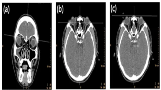
Firstly, areas of both sides of the skull were compared based on the nasal septum on the coronal plane of MPR so as to check the angle of optic foramen and MSP to have symmetry, and were recomposed on axial plane such that the section for observing right optic foramen best was set. MSP sets the line across the crista galli and nasal septum, connects the right MSP with the line formed by the right optic nerve and measures the angle between the two lines as shown in Fig. 1.
Fig. 1. The MSP was set up the basis of nasal septum as a standard at coronal plane of reconstructed image(a), The slice is selected for measurement of the angle at axial plane of reconstructed image(b), The angle between Rt. optic foramen and MSP was measured at the axial image of MPR(c).
1.2. Angle between Orbitomeatal line(OML) Line and optic foramen
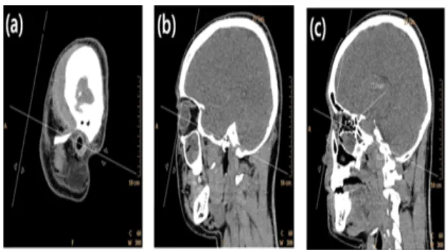
The angle of optic foramen with respect to orbitomeatal line (OML) confirmed on the sagittal plane was measured. The both sides of skull were mirrored based on nasal septum on the coronal plane of MPR which is recomposed facial 3D data using EBW workstation and were recomposed on sagittal plane so as to set the section for observing the right optic foramen best. On the corresponding section, a line connecting the external auditory meatus (EAM) and the center of orbit was drawn to set OML. And then, a line parallel to the forward tilted angle of right optic foramen was set. The angle between the
"J. Korean Soc. Radiol., Vol. 11, No. 5, October 2017"
OML and the optic nerve was measured by connecting the OML and the two lines of the right eye as shown in Fig. 2.
Fig. 2. The position of Rt. external auditory meatus (EAM) was set up at sagittal plane of reconstructed image(a), The OML was set up the line was connected the meddle of Rt. EAM and eyeball of the sagittal plane of reconstructed image(b), The angle between Rt.
optic foramen and OML was measured at the sagittal image of MPR(c).
2.3. STATISTICAL ANALYSIS
With respect to the MSP, the average and standard deviation were obtained by carrying out technical statistics with SPSS (Ver 18.0, USA, Chicago) and one-sample t-test was performed to find statistical difference from 53˚ proposed for existing imaging method.
With respect to the measurement of angle of optic foramen with respect to the OML, the average and standard deviation were obtained by carrying out technical statistics with SPSS (Ver 18.0, USA, Chicago) based on obtained data.
Ⅲ. RESULT
1. Angle of Median sagittal plane and optic foramen The image of measuring angle of right optic foramen with respect to the MSP is the same as Fig.
3. The results were as shown in Table. 1, where the angle of the optic foramen with respect to the midsagittal plane was minimum 31.1˚ to maximum 48.6˚ and was measured 39.9±4.63˚ on average. As
the result of one-sample t-test for 53˚ with respect to the existing imaging method, there was statistically significant difference (p<0.05).
Fig. 3. The angle between Rt. optic foramen and MSP was measured at the axial image of MPR.
Table 1. The results of the analysis of the angle between Rt. optic foramen and MSP.
Average±SD standard P
RTOF 39.9±4.63˚ 53˚ <0.05
*RTOF : Rt. optic foramen
2. Angle of OML and optic foramen
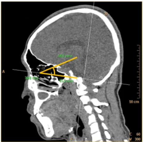
The image of measuring the angle of right optic foramen with respect to OML is the same as Fig. 5.
Fig. 4. The comparison of between 53˚ that is the original angle of Rhese method and results that is the angle MSP with Rt. optic foramen from this study.
The results were as shown Table. 2, where the angle of optic foramen with respect to OML was minimum 22.8˚ to maximum 52.7˚, and was measured 40.8 ± 6.6˚ on average.
Fig. 5. The angle between Rt. optic foramen and OML was measured at the sagittal image of MPR.
Table 2. The results of the angle between Rt. Optic foramen and OML
Location Average±SD
RTOF 40.8±6.6˚
*RTOF : Rt. optic foramen
Ⅳ. DISCUSSION
Currently, the general radiography inspection which is a basic diagnostic inspection is continuously increasing due to extension of desired life time in accordance with graying and change of medical instruments through rapid development and introduction thereof.[14,15] The general radiography is useful due to relatively short inspection time, the simple inspection, and rapid and easy check of anatomical image information through low radiography exposure. However, distortion of image can be caused due to linearity and diffusion of X-ray in general radiography or anatomical structure difference between individuals. JUNG et al. reported, in the research on orbit structures of Korean and
Caucasian, that the orbit structures of Westerner and Asian are different upon skull structure.[16] In addition, Yoon, et al. reported, in the research on manufacturing Korean skull model, that as the result of comparing the frame of Korean and Westerner, the basio-bregmatic height and auriculo- bregmatic height of Korean are higher than those of Westerner, and maximal cranial length minimal frontal breadth and facial length of Korean are lower than those of Westerner.[17] Accordingly, it was found that Asian and Westerner are different. According to the above researches, if the Korean is imaged based on Westerner, it is considered that there would be high possibility of imaging error.
The Rhese method, of which obvious inspection angle is supposed, among general radiography inspection methods is an imaging method which can diagnosis diseases of optic foramen, or inflammation of optic nerve, or optic neuritis showing Vascular Lesions and congenital diseases such as optic nerve glioma or the like, by rotating the MSP plane of the skull to form 53˚ with the imaging stage in Posteroanterior(PA) projection imaging, and supporting the head by placing chin, cheek and nose. But, even irradiating X-ray at the same angle, the optic foramen can be seen narrower if the anatomical structure is different, and the image can be distorted so as to cause difficulty in accurately checking presence of lesion on optic foramen or progress there of, especially for the inspection method needing specific and obvious observation of optic foramen such as Rhese method.
Therefore, this research compared and analyzed the result value by measuring the angle of optic foramen with respect to the MSP and OML, and the angle formed by OML and right optic foramen for patients not having abnormality of optic nerve or optic foramen. As the first result, the angle formed by the midsagittal plane and the right optic foramen was from minimum 31.1˚ to maximum 48.6˚ and was
"J. Korean Soc. Radiol., Vol. 11, No. 5, October 2017"
confirmed to 39.9±4.63˚ on average. In this research, the value has statistically significant difference from 53˚ which is the angle proposed by Rhese method.
This is because of difference between frames of Asian and Westerner as discussed above. Therefore, it is found that when applied to Asian or Korea, imaging error can be caused. As the second result, the angle formed by OML and right optic foramen was measured and the angle corresponding to the result of data analysis was from minimum 22.8˚ to maximum 52.7˚ and the average was 40.8±6.6˚. This has meaning of firstly quantifying the chin, cheek and nose which was a qualitative part expressed as making contact with the imaging stage in a text book.
But, this research was a small scale group research on 50 Koreans, and thus it cannot be the perfect average imaging angle of Rhese method for Koreans.
Ⅴ. CONCLUSION
Consequently, in this research, it was found that the optic foramen form double angles with respect to the midsagittal plane and OML, and it was found that the angle proposed in Rhese method and the angle measured in the experimentation were different in the analysis of the first angle and the angle formed by the MSP and OML. This is because of difference in anatomical structures of Westerner and Asian, and it is considered that readjustment of slope angle of various imaging methods for Asians or Koreans is needed, and this research can be used as fundamental data.
Reference
[1] J. S. Kim, J. M. Kim, Y. H. Lee, D. N. Seo, I. S.
Choi, S. R. Nam, T. S. Yoon, H. J. Kim, H. L. Mi n, J. H, S. H. Han, “National data analysis of gener al radiography projection method in medical imagin g,” Journal of the Korean Society of Radiological Te chnology, Vol. 37, No. 3, pp. 169-175, 2014.
[2] C. Cowling, “ A global overview of the changing rol es of radiographers,” The International Society of Ra
diographers and Radiological Technologists(ISRRT), V ol. 14, No. 1, pp. 29-32, 2008.
[3] J. H. Choi, C. K. Kim, W. C. Kim, S. C. Kim, “Stu dy on development in professional work of radiologic al technologists,” Korean Society of Radiological Scie nce(KSRS), Vol. 29, No. 3, pp. 197-209, 2006.
[4] G.J. Yeon, M. S. Ahn, S. J. Lee, C. M. Cho, M. A.
Yoo, H. S. Jeong, H. S. Moon, N. S. Jo, “Evaluation and conclusion of the general radiography images(part of the examination position),” Journal of The Korean Radiological Technologists Association, Vol. 29, No.
1, pp. 214-214, 2002.
[5] K. H. Yoo, D. S. Kim, S. Y. Shin, K. Y. Chwa, “A n Algorithm for Finding the Completely Visible Regi on from a Special Area,” Korean Information Science Society, Vol. 21. pp. 807-810, 1994.
[6] S. H. Son, Y. G. Song, S. K. Kim, S. W. Hong, J.
B. Kim, “A study on Projection Angles for an Optim al Image of PNS Water’s Veiw on Children,” Journal of the Korean Radiological Technologists Association, Vol. 30, No. 2, pp. 105-111, 2007.
[7] B. S. Kang et al, Radiographic Imaging, 3rd ed., Dai hak Publishing Co., Seoul, pp. 351-410. 2014.
[8] S. S. Kang et al, Textbook of Radiographic Positioni ng and Clinical Diagnosis(Volume I), 4th ed., Chung- Ku Publiching Co., Seoul, pp. 344-348, 2014.
[9] J. H. Lee, “Comparartive study of the lid anatomy b etween the occidental and the oriental,” The Journal of Soonchunhyang University, Vol. 15, No. 4, pp. 12 67-1277, 1992.
[10] H. S. Kang, “Anatomical studies of the orbital cavit y using 3D CT in Korean subjects,” Chonnam Univ ersity, 2016.
[11] J. Y. Hwang, H. Lee, M. W. Chang, S. H. Baek, T. S. Lee, “Optic Canal Location Using Computed Tomography,” The Korean Opthalmological Society, Vol. 55, No. 9, pp. 1272-1276, 2014.
[12] S. S. Cha, J. R. Jang, J. S. Lee, Y. S. Chae, C. Ba e, “The Radiological Study of Optic Canal in Korea n,” Journal of the Korean Radiological Society, Vol.
18, No. 3, pp. 421-427, 1982.
[13] S. S. Kang, C. S. Kim, S. Y. Choi, S. J. Ko, J. H.
Kim, “Evaluation of Present Curriculum for Develop ment of Dept, of Radiological Science Curriculum,”
The Journal of the Korea Contents Association, Vol.
11, No. 5, pp. 242-251, 2011.
[14] C. Schaefer-Prokop, U. Neitzel, H. W. Venema, M.
Uffmann, M. Prokop, “Digital chest radiography; an update on modern technology, dose containment and control of image quality,” European Radiology, Vol.
18, No. 9. pp. 1818-1830. 2008.
[15] K. Doi, “Diagnostic imaging over the last 50 years:
research and development in medical imaging scienc e and technology,” Physics in Medicine & Biology, Vol. 51. No. 13. pp. 5-27, 2006.
[16] H. C. Jeong, H. B. Ahn, “Comparison of Orbital An atomy in Korean and Caucasian Patients Using Com puted Tomography,” The Korean Opthalmological So ciety, Vol. 56, No. 9, pp. 1311-1315, 2015.
[17] K. H. Youn, K. S. Hu, W. C. Song, K. S. Koh, H.
J. Kim, “Fabrication of Korean Skull Model,” Korea n Journal of Physical Anthropology, Vol. 23, No. 4, pp. 229-234, 2010.
"J. Korean Soc. Radiol., Vol. 11, No. 5, October 2017"
Rhese법 촬영에서 시신경구멍의 이중 각도에 대한 연구
박상조,1 유지나,1 유명석,2 허영철1,*
1을지대학교 방사선학과
2서울대학교 보라매병원
요 약
본 연구의 목적은 한국인의 시신경구멍의 이중각도를 확인하여 Rhese법에 적용하는 것이다. 먼저 3차원 CT의 MPR 기법을 이용하여 축상면 영상에서 좌측 안구와 정중시상면 사이의 각도를 측정하였다. 두 번째 는 MPR 영상의 시상면에서 좌측 안구의 시신경구멍과 OML 사이의 각도를 측정하였다. 시신경과 정중시 상면 사이의 각도는 평균 39.9±4.63° 였고, 이는 Rhese 방법으로 제시해 오던 53° 와 달랐음을 확인하였다(p
<0.05). 시신경구멍과 OML 사이의 각도는 40.8±6.6° 였다. 결론적으로 본 연구는 현행 교과서에서 제안 된 Rhese 방법의 표준이 한국인에게 적용하기 어려움을 의미한다. 따라서 다양한 일반촬영법에서 한국 표준의 각도의 연구가 필요하고 이에 본 연구가 기초 자료로 활용될 것이라 사료된다.
중심단어: Rhese, 시신경구멍, 이중각도