Motility and Chemotaxis in the Lyme Spirochete Borrelia burgdorferi:
Role in Pathogenesis
Ah Young Yoo
1, Ho Young Kang
1and Ki Hwan Moon
2*
1Department of Microbiology, College of Natural Sciences, Pusan National University, Busan 46241, Korea
2Division of Marine Bioscience, College of Ocean Science and Technology, Korea Maritime and Ocean University, Busan 49112, Korea Received May 10, 2018 /Revised May 22, 2018 /Accepted May 24, 2018
Motility and chemotaxis are crucial for disease development in many motile pathogens, including spirochetes. In many bacteria, motility is provided by flagella rotation, which is controlled by a che- motaxis-signal-transduction system. Thus, motility and chemotaxis are inextricably linked. Spirochetes are a unique group of bacteria with distinctive flat-wave morphology and corkscrew-like locomotion.
This unusual motility pattern is believed to be important for efficient motility within the dense tissues through which these spirochetes preferentially disseminate in a host. Unlike other externally flagel- lated bacteria-where flagella are in the ambient environment-the flagella of spirochetes are enclosed by the outer membrane and thus are called periplasmic flagella or endoflagella. Although motility- and chemotaxis-associated genes are well studied in some bacteria, the knowledge of how the spi- rochete achieves complex swimming and the roles of most of the putative spirochetal chemotaxis pro- teins are still elusive. Recently, cutting-edge imaging methods and unique genetic manipulations in spirochetes have helped to unravel the mystery of motility and chemotaxis in spirochetes. These con- temporary advances in understanding the motility and chemotaxis of spirochetes in a host’s persis- tence and disease process are highlighted in this review.
Key words : Borrelia burgdorferi, chemotaxis, lyme disease, motility, spirochete
*Corresponding author
*Tel : +82-51-410-4754, Fax : +82-51-404-4750
*E-mail : [email protected]
This is an Open-Access article distributed under the terms of the Creative Commons Attribution Non-Commercial License (http://creativecommons.org/licenses/by-nc/3.0) which permits unrestricted non-commercial use, distribution, and reproduction in any medium, provided the original work is properly cited.
Journal of Life Science 2018 Vol. 28. No. 5. 627~637 DOI : https://doi.org/10.5352/JLS.2018.28.5.627
서 론
Spirochete (스피로헤타) 그룹에 속하는 세균은 타 균주와 구별하기 쉬운 특이한 형태 및 운동성을 가진다[12, 25, 26, 47]. 특히 형태학적 특성의 경우, 17세기 초기 현미경을 이용한 안톤 판 레벤후크(Antoine von Leeuwenhoek)의 관찰에서도 다른 세균 그룹과 잘 구별되었다[85]. 현재까지 많은 종류의 스피로헤타 균이 분리되었으며, 이 중 다양한 종이 질병을 유 발하는 병원성 균주이다. Borrelia burgdorferi sensu lato (s.l.)는 북미 및 유럽지역에서 흔히 발생하는 벡터-매개성 감염 질병 인 라임병(i.e. Lyme disease, Lyme borreliosis)을 유발한다 [10, 31, 37, 64]. 미국 지역 내에서 가장 빈번히 발견되는 B.
burgdorferi sensu stricto (s.s.)의 경우 주로 소아관절염의 증상
을 유발하며, 유럽 지역에서 많이 발견되는 B. garinii와 B. afze-
lii는 주로 신경 계통 감염 및 증상을 유발한다[4, 6, 84]. B.burgdorferi s.l. 이외에도 Borrelia속에 포함되는 다양한 종들이
감염성 질병의 원인균이다. B. hermsii와 B. miyamotoi의 경우
진드기-매개성 재발열(tick-borne relapsing fever) 증상을 유 발하며, B. recurrentis는 이-매개성 재발열(louse-borne relaps- ing fever) 증상을 유발한다. Leptospira는 렙토스피라증을 유발 하는 대표적인 병원성 스피로헤타로 전 세계 인구의 65% 이상 이 이 균에 감염되어 있어, 공중보건학적으로 매우 중요한 감 염성 세균이다[1, 29, 34, 87]. Treponema pallidum subspecies pal-
lidum은 성병의 일종인 매독을 유발하여 전세계적으로 공중보건학적 문제를 야기하는 또 하나의 스피로헤타 세균이다[23].
매독균 외 일부 Treponema 속의 종들(e.g. T. denticola)은 치주 질환의 원인균으로 작용하기도 한다[16]. 이처럼 스피로헤타 는 범세계적으로 다양한 질병을 유발하고 있으며, 스피로헤타 유래의 질병을 극복하기 위한 대책 마련 및 백신개발이 시급 한 실정이다. 이를 위해 스피로헤타의 병원성 인자 및 질병 유발 기작에 대한 연구가 전 세계 다양한 그룹에서 수행되고 있다.
최근의 연구에 의하면 운동성 및 주화성이 병원성 스피로헤 타의 병원성과 매우 깊은 상관관계가 있으며, 특히 Borrelia
burgdorferi의 경우 숙주와 벡터 사이의 감염 순환주기에 직접적인 영향을 미치는 것으로 보고되었다[12, 28, 38, 40, 41, 43, 65]. 본 총설에서는 대표적인 병원성 스피로헤타인 B. burgdor-
feri를 이용한 최근 연구에서 밝혀진 내용의 소개를 통해, 스피로헤타의 복잡하고 특이한 운동성과 주화성이 병원성에 미치 는 영향에 대해 설명하고자 한다.
- Review -
Fig. 1. Schematic diagram of B. burgdorferi. Longitudinal dia- gram (top) and cross-section diagram (bottom) of B.
burgdorferi. Note that periplasmic flagella overlap in the cell center and form a tightly packed ribbon that causes the outer membrane to bulge.
본 론
Borrelia burgdorferi의 형태적 특징 및 운동성
스피로헤타는 물결형태의 운동성과 조밀하게 꼬여진 나선 형태의 모양으로 다른 균들과 구별된다[12, 13, 18]. 스피로헤 타의 막 구조는 일반적인 그람음성균과 마찬가지로 외막, 펩 티도글리칸 층, 내막으로 구성된다. Leptospira종을 제외한 대 부분의 스피로헤타의 외막은 지질 이중층(lipid bilayer)으로 구성되지만, 일반적인 그람음성균이 가지는 지질다당체 (lipopolysaccharide, LPS)를 가지지 않는다[7]. 내막의 경우 일 반적인 원핵생물이 가지는 지질 이중층을 가지며, 얇은 펩티 도글리칸 층으로 둘러싸여 있다[12]. 스피로헤타의 운동성은 세포 운동 소기관 중 하나인 편모의 회전에 의해 발생한다.
대부분의 그람 음성세균의 편모는 세포 외부의 소기관으로 존재하는 외부편모이지만, 스피로헤타의 편모는 세포의 외막 과 펩티도글라이칸 층 사이의 주변세포질 공간에 존재한다.
이로 인해 현미경 하에서 스피로헤타의 편모를 관찰할 수 없 으며, 편모의 특이한 위치를 나타내고 일반 외부편모와 구분 하기 위하여 주변세포질 편모(periplasmic flagella) 또는 내부 편모(endoflagella)라 부른다. 각각의 내부편모는 스피로헤타 의 양 말단에 부착되어 세포 몸통을 감싸며 세포 중앙부에서 서로 만나 리본 형태를 이루게 된다[12]. 스피로헤타는 그 종류 에 따라 다양한 크기와 숫자의 내부편모를 가진다. Cristispira 종의 경우 0.5~3 μm 직경에, 30~180 μm 길이의 100개 이상의 내부편모가 세포 양 말단에 부착된 형태로 존재한다. 반면
Leptospira종에는 0.1 μm 직경에, 10~20 μm 길이의 내부편모가세포 양쪽 말단에 각 한 개씩만 부착된 형태로 존재한다[12, 13, 18, 25, 46].
본 총설에서는 대표적인 스피로헤타 중 하나인 B. burgdor-
feri의 주요 연구들을 중심으로 소개하고자 한다. B. burgdorferi는 약 0.3 μm 직경에 5~20 μm 길이의 세포 몸통을 가지며, 7~11개의 내부편모를 세포 양 말단에 가진다(Fig. 1) [25, 32, 36, 86]. 세포의 양 말단에 부착된 이 편모들은 유영운동을 할 때 그 회전 방향을 조절하여 세포의 운동 방향을 결정한다.
B. burgdorferi 양 말단의 내부편모가 비대칭적으로 회전할 경
우 세포는 한쪽 방향으로 유영(running or reverse running) 하며, 대칭적으로 회전할 경우 제자리에서 세포를 수축시키며 운동 방향을 바꾼다(flexing or tumbling) [13, 39]. 일반적인 그람음성세균의 외부편모와 스피로헤타의 내부편모는 아미 노산 서열과 구조 및 기능에 있어 상당한 유사성을 가진다.
하지만 내부편모의 경우 특이한 위치와 강력한 나선형태의 회전력을 부여하는 운동성으로 인해 외부편모에서는 발견되 지 않는 특이한 모터 구조를 가진다. 그 예로 내부편모의 모터 는 칼라 단백질(collar proteins)이라는 특이한 구조를 가진다.
이 칼라 단백질은 내부편모의 모터 구조에서만 발견되며, 스 피로헤타에서는 보존적으로 발견되나 외부편모를 가지는 일
반적인 그람음성세균에서는 발견되지 않는다(Fig. 2) [14, 90, 91]. 또한, 일반적인 외부편모는 세포에 운동성만 제공하지만, 내부편모의 경우 운동성뿐만 아니라 세포의 형태에 영향을 미친다[46, 77, 80]. 편모 주요 단백질인 FlaB가 결여된 돌연변 이 균주의 경우 스피로헤타의 특징적인 나선형 형태학적 구조 대신 막대형의 구조를 나타내는 것이 확인되었다[46, 77]. 또 한, 내부편모의 회전이 제대로 발생하지 못할 경우, 내부편모 가 세포 중앙부로 향하지 못하고 리본형태를 형성하지 않아 나선형의 구조를 이루지 못한다[80]. 이처럼 내부편모의 방향 성 역시 세포의 형태에 중요한 영향을 미치는데, 그 예로 내부 편모 모터 구조를 이루는 단백질 중 하나인 FliL이 결손 될 경우 편모의 방향이 세포 중앙부가 아닌 세포 말단을 향하게 되며, MotB의 결손과 마찬가지로 세로 중앙부에서 제대로 된 리본형태의 편모를 형성하지 못하여 나선형 구조를 잃게 된다 [50].
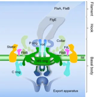
내부편모의 경우 외부편모와 동일하게 크게 기저체(basal body), 고리(hook), 필라멘트(filament) 세 부분으로 구성되어 있다(Fig. 3). 그 중 기저체는 수출장치(export apparatus), MS- ring, C-ring 스위치 복합체, 칼라 단백질, FilL, stator (MotA, MotB)로 구성되어 있다[12, 20, 36, 42, 50, 91]. 고리의 경우 기둥(rod) 단백질에 결합하며 FlgE 단백질로 이루어진다[66].
필라멘트는 주요 플라젤린 단백질인 FlaB와 보조 플라젤린 단
백질인 FlaA로 이루어진다(Fig. 4) [22, 51]. B. burgdorferi의 유
전체 분석 결과에 따르면 50개 이상의 유전자가 운동성 및
주화성에 영향을 미치며, 이는 B. burgdorferi 전체 유전자의
5~6%를 차지하는 방대한 양이다[12, 20]. 최근 4개의 cyclic-di-
GMP 대사에 관여하는 유전자가 B. burgdorferi의 운동성/주화
성에 영향을 미치는 것으로 밝혀졌다[12, 56, 60, 78, 79]. 칼라
Fig. 2. Unique collar structures are conserved in all spirochetes.
(A) External flagellar motor of Escherichia coli; (B-D) peri- plasmic flagellar motors of the spirochetes, Leptospira in- terrogans, Treponema primitia, and Borrelia burgdorferi. The distinctive collar structure is indicated using an arrow.
Because of the dynamic nature of the E. coli stator, it was not visualized by cryo-ET (A). (E, F) Schematic mod- els of the external flagellar motor of E. coli (E) and the periplasmic flagellar motor of B. burgdorferi (F). Periplas- mic flagella are distinct from the external flagella, as they are enclosed within the outer membrane and their flag- ellar motors are considerably larger and more complex.
However, the core architecture of the two flagellar types is comparable. Shared structures include the export ap- paratus, stator, the MS-ring, the C-ring, the rod connect- ing the hook with the MS-ring, hook, and filament (not shown). Unique collar structure in the periplasmic flag- ellar apparatus is shown using an orange color. IM, inner membrane; OM, outer membrane; PG, peptidoglycan layer.
Fig. 3. Schematic diagram of the B. burgdorferi flagellar motor.
The major components (the export apparatus, the rod, the stator, the P ring, the C ring, and the MS ring) are labeled. The C ring is composed of FliG, FliM, and FliN.
The “collar” is a spirochete-specific feature. The export apparatus is divided into two separate densities, al- though the boundary between the MS ring and the ex- port apparatus is not well defined. Hook is composed of FlgE, and filaments are composed of major flagellin FlaB and minor flagellin FlaA. IM, inner membrane.
단백질을 구성하는 유전자 역시 밝혀지지 않고 있었으나, 최 근 flbB 유전자가 이 구조 단백질을 이루는데 중요한 역할을 하는 것으로 알려졌다[45]. 흥미롭게도, B. burgdorferi의 운동 성 및 주화성 관련 유전자들은 일반 그람음성세균의 주요 발 현 조절인자(e.g. flhDC master transcriptional regulator)에 의 한 계층적 조절이 아닌 RNA 중합효소의 σ70 소단위에 의해 대부분 조절된다[2, 12, 15]. 뿐만 아니라, 일부 편모 유전자들 의 경우(e.g. flaB), carbon storage regulator CsrA에 의해 전사 후 단계에서 조절된다[33, 68, 81, 82].
B. burgdorferi는 절대 기생성 세균으로 자연상에서 절지동
물 벡터인 진드기와 척추동물 숙주에서만 발견된다[54, 62].
이 병원성 스피로헤타의 병원성에 미치는 운동성의 영향에 관련된 여러 연구들 중 하나에서 주요 플라젤린 단백질인 FlaB 를 결손 시킨 돌연변이주가 진드기 벡터와 실험쥐 숙주 모두 에서 병원성을 나타내지 못하는 것이 밝혀졌다[77]. 이러한 현 상이 편모 결여에 의한 현상이 아닌 것을 확인하기 위해 편모
모터인 MotB를 결손시킨 돌연변이주를 이용한 실험에서 진드 기와 실험쥐 모두에서 병원성이 결여되는 것이 밝혀졌다[80].
종합해 보면 편모의 유무에 상관없이 운동성을 나타내지 못하 는 B. burgdorferi는 병원성을 잃는 것을 알 수 있다.
B. burgdorferi의 주화성 기작
주화성은 세균이 영양물질 및 특정 환경 내 자극이 될 수
있는 화학물질의 농도구배를 인식하여 세포가 원하는 방향으
로 이동할 수 있게 해주는 신호 변환 시스템으로 원핵생물에
서 매우 보존적이다[61]. 주화성에 관한 연구는 그람음성세균
중 Escherichia coli에서, 그람양성세균 중 Bacillus subtilis에서
가장 많이 수행되었다. 두 그람음성 및 그람양성 대표 균주에
서 주화성 관련 유전자는 매우 보존적이며 비슷한 기능을 수
행하는 것으로 밝혀졌다[8, 21, 61]. E. coli의 경우 methyl-ac-
cepting chemotaxis protein receptor (MCP)라고 불리는 막
단백질이 외부 신호 수용 기관으로 작용한다. 이 신호 수용
단백질에는 두 종류의 단백질이 부착되어 있는데, 연결단백질
인 CheW와 histidine kinase인 CheA가 그것이다. CheA의 촉
매작용은 MCP에 신호인자 부착 여부에 따라 조절된다. E. coli
의 경우 MCP에 신호가 수용되지 않을 경우, CheA는 ad-
enosine triphosphate (ATP)를 이용해 자가인산화 반응을 일
Fig. 4. Model of the B. burgdorferi chemotaxis system. A simplistic chemotaxis signaling pathway of B. burgdorferi. The genome of B. burgdorferi encodes multiple homologs of several chemotaxis genes (e.g. six mcp, two cheA, three cheW, three cheY, two cheB, and two cheR genes), making it more complex than other bacteria. The role of most borrelial chemotaxis proteins are still unknown. To date, only two studies have shown that chemotaxis-specifically involving the histidine kinase cheA2 (box 1) and chemotaxis response regulator cheY3 (box 2)-are essential for the infectious life cycle of B. burgdorferi. CheY2 may serve as a regulator for a virulence determinant in B. burgdorferi (box 3). The role of cheY1 and other putative chemotaxis genes in the infectious life cycle of B. burgdorferi has not been studied until now.
으킨다[30, 61]. ATP로부터 CheA로 전달된 인산기는 다시 re- sponse regulator인 CheY로 전달되며, 인산화된 CheY-P는 편 모 스위치 단백질인 FliM과 FliN에 부착하여, 편모 모터의 회 전방향을 반시계방향에서(질주 운동성) 시계방향으로(뒹굴 기) 바꾸어 세포가 방향을 전환할 수 있도록 한다[59, 70]. 따라 서, 세포는 영양물질(화학 유인제)이 있는 새로운 곳으로 운동 방향을 전환할 수 있는 것이다. 인산화 된 CheY-P는 탈인산화 효소인 CheZ에 의해 다시 CheY로 돌아가며, 탈인산화 된 CheY는 스위치로부터 떨어져 다시 편모 모터의 회전방향을 반시계방향으로 전환시킨다[30, 61]. MCP에 화학 유인제가 부 착될 경우 CheA의 인산화효소 기능이 감쇠하게 되며, 그 결과 세포 내 CheY-P의 농도가 줄어 뒹굴기의 빈도를 줄여 세포가 한 방향으로 질주할 수 있게 해준다. 즉, 세포 내 CheY-P의 농도가 높을 경우 세포는 질주 운동을 하게 되며, CheY-P의 농도가 낮을 경우 세포는 뒹굴기를 하여 방향 전환을 하게 된다. 주화성 시스템은 메틸기에 의한 MCP의 변형에 의해서 조절되기도 하는데 이를 적응(adaptation)이라고 한다. 이러한 MCP의 변형은 methyltransferase인 CheR과 methylesterase 인 CheB에 의해 조절되며, E. coli에서 이 두 단백질이 유일한 chemoreceptor 변형 효소로 알려져 있다[74]. 이처럼 E. coli의 운동성은 5개의 MCP와 각 하나씩의 CheA, W, Y, Z, B, R로
구성된 주화성 시스템에 의해 조절된다[30, 61, 74]. 대표적인 그람양성 균주인 B. subtilis 또한 유사한 주화성 시스템을 가진 다. E. coil와 달리 B. subtilis는 10개의 MCP와 각 하나씩의 CheA, W, B, R, 그리고 탈인산화효소 CheZ 대신 동일한 역할 을 수행하는 CheC를 가진다[8, 21]. 또한, E. coli에서 발견되지 않는 CheD를 가진다. CheD는 현재까지 MCP를 탈아민화 또 는 메틸화 시키며 CheC의 탈인산화 반응을 촉진 시키는 효소 로 알려져 있다[11, 24, 35, 53]. 특이한 점은 E. coli와 달리 B.
subtilis의 경우 CheA의 인산화 작용이 MCP의 신호 수용이
있을 때 일어난다는 것이다. MCP에 화학 유인제가 부착하게 되면 CheA는 자가 인산화 반응을 통해 인산기를 획득하고 이를 CheY로 전달한다. 인산화 된 CheY-P는 편모 모터 스위 치 단백질에 결합하는데, 이 때 편모 모터가 E. coli와 반대로 반시계방향으로 회전하게 되고 세포는 질주운동을 하게 된다.
반면, CheY-P가 탈인산화 되어 CheY로 전환 후 스위치 단백
질에서 떨어지게 되면 모터는 시계방향으로 회전하여 세포가
뒹굴기를 통해 방향을 전환할 수 있게 된다[8, 21]. 즉, 두 균주
의 CheA-CheY two component 주화성 시스템의 과정은 서로
반대로 작용하나, 외부 신호에 대한 세포의 반응은 동일하다
는 것을 알 수 있다. 화학 유인제에 의해 두 세균 모두 편모
모터가 반시계 방향으로 회전하여 질주 운동을 하며, 유인제
가 없을 경우 모터는 시계방향으로 회전하여 뒹굴기를 통한 방향 전환을 한다.
원핵생물의 주화성 시스템을 이루는 유전자 및 그 단백질의 기능은 앞서 언급한 E. coli와 B. subtilis의 비교에서도 알 수 있듯이 매우 유사하며 잘 보존되어 있다. 하지만 B. burgdorferi 의 주화성 시스템은 앞서 언급한 세균의 주화성 시스템에 비 해 매우 복잡하다. B. burgdorferi의 경우 일반 편모 운동성 세균 과 달리 많은 숫자의 주화성 관련 유전자를 가진다. 앞서 소개 한 E. coli와 B. subtilis의 경우 신호수용체인 MCP를 제외한 주화성 관련 유전자가 염색체 내 하나씩 존재하는 반면, B.
burgdorferi는 총 6개의 mcp, 2개의 cheA, 3개의 cheY, 2개의 cheB, 2개의 cheR, 3개의 cheW 유전자를 가지며, 탈인산화효소
역할을 수행하는 하나의 CheX와 B. subtilis에서 발견되었던 하나의 CheD를 가지고 있다(Fig. 4) [20]. B. burgdorferi의 histi- dine kinase인 CheA1과 CheA2 두 단백질 모두 response reg- ulator를 인산화시킬 수 있는 것으로 밝혀졌다[49]. Response regulator인 CheY의 경우, CheY1과 CheY2를 제외한 CheY3 만이 in vitro상에서의 운동성에 영향을 주는 것으로 밝혀졌다 [52]. 특이한 점은, cheY2와 cheY3 두 유전자 모두 실험쥐 숙주 내에서의 B. burgdorferi의 병원성에 영향을 미치지만, cheY3 만이 진드기 벡터에서 실험쥐로의 감염 전달에 영향을 미치는 것으로 밝혀졌다[55, 87]. 또한 CheD 역시 in vitro 운동성에는 크게 영향을 미치지 못하나 진드기 벡터 및 실험쥐 숙주에서 병원성을 감소시키는 것을 확인하였다[44]. 그 외 CheW1, CheW3, CheX 단백질이 in vitro 상에서 B. burgdorferi의 주화 성에 직접적인 영향을 미치는 것으로 밝혀졌다[48, 89]. 앞서 언급한 바와 같이 B. burgdorferi의 운동성 및 주화성은 이 병원 균의 주요 질병 원인인자이지만, 현재까지 CheA2, CheY2, CheY3, CheD 이외의 B. burgdorferi의 다른 주화성 관련 유전 자들과 병원성 사이의 직접적인 상관관계는 밝혀지지 않았다 [44, 55, 83, 87]. 편모를 이용한 운동성을 가지는 다른 세균들에 비해 스피로헤타가 이처럼 많은 종류의 주화성 관련 유전자를 가지는 이유는 아직 밝혀지지 않았으나, 일부 선행 연구를 참 고하면 B. burgdorferi가 가지는 절대 기생성 생육환경에 의한 것이라 추측할 수 있다. 앞서 언급한 바와 같이 B. burgdorferi의 경우 절지동물인 진드기와 척추동물을 숙주로 가진다[54, 62].
두 숙주 내 생육 환경 및 영양공급원은 매우 다르며, 같은 진드 기 벡터 내에서도 흡혈 전과 후를 기준으로 이용되는 에너지 원이 다르다. 진화학적으로 절대 기생성 스피로헤타는 특정 숙주 내 특정 환경 조건들에 따라 서로 다른 주화성 관련 단백 질을 이용해 매우 정교하게 조절되는 주화성 시스템을 가질 것으로 예측되며, 이를 밝히기 위해 더 많은 연구와 탐색이 필요할 것이다.
B. burgdorferi의 감염 생활 주기 및 운동성과 주화성을 이용한 발병 기작
B. burgdorferi의 운동성 및 주화성은 병원성 및 생활주기와
매우 밀접한 상관관계를 가진다. 척추동물 숙주 내에서는 지 속적인 운동성을 보이지만, 절지동물 벡터인 진드기 내부에서 는 이러한 지속적인 운동성을 보이지 않는다. B. burgdorferi의 주요 질병 벡터인 등빨간긴가슴잎벌레 진드기[Blacklegged tick or Deer tick (Ixodes sapularis)]의 경우 알(egg), 유충 (larva), 약충(nymph), 성체(adult)의 총 네 단계 생활 주기를 가진다(Fig. 6) [62]. B. burgdorferi는 난소를 통한 모계 감염이 거의 일어나지 않으므로, 알에서 부화한 유충은 스피로헤타에 감염되어 있지 않은 상태이다(Fig. 5A) [63]. 유충은 약충으로 변태하기 위하여 작은 척추동물 숙주로부터 흡혈을 통해 에너 지를 공급받는다(Fig. 5B). 이 흡혈과정에서 숙주가 B. burgfor-
feri에 감염 되어 있을 경우, 진드기가 스피로헤타를 획득하게된다. 일반적으로 작은 설치류 및 조류가 B. burgdorferi의 보유 숙주로 이용될 수 있으나, 그 중 흰발붉은쥐[White footed mouse (Peromyscus leucopus)]가 B. burgdorferi의 주요 보유숙 주 역할을 한다. 그 이유는 타 설치류에 비해 털손질 습성 (self-grooming behavior)이 결여되어 있어 부착된 진드기 유 충의 제거가 원활하지 못하기 때문이라 추측하고 있다. 이렇 게 보유숙주로부터 획득된 B. burgdorferi는 진드기 유충으로 이동하여 소화관 점막에 부착한다. 이 때, B. burgdorferi의 외막 단백질 중 하나인 OspA의 발현량이 증가하게 되며, 이 OspA 는 진드기 장내 존재하는 단백질인 TROSPA (tick receptor for OspA)와 결합하여 스피로헤타가 진드기 장내에 부착하는 것을 도와준다[17, 57, 58]. 진드기의 장내에 부착된 B. burgdor-
feri는 비운동성으로 전환하여 에너지 소모를 줄이며, 흡혈 중얻은 혈액 내의 포도당을 에너지원으로 이용하여 세포분화를 통해 그 수를 늘인다. 진드기 역시 흡혈을 통해 얻은 에너지를 이용해 약충으로 변태하여 두번째 흡혈을 준비한다(Fig. 5C).
흡혈 후 땅으로 내려와 약충으로 변태한 진드기는 다시 척추 동물 숙주에 부착하여 생애 두번째 흡혈을 시작한다(Fig.
5D-1). 약충 상태의 진드기는 유충 및 성체 상태일 때 보다
매우 활동적이며, 주로 이 두번째 흡혈 과정에서 사람에게 부
착하여 흡혈과 동시에 라임병을 전파한다(Fig. 5D-2). 진드기
의 두번째 흡혈이 시작되면, 장내 부착해 있던 B. burgdorferi는
OspA의 발현을 감소시키고, 대신 OspC의 발현을 증가시킨다
[71]. 이 과정에서 TROSPA와 결합해 있던 비운동성 스피로헤
타가 떨어져 나와 다시 운동성을 가동하여 약충의 침샘으로
침투하게 된다. 침샘으로 침투한 B. burgdorferi는 흡혈시 혈액
응고를 막기 위한 침 유입과 동시에 숙주의 진피조직으로 전
달된다. 두번째 흡혈이 끝난 약충은 땅으로 내려와 성체로 변
태하여, 마지막 흡혈을 준비한다(Fig. 5E). 성체가 된 진드기는
큰 척추동물 숙주에서의 마지막 흡혈과(주로 흰꼬리 사슴
[White-tailed deer (Odocoileus virginianus)]에서 흡혈) 함께 짝
짓기를 수행한다(Fig. 5F). 짝짓기 후 수컷은 죽고 암컷은 땅으
로 내려와 알을 낳아 진드기의 생활주기를 완성한다(Fig. 5G)
Fig. 5. The enzootic life cycle of B. burgdorferi. Ixodes scapularis (also known as the blacklegged tick or deer tick) is the main vector of B. burgdorferi in the United States and has a two-year life cycle with four life stages: the eggs, and three differential developmental stages: the larva, nymph, and adult stages. The representative signs and symptoms of untreated Lyme disease were shown in the dotted line boxes on right panel.
[62].
보유숙주로부터 진드기로의 B. burgdorferi 이동은 흡혈 과 정 중에 발생하지만, 이러한 스피로헤타의 이동이 흡혈 과정 중 발생하는 혈액을 통한 수동적 현상인지 흡혈 지점 인근 숙주 조직으로부터 진드기로의 능동적 이동이 존재하는지 여 부에 관련된 연구는 이루어지지 않고 있었다. 하지만 최근 Yale 의과대학교의 한 연구그룹에서 two-photon intravital 현 미경을 이용하여 진드기의 흡혈 과정 중 발생하는 B. burgdor-
feri의 능동적인 이동을 직접 관찰하였다. 녹색형광단백질을발현하는 B. burgdorferi를 실험쥐에 주사 후 2주간 감염을 진행 시킨 후, 진드기를 통한 흡혈 과정을 관찰 한 결과 6시간 정도 의 흡혈 과정 동안 피부 조직 내에 잔존하던 스피로헤타가 진드기로 이동하는 현상을 관찰하였다[9]. 이 결과는 B. burg-
dorferi의 운동성이 절지동물 벡터 내에서 강력히 조절될 뿐만아니라, 척추동물 숙주에서 기인한 어떠한 인자에 의해서도 조절되어 벡터-숙주 사이에 스피로헤타가 이동할 때 매우 중 요한 병원성 인자로 작용함을 시사한다. 또한 숙주의 피부조 직에 존재하던 스피로헤타가 진드기의 흡혈과 동시에 정확히
흡혈지점으로 이동이 가능한 것은 진드기의 타액 내 존재하는 특정 물질에 의한 주화성에 기반할 것이라 추측되고 있다. 실 제로 in vitro 상에서 수행된 진드기 타액을 이용한 모세관 주 화성 실험을 통해 B. burgdorferi가 유인되는 것이 확인되어 위 가설을 뒷받침하지만, 진드기 타액 샘플 채취의 어려움으로 HPLC와 같은 성분 분석 실험은 아직 이루어지지 못하고 있다 [5, 72].
벡터-숙주 간 스피로헤타의 이동뿐만 아니라 숙주내에서의 병원성에도 운동성 및 주화성은 중요한 영향을 미친다. 일반 적으로 B. burgdorferi가 숙주에 감염되어 라임병을 발병할 경 우, 그 시기 및 발병증상에 따라 크게 세 단계로 구분한다.
첫번째 단계인 초기 국소 라임병(early localized Lyme dis-
ease)은 진드기 흡혈 후 3~30일 내 발생하며, 감염된 환자의
70~80%의 경우 흡혈 부위에 이동성 홍반(erythema migrans)
이 발생한다(Fig. 5. 우측 점선상자 2). 이는 흡혈부위로 침투한
B. burgdorferi가 진피조직 내에서 이동하며 나타나는 염증반응으로 스피로헤타의 이동에 맞춰 나타나는 라임병의 대표적인
병적 증상인 황소 눈 모양의 발진(bull’s-eye rash)이다[10, 31,
54]. 두번째 단계인 초기 확산 라임병(early disseminated Lyme disease)은 진드기 흡혈 후 2-8주 내 발생하며, 흡혈 부위 외 다수의 이동성 홍반 및 안면마비(Fig. 5. 우측 점선상자 1), 수막염 유사 증상과 같은 신경계 병증이 나타난다. 이러한 병 증 역시피부조직으로부터 유래한 B. burgdorferi가 운동성 및 주화성을 이용하여 림프조직 내부로 침투해 발생하는 염증 반응의 일부이다[10, 31, 54, 73]. 세번째 단계인 후기 만성 라임 병(late persistent Lyme disease)은 진드기 흡혈 후 6개월 이후 발생하며, 주로 항생제 치료 후 회복되지 못한 환자들에게서 관절염 등의 증상으로 나타난다(Fig. 5. 우측 점선상자 3). 이러 한 관절염 증상은 사이토카인 및 세포매개 면역 반응의 조절 장애로 인한 면역 관련 질환이지만, 명확한 발병 원인에 대해 서는 논쟁의 여지가 남아있다. 현재까지 크게 두가지 원인으 로 분석되는데 첫째는 항생제 치료에도 불구하고 살아남은 스피로헤타의 관절 내 집락형성에 의한 재발병 이라는 주장 과, 두번째로 B. burgdorferi의 외막 단백질의 숙주 유사성에 의한 자가면역질환 증상이라는 주장이다[3, 10, 31, 54, 67]. 후 기 만성 라임병으로 인한 관절염 환자의 관절활액에서 PCR (polymerase chain reaction, 중합효소 연쇄반응)을 통한 B.
burgdorferi의 DNA는 탐지되었지만, 실제 관절활액 및 조직
배양을 통한 B. burgdorferi의 재배양은 확인하지 못한 상태이 다[3, 75]. 또한, 일부 연구 그룹에서는 특정 스트레스 상황(e.g.
항생제 처리, 영양분 고갈 등)에서 스피로헤타가 동그랗게 말 려(round body form) 운동성 및 에너지 소모를 줄이고 스스로 를 외부 환경환경부터 격리시키는 일종의 동면상태를 가진다 는 보고도 있으나, 아직은 논쟁의 대상이 되고 있는 발견이다 [19, 69]. 따라서, B. burgdorferi가 round body 형성을 통해 항 생제로부터 살아 남아 이후 재발병으로 이어진다는 주장도 아 직은 확인되지 못하고 있다. 또한, B. burgdorferi의 OspA
165-173항원결정인자 부위의 경우 human lymphocyte function asso- ciated antigen-1 (hLFA-1)과 서열 유사성을 나타내지만, 이로 인한 실제 자가면역질환 증상이 일어나는지 역시 밝혀지지 않은 상태이다[27, 76].
B. burgdorferi는 세포외 병원체(extracellular pathogen)로
앞서 설명한 바와 같이 숙주의 면역체계를 자극하여 염증반응 등을 유발해 병적 증상을 일으킨다. 또한, 피부, 방광, 신경계, 관절 등 특정 부위에 집락을 형성하는 현상을 보이는데 이는 주화성과 크게 관련 있을 것으로 추측된다. 실제로 in vitro 실험을 통해 B. burgdorferi의 주화성 신호 수용 단백질인 methyl accepting chemotaxis proteins (MCPs)이 신경전달물질로 많 이 이용되는 glutamate 및 glycine, 관절 내 많이 존재하는 glu- cosamine, N-acetylglucosamine, 그리고 진드기의 장 내에서 발견되는 glycerol 및 chitosan과 결합하며, 이 물질들에 의해
B. burgdorferi가 유인되는 것이 밝혀졌다[5, 48]. 위 결과들을종합해 볼 때, B. burgdorferi의 운동성 및 주화성은 절지동물 벡터와 척추동물 숙주 사이의 전파에 중요한 역할을 하여 감
염 생활주기를 완성하는 역할을 할 뿐만 아니라, 숙주 내에서 특정 조직으로의 이동 및 질병 유발에도 중요한 역할을 수행 하는 것을 알 수 있다.
결 론
스피로헤타의 경우 나선형의 특징적인 형태를 가지며, 편모 가 세포 외부가 아닌 내부에 존재하고, 세포 양 말단에 위치하 는 이 내부편모의 회전방향에 의해 운동방향이 결정된다. 또 한, 외부편모를 가지는 세균에서는 발견되지 않는 특징적인 편모모터 구조를 가지며, 병원성을 가지기 위해서는 운동성 및 주화성이 필수적으로 요구된다. 지난 수년 간의 유전자 조 작 기술의 발달로 인해 B. burgdorferi의 운동성 및 주화성에 관여하는 일부 인자들이 병원성 인자로써 작용할 수 있음이 밝혀졌다. 하지만 아직까지 많은 유전자들의 기능이 밝혀지지 않았으며, 특히 이 병원성 스피로헤타에서 내부편모의 조립과 정과 서로 다른 숙주 내에서의 운동성과 주화성의 조절 등에 관한 여러 연구가 결여되어 있다. B. burgdorferi의 운동성 및 주화성에 대한 심화 연구를 통해 아직까지 밝히지 못한 이 특이한 세균의 발병 기작을 이해하는 것은 배양이 불가능한
T. pallidum 및 다른 병원성 스피로헤타들의 특성을 찾는데 중요한 정보를 제공할 수 있다.
감사의 글
이 논문은 2017학년도 한국해양대학교 학술연구지원사업 신진교수정착연구비의 지원을 받아 수행된 연구임. 이 논문의 일부 그림들(Fig. 2, Fig. 3, Fig. 4)은 East Carolina University 의 Md A. Motaleb 교수와 Yale University의 Jun Liu 교수로 부터 제공 받음.
References
1. Adler, B. and de la Pena Moctezuma, A. 2010. Leptospira and leptospirosis. Vet. Microbiol. 140, 287-296.
2. Aldridge, P. and Hughes, K. T. 2002. Regulation of flagellar assembly. Curr. Opin. Microbiol. 5, 160-165.
3. Arvikar, S. L. and Steere, A. C. 2015. Diagnosis and treat- ment of Lyme arthritis. Infect. Dis. Clin. North. Am. 29, 269-280.
4. Bacon, R. M., Kugeler, K. J. and Mead, P. S. Centers for Disease and Prevention (CDC). 2008. Surveillance for Lyme disease--United States, 1992-2006. MMWR. Surveill. Summ.
57, 1-9.
5. Bakker, R. G., Li, C., Miller, M. R., Cunningham, C. and Charon, N. W. 2007. Identification of specific chemo- attractants and genetic complementation of a Borrelia burg- dorferi chemotaxis mutant: flow cytometry-based capillary tube chemotaxis assay. Appl. Environ. Microbiol. 73, 1180-
1188.
6. Balmelli, T. and Piffaretti, J. C. 1995. Association between different clinical manifestations of Lyme disease and differ- ent species of Borrelia burgdorferi sensu lato. Res. Microbiol.
146, 329-340.
7. Ben-Menachem, G., Kubler-Kielb, J., Coxon, B., Yergey, A.
and Schneerson, R. 2003. A newly discovered cholesteryl galactoside from Borrelia burgdorferi. Proc. Natl. Acad. Sci.
USA 100, 7913-7918.
8. Bischoff, D. S. and Ordal, G. W. 1992. Bacillus subtilis chemo- taxis: a deviation from the Escherichia coli paradigm. Mol.
Microbiol. 6, 23-28.
9. Bockenstedt, L. K., Gonzalez, D., Mao, J., Li, M., Belperron, A. A. and Haberman, A. 2014. What ticks do under your skin: two-photon intravital imaging of Ixodes scapularis feed- ing in the presence of the lyme disease spirochete. Yale. J.
Biol. Med. 87, 3-13.
10. Bockenstedt, L. K. and Wormser, G. P. 2014. Review: un- raveling Lyme disease. Arthritis. Rheumatol. 66, 2313-2323.
11. Chao, X., Muff, T. J., Park, S. Y., Zhang, S., Pollard, A. M., Ordal, G. W., Bilwes, A. M. and Crane, B. R. 2006. A re- ceptor-modifying deamidase in complex with a signaling phosphatase reveals reciprocal regulation. Cell 124, 561-571.
12. Charon, N. W., Cockburn, A., Li, C., Liu, J., Miller, K. A., Miller, M. R., Motaleb, M. A. and Wolgemuth, C. W. 2012.
The unique paradigm of spirochete motility and chemotaxis.
Annu. Rev. Microbiol. 66, 349-370.
13. Charon, N. W. and Goldstein, S. F. 2002. Genetics of motility and chemotaxis of a fascinating group of bacteria: the spirochetes. Annu. Rev. Genet. 36, 47-73.
14. Chen, S., Beeby, M., Murphy, G. E., Leadbetter, J. R., Hen- drixson, D. R., Briegel, A., Li, Z., Shi, J., Tocheva, E. I., Müller, A., Dobro, M. J. and Jensen, G. J. 2011. Structural diversity of bacterial flagellar motors. EMBO. J. 30, 2972-2981.
15. Chevance, F. F. and Hughes, K. T. 2008. Coordinating as- sembly of a bacterial macromolecular machine. Nat. Rev.
Microbiol. 6, 455-465.
16. Dashper, S. G., Seers, C. A., Tan, K. H. and Reynolds, E.
C. 2011. Virulence factors of the oral spirochete Treponema denticola. J. Dent. Res. 90, 691-703.
17. de Silva, A. M., Telford, S. R. 3rd, Brunet, L. R., Barthold, S. W. and Fikrig, E. 1996. Borrelia burgdorferi OspA is an arthropod-specific transmission-blocking Lyme disease vac- cine. J. Exp. Med. 183, 271-275.
18. Dombrowski, C., Kan, W., Motaleb, M. A., Charon, N. W., Goldstein, R. E. and Wolgemuth, C. W. 2009. The elastic basis for the shape of Borrelia burgdorferi. Biophys. J. 96, 4409- 4417.
19. Feng, J., Shi, W., Zhang, S., Sullivan, D., Auwaerter, P. G.
and Zhang, Y. 2016. A drug combination screen identifies drugs active against amoxicillin-induced round bodies of in vitro Borrelia burgdorferi persisters from an FDA drug library.
Front. Microbiol. 7, 743.
20. Fraser, C. M., Casjens, S., Huang, W. M., Sutton, G. G., Clayton, R., Lathigra, R., White, O., Ketchum, K. A., Dodson,
R., Hickey, E. K., Gwinn, M., Dougherty, B., Tomb, J. F., Fleischmann, R. D., Richardson, D., Peterson, J., Kerlavage, A. R., Quackenbush, J., Salzberg, S., Hanson, M., van Vugt, R., Palmer, N., Adams, M. D., Gocayne, J., Weidman, J., Utterback, T., Watthey, L., McDonald, L., Artiach, P., Bowman, C., Garland, S., Fuji, C., Cotton, M. D., Horst, K., Roberts, K., Hatch, B., Smith, H. O. and Venter, J. C. 1997.
Genomic sequence of a Lyme disease spirochaete, Borrelia burgdorferi. Nature 390, 580-586.
21. Garrity, L. F. and Ordal, G. W. 1995. Chemotaxis in Bacillus subtilis: how bacteria monitor environmental signals. Phar- macol. Ther. 68, 87-104.
22. Ge, Y. and Charon, N. W. 1997. FlaA, a putative flagellar outer sheath protein, is not an immunodominant antigen associated with Lyme disease. Infect. Immun. 65, 2992-2995.
23. Giacani, L. and Lukehart, S. A. 2014. The endemic trepone- matoses. Clin. Microbiol. Rev. 27, 89-115.
24. Glekas, G. D., Plutz, M. J., Walukiewicz, H. E., Allen, G.
M., Rao, C. V. and Ordal, G. W. 2012. Elucidation of the multiple roles of CheD in Bacillus subtilis chemotaxis. Mol.
Microbiol. 86, 743-756.
25. Goldstein, S. F., Buttle, K. F. and Charon, N. W. 1996.
Structural analysis of the Leptospiraceae and Borrelia burg- dorferi by high-voltage electron microscopy. J. Bacteriol. 178, 6539-6545.
26. Goldstein, S. F., Charon, N. W. and Kreiling, J. A. 1994.
Borrelia burgdorferi swims with a planar waveform similar to that of eukaryotic flagella. Proc. Natl. Acad. Sci. USA. 91, 3433-3437.
27. Guerau-de-Arellano, M. and Huber, B. T. 2002. Development of autoimmunity in Lyme arthritis. Curr. Opin. Rheumatol.
14, 388-393.
28. Guyard, C., Raffel, S. J., Schrumpf, M. E., Dahlstrom, E., Sturdevant, D., Ricklefs, S. M., Martens, C., Hayes, S. F., Fischer, E. R., Hansen, B. T., Porcella, S. F. and Schwan, T. G. 2013. Periplasmic flagellar export apparatus protein, FliH, is involved in post-transcriptional regulation of FlaB, motility and virulence of the relapsing fever spirochete Borrelia hermsii. PLoS One 8, e72550.
29. Haake, D. A. and Levett, P. N. 2015. Leptospirosis in hu- mans. Curr. Top. Microbiol. Immunol. 387, 65-97.
30. Hazelbauer, G. L. 2012. Bacterial chemotaxis: the early years of molecular studies. Annu. Rev. Microbiol. 66, 285-303.
31. Herzer, P., Fingerle, V., Pfister, H. W. and Krause, A. 2014.
[Lyme borreliosis]. Internist (Berl). 55, 789-802; quiz 803-804.
32. Hovind-Hougen, K. 1984. Ultrastructure of spirochetes iso- lated from Ixodes ricinus and Ixodes dammini. Yale. J. Biol.
Med. 57, 543-548.
33. Karna, S. L., Sanjuan E., Esteve-Gassent, M. D., Miller, C.
L., Maruskova, M. and Seshu, J. 2011. CsrA modulates levels of lipoproteins and key regulators of gene expression critical for pathogenic mechanisms of Borrelia burgdorferi. Infect.
Immun. 79, 732-744.
34. Ko, A. I., Goarant, C. and Picardeau, M. 2009. Leptospira:
the dawn of the molecular genetics era for an emerging zoo- notic pathogen. Nat. Rev. Microbiol. 7, 736-747.
35. Kristich, C. J. and Ordal, G. W. 2002. Bacillus subtilis CheD is a chemoreceptor modification enzyme required for che- motaxis. J. Biol. Chem. 277, 25356-25362.
36. Kudryashev, M., Cyrklaff, M., Baumeister, W., Simon, M.
M., Wallich, R. and Frischknecht, F. 2009. Comparative cryo- electron tomography of pathogenic Lyme disease spirochetes.
Mol. Microbiol. 71, 1415-34.
37. Kuehn, B. M. 2013. CDC estimates 300,000 US cases of Lyme disease annually. JAMA. 310, 1110.
38. Lambert, A., Picardeau, M., Haake, D. A., Sermswan, R. W., Srikram, A., Adler, B. and Murray, G. A. 2012. FlaA pro- teins in Leptospira interrogans are essential for motility and virulence but are not required for formation of the flagellum sheath. Infect. Immun. 80, 2019-2025.
39. Li, C., Bakker, R. G., Motaleb, M. A., Sartakova, M. L., Cabello, F. C. and Charon, N. W. 2002. Asymmetrical flag- ellar rotation in Borrelia burgdorferi nonchemotactic mutants.
Proc. Natl. Acad. Sci. USA. 99, 6169-6174.
40. Liao, S., Sun, A., Ojcius, D. M., Wu, S., Zhao, J. and Yan, J. 2009. Inactivation of the fliY gene encoding a flagellar mo- tor switch protein attenuates mobility and virulence of Leptospira interrogans strain Lai. BMC. Microbiol. 9, 253.
41. Lin, T., Gao L., Zhang, C., Odeh, E., Jacobs, M. B., Coutte, L., Chaconas, G., Philipp, M. T. and Norris, S. J. 2012.
Analysis of an ordered, comprehensive STM mutant library in infectious Borrelia burgdorferi: insights into the genes re- quired for mouse infectivity. PLoS One 7, e47532.
42. Liu, J., Lin, T., Botkin, D. J., McCrum, E., Winkler, H. and Norris, S. J. 2009. Intact flagellar motor of Borrelia burgdorferi revealed by cryo-electron tomography: evidence for stator ring curvature and rotor/C-ring assembly flexion. J. Bacteriol.
191, 5026-5036.
43. Lux, R., Miller, J. N., Park, N. H. and Shi, W. 2001. Motility and chemotaxis in tissue penetration of oral epithelial cell layers by Treponema denticola. Infect. Immun. 69, 6276-6283.
44. Moon, K. H., Hobbs, G. and Motaleb, M. A. 2016. Borrelia burgdorferi CheD Promotes Various Functions in Chemotaxis and the Pathogenic Life Cycle of the Spirochete. Infect.
Immun. 84, 1743-1752.
45. Moon, K. H., Zhao, X., Manne, A., Wang, J., Yu, Z., Liu, J. and Motaleb, M. A. 2016. Spirochetes flagellar collar pro- tein FlbB has astounding effects in orientation of peri- plasmic flagella, bacterial shape, motility, and assembly of motors in Borrelia burgdorferi. Mol. Microbiol. 102, 336-348.
46. Motaleb, M. A., Corum, L., Bono, J. L., Elias, A. F., Rosa, P., Samuels, D. S. and Charon, N. W. 2000. Borrelia burgdor- feri periplasmic flagella have both skeletal and motility functions. Proc. Natl. Acad. Sci. USA. 97, 10899-10904.
47. Motaleb, M. A., Liu, J. and Wooten, R. M. 2015. Spirochetal motility and chemotaxis in the natural enzootic cycle and development of Lyme disease. Curr. Opin. Microbiol. 28, 106- 113.
48. Motaleb, M. A., Miller, M. R., Li, C., Bakker, R. G., Goldstein, S. F., Silversmith, R. E., Bourret, R. B. and Charon, N. W. 2005. CheX is a phosphorylated CheY phos- phatase essential for Borrelia burgdorferi chemotaxis. J.
Bacteriol. 187, 7963-7969.
49. Motaleb, M. A., Miller, M. R., Li, C. and Charon, N. W.
2007. Phosphorylation assays of chemotaxis two-component system proteins in Borrelia burgdorferi. Methods. Enzymol. 422, 438-447.
50. Motaleb, M. A., Pitzer, J. E., Sultan, S. Z. and Liu, J. 2011.
A novel gene inactivation system reveals altered periplasmic flagellar orientation in a Borrelia burgdorferi fliL mutant. J.
Bacteriol. 193, 3324-3331.
51. Motaleb, M. A., Sal, M. S. and Charon, N. W. 2004. The decrease in FlaA observed in a flaB mutant of Borrelia burg- dorferi occurs posttranscriptionally. J. Bacteriol. 186, 3703- 3711.
52. Motaleb, M. A., Sultan, S. Z., Miller, M. R., Li, C. and Charon, N. W. 2011. CheY3 of Borrelia burgdorferi is the key response regulator essential for chemotaxis and forms a long-lived phosphorylated intermediate. J. Bacteriol. 193, 3332-3341.
53. Muff, T. J. and Ordal, G. W. 2007. The CheC phosphatase regulates chemotactic adaptation through CheD. J. Biol.
Chem. 282, 34120-34128.
54. Murray, T. S. and Shapiro, E. D. 2010. Lyme disease. Clin.
Lab. Med. 30, 311-328.
55. Novak, E. A., Sekar, P., Xu, H., Moon, K. H., Manne, A., Wooten, R. M. and Motaleb, M. A. 2016. The Borrelia burg- dorferi CheY3 response regulator is essential for chemotaxis and completion of its natural infection cycle. Cell. Microbiol.
18, 1782-1799.
56. Novak, E. A., Sultan, S. Z. and Motaleb, M. A. 2014. The cyclic-di-GMP signaling pathway in the Lyme disease spi- rochete, Borrelia burgdorferi. Front. Cell. Infect. Microbiol. 4, 56.
57. Ohnishi, J., Piesman, J. and de Silva, A. M. 2001. Antigenic and genetic heterogeneity of Borrelia burgdorferi populations transmitted by ticks. Proc. Natl. Acad. Sci. USA. 98, 670-675.
58. Pal, U., Li, X., Wang, T., Montgomery, R. R., Ramamoorthi, N., Desilva, A. M., Bao, F., Yang, X., Pypaert, M., Pradhan, D., Kantor, F. S., Telford, S., Anderson, J. F. and Fikrig, E.
2004. TROSPA, an Ixodes scapularis receptor for Borrelia burgdorferi. Cell 119, 457-468.
59. Park, S. Y., Lowder, B., Bilwes, A. M., Blair, D. F. and Crane, B. R. 2006. Structure of FliM provides insight into assembly of the switch complex in the bacterial flagella motor. Proc.
Natl. Acad. Sci. USA. 103, 11886-11891.
60. Pitzer, J. E., Sultan, S. Z., Hayakawa, Y., Hobbs, G., Miller, M. R. and Motaleb, M. A. 2011. Analysis of the Borrelia burg- dorferi cyclic-di-GMP-binding protein PlzA reveals a role in motility and virulence. Infect. Immun. 79, 1815-1825.
61. Porter, S. L., Wadhams, G. H. and Armitage, J. P. 2011.
Signal processing in complex chemotaxis pathways. Nat.
Rev. Microbiol. 9, 153-165.
62. Radolf, J. D., Caimano, M. J., Stevenson, B. and Hu, L. T.
2012. Of ticks, mice and men: understanding the dual-host lifestyle of Lyme disease spirochaetes. Nat. Rev. Microbiol.
10, 87-99.
63. Rollend, L., Fish, D. and Childs, J. E. 2013. Transovarial
transmission of Borrelia spirochetes by Ixodes scapularis: a summary of the literature and recent observations. Ticks.
Tick. Borne. Dis. 4, 46-51.
64. Rosa, P. A., Tilly, K. and Stewart, P. E. 2005. The burgeoning molecular genetics of the Lyme disease spirochaete. Nat.
Rev. Microbiol. 3, 129-143.
65. Rosey, E. L., Kennedy, M. J. and Yancey, R. J. Jr. 1996. Dual flaA1 flaB1 mutant of Serpulina hyodysenteriae expressing periplasmic flagella is severely attenuated in a murine mod- el of swine dysentery. Infect. Immun. 64, 4154-4162.
66. Sal, M. S., Li, C., Motalab, M. A., Shibata, S., Aizawa, S. and Charon, N. W. 2008. Borrelia burgdorferi uniquely regulates its motility genes and has an intricate flagellar hook-basal body structure. J. Bacteriol. 190, 1912-1921.
67. Sanchez, J. L. 2015. Clinical manifestations and treatment of lyme disease. Clin. Lab. Med. 35, 765-778.
68. Sanjuan, E., Esteve-Gassent, M. D., Maruskova, M. and Seshu, J. 2009. Overexpression of CsrA (BB0184) alters the morphology and antigen profiles of Borrelia burgdorferi.
Infect. Immun. 77, 5149-5162.
69. Sapi, E., Kaur, N., Anyanwu, S., Luecke, D. F., Datar, A., Patel, S., Rossi, M. and Stricker, R. B. 2011. Evaluation of in-vitro antibiotic susceptibility of different morphological forms of Borrelia burgdorferi. Infect. Drug. Resist. 4, 97-113.
70. Sarkar, M. K., Paul, K. and Blair, D. 2010. Chemotaxis signal- ing protein CheY binds to the rotor protein FliN to control the direction of flagellar rotation in Escherichia coli. Proc.
Natl. Acad. Sci. USA. 107, 9370-9375.
71. Schwan, T. G. and Piesman, J. 2000. Temporal changes in outer surface proteins A and C of the lyme disease-asso- ciated spirochete, Borrelia burgdorferi, during the chain of in- fection in ticks and mice. J. Clin. Microbiol. 38, 382-388.
72. Shih, C. M., Chao, L. L. and Yu, C. P. 2002. Chemotactic migration of the Lyme disease spirochete (Borrelia burgdor- feri) to salivary gland extracts of vector ticks. Am. J. Trop.
Med. Hyg. 66, 616-621.
73. Smith, R. P., Schoen, R. T., Rahn, D. W., Sikand, V. K., Nowakowski, J., Parenti, D. L., Holman, M. S., Persing, D.
H. and Steere, A. C. 2002. Clinical characteristics and treat- ment outcome of early Lyme disease in patients with micro- biologically confirmed erythema migrans. Ann. Intern. Med.
136, 421-428.
74. Sourjik, V. and Wingreen, N. S. 2012. Responding to chem- ical gradients: bacterial chemotaxis. Curr. Opin. Cell. Biol. 24, 262-268.
75. Steere, A. C., Duray, P. H. and Butcher, E. C. 1988. Spiroche- tal antigens and lymphoid cell surface markers in Lyme synovitis. Comparison with rheumatoid synovium and ton- sillar lymphoid tissue. Arthritis. Rheum. 31, 487-495.
76. Steere, A. C., Gross, D., Meyer, A. L. and Huber, B. T. 2001.
Autoimmune mechanisms in antibiotic treatment-resistant lyme arthritis. J. Autoimmun. 16, 263-268.
77. Sultan, S. Z., Manne, A., Stewart, P. E., Bestor, A., Rosa, P. A., Charon. N. W. and Motaleb, M. A. 2013. Motility is crucial for the infectious life cycle of Borrelia burgdorferi.
Infect. Immun. 81, 2012-2021.
78. Sultan, S. Z., Pitzer, J. E., Boquoi, T., Hobbs, G., Miller, M.
R. and Motaleb, M. A. 2011. Analysis of the HD-GYP do- main cyclic dimeric GMP phosphodiesterase reveals a role in motility and the enzootic life cycle of Borrelia burgdorferi.
Infect. Immun. 79, 3273-3283.
79. Sultan, S. Z., Pitzer, J. E., Miller, M. R. and Motaleb, M.
A. 2010. Analysis of a Borrelia burgdorferi phosphodiesterase demonstrates a role for cyclic-di-guanosine monophosphate in motility and virulence. Mol. Microbiol. 77, 128-142.
80. Sultan, S. Z., Sekar, P., Zhao, X., Manne, A., Liu, J., Wooten, R. M. and Motaleb, M. A. 2015. Motor rotation is essential for the formation of the periplasmic flagellar ribbon, cellular morphology, and Borrelia burgdorferi persistence within Ixodes scapularis tick and murine hosts. Infect. Immun. 83, 1765-1777.
81. Sze, C. W. and Li, C. 2011. Inactivation of bb0184, which encodes carbon storage regulator A, represses the infectivity of Borrelia burgdorferi. Infect. Immun. 79, 1270-1279.
82. Sze, C. W., Morado, D. R., Liu, J., Charon, N. W., Xu, H.
and Li, C. 2011. Carbon storage regulator A (CsrA(Bb)) is a repressor of Borrelia burgdorferi flagellin protein FlaB. Mol.
Microbiol. 82, 851-864.
83. Sze, C. W., Zhang, K., Kariu, T., Pal, U. and Li, C. 2012.
Borrelia burgdorferi needs chemotaxis to establish infection in mammals and to accomplish its enzootic cycle. Infect.
Immun. 80, 2485-2492.
84. van Dam, A. P., Kuiper, H., Vos, K., Widjojokusumo, A., de Jongh, B. M., Spanjaard, L., Ramselaar, A. C., Kramer, M. D. and Dankert, J. 1993. Different genospecies of Borrelia burgdorferi are associated with distinct clinical manifes- tations of Lyme borreliosis. Clin. Infect. Dis. 17, 708-717.
85. van Leeuwenhoek, A. 1684. An abstract of a letter from Mr.
Anthony Leevvenhoeck at Delft, dated Sep. 17. 1683.
Containing some microscopical observations, about animals in the scurf of the teeth, the substance call'd worms in the nose, the cuticula consisting of scales. Philos. Trans. 14, 568-574.
86. Xu, H., Raddi, G., Liu, J., Charon, N. W. and Li, C. 2011.
Chemoreceptors and flagellar motors are subterminally lo- cated in close proximity at the two cell poles in spirochetes.
J. Bacteriol. 193, 2652-2656.
87. Xu, H., Sultan, S., Yerke, A., Moon, K. H., Wooten, R. M.
and Motaleb, M. A. 2017. Borrelia burgdorferi CheY2 is dis- pensable for chemotaxis or motility but crucial for the in- fectious life cycle of the spirochete. Infect. Immun. 85, e00264-16.
88. Xue, F., Yan, J. and Picardeau, M. 2009. Evolution and pathogenesis of Leptospira spp.: lessons learned from the genomes. Microbes Infect. 11, 328-333.
89. Zhang, K., Liu, J., Tu, Y., Xu, H., Charon, N. W. and Li, C. 2012. Two CheW coupling proteins are essential in a che- mosensory pathway of Borrelia burgdorferi. Mol. Microbiol. 85, 782-794.
90. Zhao, X., Norris, S. J. and Liu, J. 2014. Molecular architecture of the bacterial flagellar motor in cells. Biochemistry 53, 4323-4333.
초록:라임병 원인 스피로헤타
Borrelia burgdorferi의 운동성과 주화성: 발병기전에서의 역할
유아영
1․강호영
1․문기환
2*
(1부산대학교 자연과학대학 미생물학과, 2한국해양대학교 해양과학기술대학 해양생명과학부)
운동성 및 주화성은 스피로헤타를 포함한 많은 병원성 세균의 병원인자로 작용한다. 라임병은 스피로헤타인
Borrelia burgdorferi에 의해 발병하며, 검은다리 참진드기에 물린 상처를 통해 사람에게 전염되는 미국 및 유럽 내에서 가장 유행하는 벡터-매개성 질병이다. Borrelia를 포함한 스피로헤타 균들은 다른 일반적인 편모를 가지는 균들과 달리 주변세포질에 그 편모를 가지며, 운동성이 결여된 돌연변이주의 경우 야생주와 같은 병원성을 가지 지 못한다고 알려져 있다. 또한 대장균에 비해 더욱 다양한 종류의 주화성 관련 유전자를 지니고 있어, 편모를 통한 이 균의 운동성이 매우 복잡한 메커니즘을 가질 것으로 예상할 수 있다. 최근 초저온 전자현미경 및 새로운 유전자 조작기술의 발달로 인해 베일에 싸여 있던 스피로헤타의 운동성 및 주화성, 특이한 편모의 구조가 밝혀지 고 있다. 본 리뷰 논문에서는 이러한 최첨단 기술의 이용으로 현재까지 밝혀진 Borrelia burgdorferi의 새로운 편모 모터 구조를 소개하고, 균의 병원성과 운동성 및 주화성의 상관관계에 대해 설명하고자 한다.
91. Zhao, X., Zhang, K., Boquoi, T., Hu, B., Motaleb, M. A., Miller, K. A., James, M. E., Charon, N. W., Manson, M. D., Norris, S. J., Li, C. and Liu, J. 2013. Cryoelectron tomog
raphy reveals the sequential assembly of bacterial flagella in Borrelia burgdorferi. Proc. Natl. Acad. Sci. USA. 110, 14390-
14395.