—1—
서 론
양볼락과(Scorpaenidae) 어류는 형태와 체색이 유사한 종 이 많고, 초기 자어시기에 형태를 구분하기 어려우며, 산업 적으로 중요한 종이 많은 것으로 알려져 있다(Kim et al., 1993). 전 세계 56속 400종이 알려져 있고, 국내에는 19속 43종이 분포하는 것으로 보고되어 있으며, 황해볼락(Seb- astes koreanus)은 쏨뱅이목 (Scorpaeniformes) 양볼락과에 속하는 어류로 우리나라 서해안에 서식한다(NFRDI, 2004;
김 등, 2005).
양볼락과 어류에 대한 국내 연구로는 볼락 S. inermis의 생식과 체내 자어발달(이와 김, 1992), 부화자어 형태(Kim and Han, 1993), 골격발달(Kim et al., 1993), 불볼락 S. thomp- soni과 개볼락 S. pachycephalus의 자어 형태발달(Han et al., 1996), 조피볼락 S. schlegelii의 종묘생산에 관한 연구(김과 이, 1991), 초기생활사(Kim and Han, 1991) 및 자치어 골격
발달(변 등, 2012) 등이 수행되었고, 국외 연구로는 북태평양 볼락류의 초기생활사(Kendall and Lenarz, 1986), 일본산 조 피볼락의 초기생활사 (Nagasawa and Kobayashi, 1995), S.
bastes rastrelliger의 자치어 성장(Laidig and Sakuma, 1998), S. saxiocola의 자치어 성장(Laidig and Sakuma, 1996), 양볼 락과 어류의 자치어 발달(Moser, 1974), 조피볼락의 자치어 (Hoshiai, 1977), 조피볼락 자어의 골격발달 (Omori et al., 1996), 좀볼락 S. minor의 자치어(Nagasawa, 1993) 및 양볼 락과 어류 3종의 자치어 발달(Richardson and Laroche, 1979) 등이 수행되었다.
조피볼락, 불볼락 및 볼락은 우리나라 어류양식에서 경제 성이 높은 어종으로 손꼽히지만 최근 소비가 점차 감소하 고 있어 새로운 양식 대상품종 개발이 필요하다(NFRDI, 2007). 황해볼락의 어획 시기는 겨울철로 알려져 있고, 양볼 락과 어류 대부분의 산출시기는 겨울에서 봄철 사이로 어획 시기와 산출시기가 겹친다는 점에서 지속적인 남획에 의해 자원량이 감소하고 있을 것으로 보고 있다(Kim and Lee, 1994; NFRDI, 2004). 따라서 본 연구에서는 사육을 통한 황 해볼락 어미의 산출시기를 밝히고, 자치어 형태발달을 조사
황해볼락(Sebastes koreanus) 자치어의 형태 및 골격발달
박재민∙조재권1∙한 현2∙한경호2,*
경상북도 토속어류산업화센터, 1남서해수산연구소 해역산업과, 2전남대학교 양식생물학전공
Morphological and Skeletal Development and Larvae and Juvenile of Sebastes koreanus (Pisces:
Scorpaenidae)by Jae Min Park, Jae Kwon Cho1, Hyun Han2and Kyeong Ho Han2,* (Gyeongsangbuk-do Native Fish Business Center, Uiseong 769-921, Korea; 1Southwest Sea Fisheries Research Institute, NFRDI, Yeosu 556-823, Korea;
2Chonnam National University, Department of Aqualife Science, Yeosu 550-749, Korea)
ABSTRACT The morphological and skeletal development and larvae and juvenile of Sebastes koreanus were studied. The Sebastes koreanus were caught at Yeosu-si, Jeolla-namdo from March in 2014. Larvae beared at water temperature of 13.5~~15.5�C (mean 14.5±0.1�C). The just beared larvae were 6.38~~6.43 mm (mean 6.40±0.02 mm, n==5) in total length and their mouth and anus were already opened. They began to eat rotifer and transformed to postlarva stage. 5 days after bearing postlarvae was measured 6.45~~6.49 mm (mean 6.47±0.02 mm) in total length. 15 days after bearing postlarvae was measured 6.55~~6.72 mm (mean 6.64±0.08 mm) in total length. 60 days after bearing juvenile was measured 15.5~~20.0 mm (mean 17.7±2.25 mm) in total length with dorsal fin rays X IV-12~~13; anal fin rays III-7; caudal fin rays 16.
Key words : Juvenile, larvae, skeleton, Sebastes koreanus
*Corresponding author: Kyeong Ho Han Tel: 82-61-659-7163 Fax: 82-61-659-7169, E-mail: [email protected]
http://www.fishkorea.or.kr
하여 자원보호 및 분류학적 연구를 위한 기초자료로 이용 하고자 한다.
재료 및 방법
1. 실험어 관리 및 자어사육
실험에 사용된 어미(Fig. 1)는 2014년 3월 전남 여수시 주 변 해역에서 정치망으로 어획한 개체 5마리(전장 13.5~
15.5 cm, 평균 14.0±0.5 cm)를 실험실로 운반 후 폴리프로 필렌(Polypropylene, PP) 재질의 1 t 원형수조(50×100×100 cm)에 수용하여 유수식으로 사육하였다. 사육수온은 자동 수온조절장치를 이용하여 13.5~15.5�C (평균 14.5±1.0�C) 범위로 유지시켜 주었고, 염분은 32.5~33.5 psu (평균 33.0
±0.5 psu) 범위였다.
어미는 원형수조 내부에 사각형으로 된 분리망(40×30×
15 cm)을 설치하여 수용하였으며, 자연산출을 유도하였다.
산출된 자어는 지수식으로 사육하였고, 해산 클로렐라를 mL 당 100~200 cell을 공급하여 물 만들기를 하였으며, 부화 후 2일째부터 기수산 로티퍼(Brachionus rotundiformis)를 mL 당 3~5개체가 유지되도록 공급하였다.
부화 후 20일째부터는 알테미아를 mL당 5~10개체 공급 하였고, 부화 후 30일째부터 배합사료(Love larva, Japan)를 혼합 공급하였으며, 유수식으로 사육하였다.
2. 자치어 외부형태 및 골격발달
자치어의 외부 형태발달을 관찰하기 위해 부화 직후부터 1일마다 5마리씩 채취하여 마취제(MS-222, Tricaine metha- ne sulfonate: Sandoz)로 마취시킨 후 실체현미경(Nikon JP
SMZ800)하에서 관찰 및 사진촬영을 하였고, 만능투영기 (Nikon JP V-12B)를 이용하여 어체의 각 부위를 0.01 mm까 지 측정하였다. 자치어의 발달단계는 Kendall (1984)에 따라 구분하였다.
골격 형태발달을 관찰하기 위해 채취한 자치어를 10% 중 성포르말린에 고정하였고, Walker and Kimmel (2006)의 이중 염색법에 의해 염색하였으며, 이후 KOH 0.1%와 Glycerol 50%에 보존하여 실체현미경을 이용, 관찰 및 스케치를 하 였다. 골격의 각 부위 명칭은 Kim and Han (1991)에 따랐다.
결 과
1. 자치어 외부형태발달
산출 직후의 전기자어는 전장 6.38~6.43 mm (평균 6.40
±0.02 mm, n==5)로 입과 항문이 열려 있었고, 아래턱은 위 턱보다 길었으며, 난황은 대부분 흡수된 상태였다. 모든 지 느러미는 막으로 되어 있었고, 소화관 위쪽에 나뭇가지 모 양의 흑색소포가 침착되어 있었으며, 이 시기의 항문 위치 는 몸길이의 45.3%로 정중앙보다 앞쪽에 위치하였다(Fig.
2A).
산출 후 5일째 후기자어는 전장 6.45~6.49 mm (평균 6.47
±0.02 mm)로 난황을 모두 흡수하였고, 전새개골의 위쪽과 아래쪽에는 2개의 극이 형성되었다. 꼬리지느러미는 부채꼴 모양으로 분화하였고, 12개의 줄기가 형성되기 시작하였다.
흑색소포는 눈과 소화관 위쪽 및 등 부분에 침착되었고, 꼬 리지느러미에 줄기가 형성되기 시작하였다. 이 시기의 항문 의 위치는 몸길이의 47.0%였다(Fig. 2B).
산출 후 12일째 후기자어는 전장 6.55~6.72 mm (평균 6.64±0.08 mm)로 척색말단이 45�로 휘어졌고, 꼬리지느러 Fig. 1.Lateral view of Sebastes koreanus. Scale bar==1.0 cm.
미 줄기는 15개로 증가하였으며, 아가미 뚜껑부 전새개골과 주상악골에는 흑색소포가 침착하였다(Fig. 2C).
산출 후 20일째 후기자어는 전장 6.56~7.77 mm (평균 7.16±0.49 mm)로 모든 지느러미에 줄기가 형성되었고, 등 지느러미의 줄기 수는 9개, 뒷지느러미는 8개, 가슴지느러미 는 12개 형성되었고, 꼬리지느러미는 8++8==16개 형성되었 다. 눈과 소화관 위쪽에 침착된 나뭇가지 모양의 흑색소포 는 넓게 침착되었고, 꼬리지느러미 뒷 가장자리에는 6개의 흑색소포가 침착되었다. 아가미 뚜껑부의 전새개골에는 6개 의 나뭇가지 모양 흑색소포가 침착되었고, 3개의 극이 형성 되었다. 이 시기의 항문 위치는 몸길이의 50.3%로 정중앙 에 위치하였다(Fig. 2D).
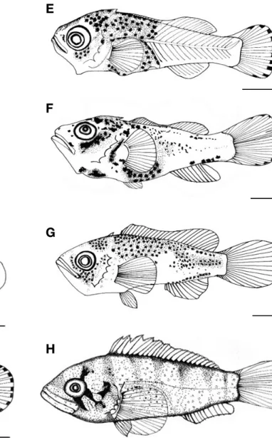
산출 후 31일째 후기자어는 전장 8.09~8.84 mm (평균 8.46±0.31 mm)로 막으로 연결되어 있던 등, 배, 꼬리지느러 미가 완전히 분리되었고, 각 지느러미 줄기 수는 등지느러 미가 14개로 증가하였고, 뒷지느러미는 10개로 증가하였으 며, 가슴지느러미는 16개로 증가하였다. 꼬리기저부의 흑색 소포는 소실되었고, 몸통 가운데 척추골을 따라 흑색소포가 침착되었으며, 두부와 몸통 중앙부에는 옅은 노란색의 색소 포가 침착되었다(Fig. 3E).
산출 후 38일째 후기자어는 전장 10.7~11.8 mm (평균 11.2±0.45 mm)로 등지느러미에 극조가 형성되었고, 극조 수는 11개였다. 뒷지느러미의 극조 수는 2개로 증가하였고, 흑색소포는 소화관 위쪽과 몸통 중앙 부분에 넓게 침착하 Fig. 2.The larvae and juvenile development of Sebastes koreanus.
A: 6.40 mm in total length (TL) just beared larvae; B: 6.50 mm in TL, 5 days after bearing; C: 6.57 mm in TL, 12 days after bearing; D: 7.16 mm in TL, 20 days after bearing. Scale bars==1.0 mm.
A
B
C
D
Fig. 3.The larvae and juvenile development of Sebastes koreanus. E:
8.46 mm in TL, 31 days after bearing; F: 11.2 mm in TL, 38 days after bearing; G: 14.5 mm in TL, 45 days after bearing; H: 17.7 mm in TL, 60 days after bearing. Scale bars=1.0 mm.
E
F
G
H
였다(Fig. 3F).
산출 후 45일째 치어는 전장 12.1~15.5 mm (평균 14.5±
1.56 mm)로 등지느러미의 극조 수는 14개로 형성되었고, 연조는 12개로 증가하였다. 뒷지느러미는 극조 3개, 연조 7 개로 증가하였고, 가슴지느러미는 17개로 증가하였다. 흑색 소포는 꼬리기저부와 상부 및 중앙부에 침착되었고, 전새개 골에는 5개의 극이 형성되었다. 이 시기에는 큰 개체가 작 은 개체를 공격하거나 포식하는 공식현상이 일어났고, 아래 턱의 치골에는 이빨이 형성되어 있었다. 이 시기의 항문 위 치는 몸길이의 46.9%로 정중앙으로부터 앞쪽에 위치하였다 (Fig. 3G).
산출 후 60일째 치어는 전장 15.5~20.0 mm (평균 17.7±
2.25 mm)로 등지느러미의 위치는 두부 끝에서 뒷지느러미 끝까지였고, 가슴지느러미의 길이는 항문까지 위치하였으며, 아가미 뚜껑부에는 3개의 줄무늬가 형성되었다. 체색은 연 한 갈색을 띄었고, 검은색 반점이 넓게 산재해 있었으며, 몸 통에는 4개의 가로띠 줄무늬가 형성되었다(Fig. 3H).
2. 자치어 골격형태발달
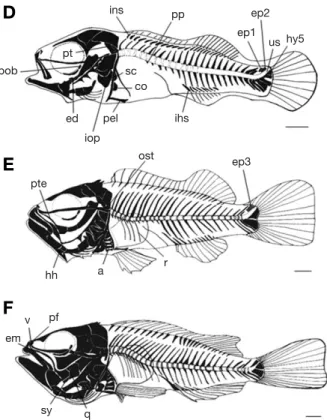
황해볼락의 자치어 골격형태 발달 과정은 다음과 같았다 (Figs. 4, 5). 산출 직후의 전기자어는 전장 6.40 mm로 두개 골을 구성하는 액골(frontal), 상후두골(supraoccipital), 상이 골(epiotic), 기저후두골(basioccipital) 및 선형의 부설골(paras- phenoid)이 골화하였다. 새개부에는 전새개골(preopercle)이 골화하였고, 턱을 지지하는 악골은 섭이와 관계하여 위턱에 는 전상악골(premaxillary) 위쪽에 주상악골(maxillary)과 아 래턱에 치골(dentary)이 골화하였다. 치골 뒤에는 관절골 (articular)이 있고, 설궁부에는 각설골 (ceratohyal), 상설골 (epihyal) 및 3개의 새조골(branchiostegal rays)이 골화하였 다. 견대부에는 쇄골(clavicle)과 후쇄골(postclavicle)이 골 화하였고, 척추골(vertebrae) 중 복추골에는 10~12개의 신 경극(neural spine)과 미추골에는 12~15개의 신경극 및 10
~15개의 혈관극(hemal spine)이 골화하였다(Fig. 4A).
산출 5일째 후기자어는 전장 6.50 mm로 두개골 중 노정 Fig. 4. Development stage of larvae and juvenile of skeleton in
Sebastes koreanus. A: Just beared larvae, 6.40 mm in total length (TL);
B: Postlarvae, 5 days after bearing, 6.50 mm in TL; C: Postlarvae, 12 days after bearing, 6.57 mm in TL. ar, articular; as, alisphenoid; an, angular; bo, basioccipital; br, branchiostegal; ch, ceratohyal; cl, clav- icle; d, dentary; ec, ectopterygoid; eh, epihyal; et, epiotic; f, frontal;
gh, glossohyal; hm, hyomandibular; hs, hemal spine; hy, hypural; ih, interhyal; mx, maxillary; n, nasal; ns, neural spine; nc, notochord; op, opercle; pa, parietal; ph, parhypural; ps, parasphenoid; pcl, postclav- icle; po, preopercle; pmx, premaxillary; sob, suborbital; soc, supraoc- cipital; sop, subopercle (Shown in Table 1, 2). Scale bars==1.0 mm.
A
B
C
Fig. 5. Development stage of larvae and juvenile of skeleton in Sebastes koreanus. D: Postlarvae, 38 days after bearing, 11.2 mm in TL; E: Juvenile, 45 days after bearing, 14.5 mm in TL; F: Juvenile, 60 days after bearing, 17.7 mm in TL. a, actinost; co, coracoid; ed, endopterygoid; ep, epural; em, ethmoid; exo, exoccipital; hh, hypohyal;
ihs, interhemal spine; iop, interopercle; ins, interneural spine; ost, opisthotic; pel, pelvic girdle; pf, prefrontal; pob, preobital; pp, para- pophysis; pte, pterygoid; pt, pterotic; q, quadrate; r, rib; sc, scapula;
sy, symplectic; us, urostyle; v, vomer (Shown in Table 1, 2). Scale bars==1.0 mm.
D
E
F
f soc
et bo ns
pmx mx
d ar ch br
eh pcl as pa
n
ec
gh ih
op hm
ph hy1hy2
hy3hy4
exo
sob sop an
cl hs po nc ps
ins pp
ep1 ep2
hy5
pob
ed iop
pel sc
co
ihs
ost ep3
pte
hh a r
v pf em
sy q
pt
us
골(parietal)과 익설골(alisphenoid)이 골화하였고, 새개부에 는 주새개골 (opercle)이 골화하였다. 구개부에는 설악골 (hyomandibular), 외익상골(ectopterygoid)이 골화하였고, 설 궁부에는 인설골(glossohyal) 및 간설골(interhyal)이 골화하 였다. 미골부에는 4개의 하미축골(hypural bone)과 1개의 준 하미축골(parhypural bone)이 동시에 골화하였다(Fig. 4B).
산출 12일째 후기자어는 전장 6.57 mm로 두개골 중 외후 두골(exoccipital)이 골화하였고, 안하골(suborbital)과 구개 부의 각골(angular)이 골화하였으며, 새개부의 전새개골은 3 개의 극이 형성되었다. 주새개골 아래에는 하새개골(subop- ercle)이 골화하였고, 척추골에는 10개의 복추골과 16개의 미추골이 골화하였다(Fig. 4C).
산출 38일째 후기자어는 전장 11.2 mm로 두개골에는 기 저후두골 위에 익이골 (pterotic)과 비골(nasal) 및 안전골 (preobital)이 골화하였고, 새개부에는 간새개골(interopercle) 이 골화하였다. 구개부에는 내익상골(endopterygoid)이 골화 하였다. 견대부에는 견갑골(scapula) 및 오훼골(coracoid)이 골화하였고, 요대부는 배지느러미를 지지하는 요대골(pelvic girdle bone)이 골화하였다. 척추골 중 4개의 측돌기(para- pophysis)가 형성되었고, 25개의 신경간극(interneural spine) 과 8개의 혈관간극(interhemal spine)이 형성되었다. 미골부
에는 하미축골이 5개로 증가하였고, 미부봉상골(urostyle)이 뚜렷하게 골화하였으며, 동시에 2개의 상미축골(epural bone) 이 골화하였다(Fig. 5D).
산출 45일째 치어는 전장 14.5 mm로 두개골 중 후이골 (opisthotic)이 골화하였고, 구개부에 익상골(pterygoid)이 골 화하였다. 설궁부에는 하설골(hypohyal)이 골화하였고, 새조 골이 6개로 증가하였다. 견대부에는 4개의 사출골(actinost) 이 골화하였고, 척추골에는 4개의 늑골(rib)이 골화하였다.
미골부는 상미축골이 3개로 증가하였고, 5개의 하미축골은 3개가 합쳐졌으며, 나머지 2개도 합쳐지면서 골화하였다 (Fig. 5E).
산출 60일째 치어는 전장 17.7 mm로 두개골 앞쪽에 전액 골(prefrontal), 사골(ethmoid) 및 서골(vomer)이 골화하였다.
구개부에는 방골(quadrate)과 접속골(symplectic)이 골화하 였다(Fig. 5F).
고 찰
황해볼락, 조피볼락 및 볼락은 수정란의 발생이 난소 내에 서 진행되고, 산란시기에 부화자어를 산출하는 난태성 어류 Table 1.The developmental process of cranium, shoulder girdle and caudal skeleton of Sebastes koreanus
Days after bearing 0 5 12 20 25 38 45 50 60
parasphenoid frontal supraoccipital epiotic basioccipital parietal alisphenoid
Cranium nasal
suborbital exoccipital preobital pterotic opisthotic prefrontal ethmoid vomer clavicle postclavicle Shoulder girdle scapula
coracoid pelvic girdle actinost
hypural 1~4th
5th Caudal skeleton parhypural
urostyle
epural 1~2th
3rd
에 속한다. 그러나 다른 양볼락과 어류인 미역치 Hypodytes rubripinnis, 쑤기미 Inimicus japonicus 및 쭈굴감팽 Scorpae-
na miostoma등은 산란을 하는 난생 어류로 같은 과에 속하
지만 생식생태에서 차이를 보인다(Kim and Han, 1993; 김
등, 2005).
황해볼락의 산출시기는 본 연구에서 자연산출을 유도한 결과 3월에 산출하는 것을 확인할 수 있었다. 다른 양볼락 과 어류와 산출시기를 비교해 보면 조피볼락(Kim and Han, Table 2.The developmental process of visceral and vertebrae skeleton of Sebastes koreanus
Days after bearing 0 5 12 20 25 38 45 50 60
Upper jaw premaxillary maxillary dentary Lower jaw articular
angular ceratohyal epihyal Hyoid arch branchiostegal
glossohyal interhyal
Visceral skeleton hypohyal
hyomandibular ectopterygoid Palate endopterygoid
pterygoid symplectic quadrate preopercle Opercular opercle
subopercle interopercle notochord neural spine hemal spine
Vertebrae parapophysis
interneural spine interhemal spine rib
Table 3.Comparison chracters of the eggs and beared larvae characters in the species Scorpaenidae
Species Egg size (mm) Newly beared Time of bearing Larvae myotomes Reference
larvae (mm) (*WT)
Sebastes koreanus 6.41 24 Present study
S. hubbsi 4.40 Shojima, 1958
S. inermis 1.20~1.35 3.15 53 hrs
20~21 Kim and Han, 1993 (12.3�C)
S. thompsoni 1.30 3.07 26~27 Han et al., 1996
S. thompsoni 4.30 Nagasawa and Kobayashi, 1995
S. pachycephalus 1.45 4.99 26~27 Han et al., 1996
S. oblongus 1.62 5.53 676 hrs
26 Byun, 1994
(13.6�C)
S. minor 4.75 Nagasawa, 1993
S. dlli 5.10 Kendall and Lenarz, 1986
S. saxicola 4.70 Laidig and Sakuma, 1996
S. rastrelliger 4.60 Laidig and Sakuma, 1998
S. schlegelii 1.20~1.50 5.52 134 hrs
26~28 Kim and Han, 1991 (16.7�C)
S. schlegelii 6.72 Hoshiai, 1977
*WT: Water temperature
1991)과 쏨뱅이(김 등, 1997)는 3월에 산출하여 황해볼락과 시기가 비슷하였고, 볼락(Kim et al., 1993)은 1월로 산출시 기가 다소 빨라 차이를 보였다.
산출 직후 자어의 전장을 비교한 결과 황해볼락은 평균 6.41±0.02 mm였고, 우럭볼락 Sebastes hubbsi (Shojima, 1958) 전장 4.40 mm, 볼락(Kim and Han, 1993) 전장 3.15 mm, 불볼락(Han et al., 1996) 전장 3.07 mm, 개볼락(Han et al., 1996) 전장 4.99 mm, 황점볼락(Byun, 1994) 전장 5.53 mm, 일본산 불볼락(Nagasawa and Kobayashi, 1995) 전장 4.30 mm, 좀볼락(Nagasawa, 1993) 전장 4.75 mm, 조피볼락(Kim and Han, 1991) 전장 5.52 mm, S. dlli (Kendall and Lenarz, 1986) 전장 5.10 mm, S. saxicola (Laidig and Sakuma, 1996) 전장 4.70 mm, S. rastrelliger (Laidig and Sakuma, 1998) 전장 4.60 mm보다는 길었으나 일본산 조피볼락(Hoshiai, 1977) 전장 6.72 mm보다는 짧았다.
산출 직후 자어의 근절 수를 비교해 보면 황해볼락은 24 개였고, 볼락(Kim and Han, 1993) 20~21개보다는 많았고, 불볼락(Han et al., 1996)은 부화 후 17~20일에 26~27개, 개볼락(Han et al., 1996) 26~27개, 황점볼락(Byun, 1994) 26 개, 조피볼락(Kim and Han, 1991) 26~28개보다는 적었다 (Table 3).
흑색소포의 형성과 형태 및 위치는 자치어 시기의 어류를 동정하는 데 중요한 형질로 이용된다(Kim and Han, 1991;
Kim and Han, 1993). 황해볼락의 산출자어는 소화관 위쪽에 8~11개의 반점 모양 흑색소포가 침착되어 있었고, 우럭볼 락(Shojima, 1958)은 체측의 전면에 침착되어 있으며, 볼락 (Kim and Han, 1993)은 나뭇가지 모양의 흑색소포가 후두부,
두정부 및 기저와 미골 중앙의 등 쪽과 배 쪽 가장자리에 분포하였다. 황점볼락(Fujita, 1958; Byun, 1994)은 눈과 두정 부, 후두부, 체측의 난황과 복추골의 윗부분 및 미부의 중앙 부분에 위치하였고, 조피볼락(Kim and Han, 1991)의 부화자 어는 소화관 위쪽과 두정부, 미부중앙, 등 및 배 쪽에 분포 하였으며, 불볼락(Han et al., 1996)은 두정부와 직장 및 미 부 중앙에 분포하였다. 개볼락(Han et al., 1996)은 두정부, 가슴지느러미 위쪽 및 직장에 분포하여 종마다 차이를 보 였다(Table 5).
황해볼락의 각 부위별 지느러미 줄기 수를 조사한 결과 (Table 4) 등지느러미 극조 14개, 연조 12~13개, 가슴지느러 미 연조 16개, 뒷지느러미 극조 3개, 연조가 7개로 Kim and Lee (1994)의 연구 결과에서 뒷지느러미 연조 수가 5~6개 인 것과 비교하였을 때 차이를 보였고, 기조수가 같은 우럭 볼락과 유사하여 혼란이 지속될 수 있으나 산출자어의 크기 및 흑색소포 위치가 다르다는 점에서 본 연구에 사용된 실 험어는 우럭볼락(Shojima, 1958)보다 황해볼락으로 보는 것 이 타당한 것으로 생각된다.
볼락(Kim and Han, 1993), 황점볼락(Fujita, 1958; Byun, 1994), 조피볼락(Kim and Han, 1991), 불볼락(Han et al., 1996) 및 개볼락(Han et al., 1996)은 미부중앙, 척추 등 쪽과 배 쪽 및 소화관 위쪽의 흑색소포 분포가 동일하다는 점에서 구분 이 어려우나(Sasaki, 1974) 황해볼락의 산출자어는 소화관 위쪽에만 반점모양의 흑색소포가 침착되어 있다는 점에서 다른 종과의 구분이 용이할 것으로 생각된다.
조피볼락과 볼락 및 쏨뱅이는 부화 직후부터 턱을 구성하 는 치골, 전상악골 및 주상악골의 골화가 가장 먼저 동시에 Table 5.Comparison of larvae melanophore distribution in Scorpaenidae fishes (present: ++; absent: -)
Species
Melanophore distribution
Just beared Parietal Rectum Occipital Caudal Ventral Lateral
larvae (mm) abdominal
Sebastes koreanus (Present syudy) 6.41 - ++ - - - -
Sebastes hubbsi (Shojima, 1958) 4.40 - - - - - ++
S. inermis (Kim and Han, 1993) 3.15 ++ ++ ++ ++ - -
S. thompsoni (Han et al., 1996) 3.07 ++ ++ - - - -
S. pachycephalus (Han et al., 1996) 4.99 - ++ ++ ++ ++ -
S. oblongus (Byun, 1994) 5.53 ++ ++ ++ ++ ++ ++
S. schlegelii (Kim and Han, 1991) 5.52 ++ ++ - ++ - -
Table 4.The number of stem with every part of fin which is related to development of Sebastes koreanus
Fins types Days after bearing
0 5 12 20 31 38 45 60
pectoral fin 0 0 0 12 16 16 17 17
dorsal fin 0 0 0 9 XI++12~13 XI++12~13 X IV++12~13 X IV++12~13
anal fin 0 0 0 8 8 II, 7 III, 7 III, 7
caudal fin 0 12 15 16 16 16 16 16
이루어지며, 이것은 먹이섭취와 관련하여 생존율을 높이기 위한 요인으로 보인다.
두부의 골격 중 구개부의 골격은 산출 5일째부터 설악골 및 외익상골 순으로 골화가 진행되며, 산출 60일째에는 방 골과 접속골이 동시에 골화되면서 구개부의 골격이 완성된 다. 조피볼락(Kim and Han, 1991), 쏨뱅이(김 등, 1997), 황점 볼락(변 등, 2012) 및 점농어 Lateolabrax maculates (강 등, 2012)의 새개부에서는 전새개골과 주새개골이 동시에 골화 하지만 황해볼락은 산출 직후 전새개골이 골화한 뒤 산출 5 일째에 주새개골이 골화한다는 점에서 다른 경향을 보였다.
척추의 골화는 전기자어 시기에 이루어지는데 황해볼락 은 볼락(Kim and Han, 1993), 조피볼락(Kim and Han, 1991) 및 쏨뱅이(김 등, 1997) 등과 마찬가지로 복추골에서 미추 골 쪽으로 골화가 진행되며, 복추골의 골화가 완료되기 전 에 미부봉상골이 골화를 시작한다는 점에서 볼락(Kim and Han, 1993)의 척추골화와 유사하였다.
반면 미부의 추체가 골화된 후에 미부봉상골이 골화하는 조피볼락(Kim and Han, 1991)과는 다른 경향을 보였다. 또 한 황해볼락은 척추골이 골화하기 전에 신경간극과 혈관간 극이 먼저 골화하였다. 이와 같은 경향은 꽁치 Cololabis saira (Fujita and Oozeki, 1994)와 복섬 Takifugu niphobles (Fujita, 1992)에서 나타났고, 황점볼락(변 등, 2012)은 황해 볼락의 추체 발달과 유사하였으나, 복추골 앞쪽의 신경극 골화가 먼저 시작된 뒤 혈관극과 모든 추체가 동시에 골화 되어 차이를 보였다. 황해볼락은 대부분의 어류들과 같이 추체의 골화가 진행된 뒤 신경간극 및 혈관간극이 형성되 는 것과 달리 신경간극과 혈관간극이 추체보다 먼저 골화 하기 때문에 이 같은 발달양상을 밝히기 위해서는 향후 해 산어류의 초기 골격발달에 대한 지속적인 연구를 통하여 유 연관계를 밝힐 필요가 있을 것으로 생각된다. 자치어의 골 격발달 연구는 종의 동정뿐만 아니라 성어의 골격특성에도 중요한 자료가 될 수 있으므로 구체적이고 체계적인 연구 가 필요하다. 황해볼락은 다른 양볼락과 어류의 기조 수에 서 차이를 보여 쉽게 구분이 되고, 본 연구를 통해 황해볼 락의 뒷지느러미 연조 수가 기존의 5~6개에서 7개까지 형 성되는 것이 관찰되면서 유사종과의 구분이 보다 명확하게 되어질 것으로 판단된다. 양볼락과 어류는 서식장소에 따라 형태적 차이가 있을 수 있어 초기형태변화에 대한 다른 종 과의 분류를 위해서는 초기생활사에 대한 연구가 지속적으 로 이루어져야 할 것으로 생각된다.
요 약
본 연구는 황해볼락 자치어의 형태 및 골격발달을 관찰 하여 분류학적 연구의 기초자료로 이용하고자 실시하였다.
실험에 이용된 어미는 2014년 3월 전라남도 여수시 주변 해역에서 포획한 것을 이용하였다. 산출된 자어의 사육수온 은 13.5~15.5�C (평균 14.5±0.1�C)였다. 산출 직후의 후기 자어는 전장 6.38~6.43 mm (평균 6.40±0.02 mm, n==5)로 입과 항문이 열려 있었고 먹이를 섭취하기 시작하였다. 산 출 후 5일째 후기자어는 전장 6.45~6.49 mm (평균 6.47±
0.02 mm)였다. 산출 후 15일째 후기자어는 6.55~6.72 mm (평균 6.64±0.08 mm)였다. 산출 후 60일째 치어는 전장 15.5~20.0 mm (평균 17.7±2.25 mm)로 이 시기에는 등지느 러미 극조 14개, 연조 12개였고, 뒷지느러미 극조 3개, 연조 7개였으며, 꼬리지느러미 줄기 수는 16개였다.
인 용 문 헌
강충배ㆍ명정구ㆍ김용억ㆍ김형철. 2012. 한국산 점농어(Lateo- labrax maculates) 자치어의 골격발달과 비늘형성. 한국수 산과학회지, 45: 271-282.
김백균ㆍ이종화. 1991. 조피볼락(Sebastes schlegeli)의 종묘생산에 관한 연구. 순천향대학교 논문집, 48: 847-857.
김용억ㆍ한경호ㆍ강충배ㆍ김진구ㆍ변순규. 1997. 쏨뱅이 Seba- stiscus marmoratus초기생활사에 관한 연구 2. 산출 자치 어의 외부형태 및 골격 발달. 한국어류학회지, 9: 186-194.
김익수ㆍ최 윤ㆍ이충렬ㆍ이용주ㆍ김병직ㆍ김지현. 2005. 한국어 류대도감. (주)교학사, pp. 210-226.
변순규ㆍ강충배ㆍ명정구ㆍ차반석ㆍ한경호ㆍ정춘구. 2012. 황점볼 락(Sebastes oblongus) 자치어의 골격발달. 한국어류학회 지, 24: 67-76.
이택열ㆍ김성연. 1992. 난태생 경골어류 볼락, Sebastes inermis의 생식과 체내자어발달. 한국수산과학회지, 25: 413-431.
Byun, S.G. 1994. Egg development and morphology of larvae and juveniles of Sebastes oblongus. M. Sc. Nat. Fish. Busan Univ., pp. 1-20. (in Korean)
Fujita, S. 1958. On the egg development and larval stages of a vivi- parous Scorpanidae fish Sebastes oblongus. Bull. Japan Soc.
Sci. Fish, 24: 475-479.
Fujita, K. 1992. Development of the caudal skeleton in the Tetrao- dontid Fish, Takifugu niphobles. Japan J. Ichthyol., 38: 438- 440.
Fujita, K. and Y. Oozeki. 1994. Development of the caudal skeleton in the saury, Cololabis saira. Japan J. Ichthyol., 41: 334- 337.
Han, K.H., Y.U. Kim and C.M. Kim. 1996. Description of egg and larvae of two species of rockfishes (Scorpaenidae: Sebastes) in Korean waters. Kor. J. Ichthyol., 8: 1-9. (in Korean) Hoshiai, G. 1977. Larvae and juvenile of the Scorpaenid fish Sebas-
tes schlegeli. Japan J. Ichthyol., 24: 35-42.
Kendall, A.W. Jr., E.H. Ahlstrom and H.G. Moser. 1984. Early life history stages of fishes and their characters. In: Moser, H.G.
et al. (eds.). Ontogeny and Systematics of Fishes. Am. Soc.
Ichthyol. Herpetol., Spec. Publ., 1: 11-22, Allen Press, Law- rence, KS.
Kendall, A.W. and W.H. Lenarz. 1986. Status of early life history studies of northeast pacific rockfishes. Proc. Int. Rockfish Symp. Alaska Sea Grant Rpt., 87: 99-128.
Kim, I.S. and W.O. Lee. 1994. A new species of the genus Sebastes (Pisces; Scorpaenidae) from the yellow sea. Kor. J. Zool., 37: 409-415. (in Korean)
Kim, Y.U. and K.H. Han. 1991. The early life history of rockfish Sebastes schlegeli. Kor. J. Ichthyol., 3: 67-83. (in Korean) Kim, Y.U. and K.H. Han. 1993. The early life history of the rockfish
Sebastes inermis 1. Egg development and morphology of larvae by artificial treatment in aquarium. Bull. Kor. Fish Soc., 26: 458-464. (in Korean)
Kim, Y.U., K.H. Han and S.K. Byun. 1993. The early life history of the rockfish Sebastes inermis 2. Morphological and skeletal development of larvae and juveniles. Bull. Kor. Fish Soc., 26: 465-476. (in Korean)
Laidig, T.E. and K.M. Sakuma. 1996. Description of pelagic larval and juvenile stripetail rockfish Sebastes saxicola (family Scorpaenidae) with an examination of larva growth. Fish Bull., 94: 289-299.
Laidig, T.E. and K.M. Sakuma. 1998. Description of pelagic larval and juvenile grass rockfish Sebastes rastrelliger (family Scorpaenidae) with an examination of age and growth. Fish Bull., 96: 788-796.
Moser, H.G. 1974. Development and distribution of larvae and juve- niles of Sebastolobus (Pisces; Family Scorpaenidae). Fish Bull., 72: 865-884.
Nagasawa, T. 1993. Planktonic larvae and pelagic juveniles of the rockfish Sebastes minor (Scorpaenidae). Japan J. Ichthyol., 40: 87-97.
Nagasawa, T. and T. Kobayashi. 1995. The early life history of the rockfish Sebastes thompsoni (Scorpaenidae) in the sea of Japan. Japan J. Ichthyol., 41: 385-396.
NFRDI (National Fisherise Research and Development Institute).
2004. Commercial fishes of the coastal offshore waters in Korea, 2: 98-102.
NFRDI (National Fisherise Research and Development Institute).
2007. Standard manual of black rockfish culture, pp. 2-20.
Omori, M., Y. Sugawara and H. Honda. 1996. Morphogenesis in hatchery reared larvae of the black rockfish Sebastes schle- geli and its relationship to the development of spawning and feeding functions. Japan J. Ichthyol., 43: 267-282.
Richardson, S.L. and W.A. Laroche. 1979. Development and occur- rence of larvae and juveniles of the rockfishes Sebastes crameri, Sebastes pinniger, and Sebastes helvomacuratus (Family Scorpaenidae) off oregon. Fish Bull., 77: 1-46.
Sasaki, T. 1974. On the larvae of three species of rockfish (Genus:
Sebastes) in hokkaido. Bull. Fac. Fish Hokkaido Univ., 25:
169-173.
Shojima, Y. 1958. Studies on the laval stage of Japanese fish Vol. 1.
The Faculty of Agriculture of Kiushu Univ. Fish. Sci. class No. 2, p. 86.
Walker, M.B. and C.B. Kimmel. 2007. A two-color acid-free carti- lage and bone stain for zebrafish larvae. Biotechnic and Histochemistry, 82: 23-28.