© Copyright
Keimyung University School of Medicine 2015 계명의대학술지 제34권 2호 161
Keimyung Med J
Vol. 34, No. 2, December, 2015
Department of Thoracic and Cardiovascular Surgery, Kyungpook National University School of Medicine, Kyungpook National University Hospital, Daegu, Korea
Schwannomas of the brachial plexus are rare. Although the tumor size is small, neurological symptoms occur due to the mass effect of the tumor within the brachial plexus. In our case, the patient did not recognize the symptoms until the tumor diameter increased to 10 cm because the mass was located at the inferior border of the brachial plexus and had grown up to the chest wall. The mass was identified at the medial cord of the right brachial plexus arising from the C8 and T1 origin. A complete excision was performed. The patient recovered without any neurologic symptoms.
Key Words: Brachial plexus, Schwannoma, Surgery
Introduction
Schwannomas may originate from any cranial nerve and peripheral nerve system. This tumor is one of the most common neurogenic tumors of the thorax, but schwannomas arising from the brachial plexus are rare [1-3]. According to a few of literatures, most schwannomas in the brachial plexus are generally found without symptoms and the sizes of the tumors are small [4-7]. In this case, we met a patient with a large sized schwannoma in the brachial plexus and the mass was successfully removed without long term neurologic sequelae.
Case Report
A 48-year-old woman visited the hospital complaining of occasional tingling sensations and paresthesia of the right arm for 4 years. The symptoms had become more aggravated in the two months preceding the visit, which prompted her to seek treatment. Upon
Received: September 10, 2015 Revised: October 2, 2015 Accepted: October 7, 2015
Corresponding Author: Deok Heon Lee, M.D., Department of Thoracic and Cardiovascular Surgery, Kyungpook National University School of Medicine, 130 Dongdeok-ro, Jung-gu, Daegu 41944, Korea Tel: +82-53-200-6589
E-mail: [email protected]
• The authors report no conflict of interest in this work.
Jong Uk Lee, M.D., Deok Heon Lee, M.D.
A Surgical Resection of Giant Schwannoma in the Brachial Plexus
162
계명의대학술지 제34권 2호 2015
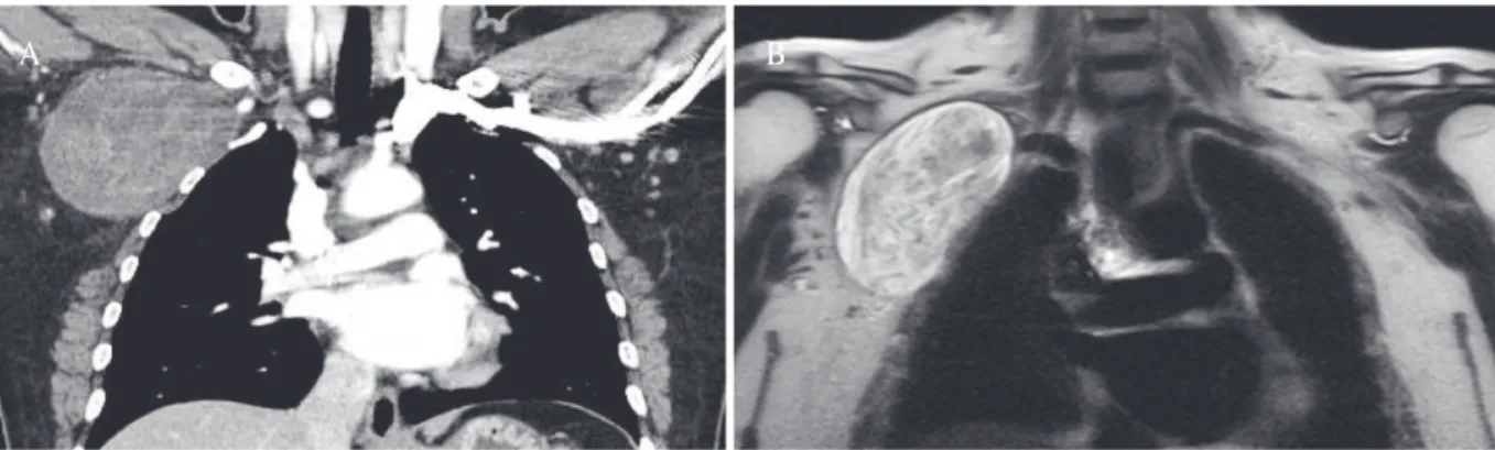
Fig. 1. (A) Chest computed tomography reveals a homogeneous mass, measuring 10 × 6 × 7 cm, located around the right brachial plexus area. (B) Contrast enhanced T2-weighted magnetic resonance image shows a well- capsulated heterogeneous mass just beneath the right brachial plexus.
A B
physical examination, a solid mass was palpable in the axillary and on the supraclavicular areas. When the mass was pressed, noticeable pain and tingling sensations appeared.
A chest computed tomography scan revealed a well-defined and oval-shaped homogeneous mass measuring 10 cm × 6 cm × 7 cm located between the coracobrachialis muscle and subscapularis muscle on the infraclavicular area. A magnetic resonance imaging further showed a well-capsulated heterogeneous mass on the brachial plexus area in the T2-weighted image signal (Fig. 1). A percutaneous needle biopsy of the mass was conducted to assess whether the tumor was a malignant peripheral nerve sheath tumor and the mass was diagnosed as a schwannoma.
Due to the large mass size, the surgery was conducted with the assistance of surgical loupe magnification and headlight illumination rather than with a microscope. The infraclavicular skin incision was made in the deltopectoral groove. During the exposure, the pectoralis major was partially divided and the mass was revealed in the pectoralis minor space. A careful dissection around the mass was performed and the mass was identified on the inferior border at the medial cord of the right brachial plexus arising from the C8 and T1 origin. Using bipolar
electrocautery and scissors, the epineurium was longitudinally incised about 7 cm in length at the prominent portion of the tumor. The yellowish, greasy, and hypervascularized tumor was exposed in the epineurium of the medial cord. It was then completely extirpated without the use of electrocautery in order to minimize damage to nerve fascicles. And then the incised epineurium was sparsely closed with absorbable suture materials.
Gross inspection characterized the mass as firm, round, translucent, and well capsulated.
Histopathologic examination revealed a parallel arrangement of elongated tumor cells forming palisades (Antoni A type) and loose cellular parts (Antoni B type) (Fig. 2B). As a result, the tumor was pathologically diagnosed as a benign schwannoma.
The tingling sensation and pain of the right arm substantially improved within the first month after surgery. Minor difficult in performing pinching motion using the thumb- and index fingers persisted.
At 18 months post-surgery, fine movement of the right hand had almost fully recovered.
Discussion
Schwannomas is the most frequent and largest
163
A Surgical Resection of Giant Schwannoma in the Brachial Plexus
tumors in the category of benign nerve tumors.
They originate from cells that have a basement membrane resembling that of a Schwann cell [1].
They can be found anywhere in the body which is innervated by a cranial nerve, sympathetic nerve, or peripheral nerve. Tumors of the brachial plexus are relatively rare, occurring in 0.3-0.4/100000 person per year [2]. Schwannomas usually present as slow growing masses which can get locally destructive if allowed to grow. Benign schwannomas rarely show malignant transformation [3].
According to published reports, schwannomas on the brachial plexus are usually smaller than 5 cm in size and are generally painless. However, they may present with radiculopathy or paresthesia secondary to adjacent nerve compression [4]. Cho et al. [5] reported 3 cm × 2 cm × 1 cm schwannoma of the right brachial plexus in the inferior trunk, which evoked dysesthetic pain in the supraclavicular area and right forearm of the C6 and C7 dermatomes.
Patel et al. [6] reported a case of a schwannoma of 4-5 cm in size arising from the left brachial plexus and presenting as monoparesis of the left upper limb. Ahn et al. [7] described a case of a schwannoma in the brachial plexus with a size of only 2 cm × 1 cm where the patient complained of
radiating pain in the third and forth fingers of the right hand. When the tumor is located between the cords of the brachial plexus, clinical symptoms appear early even the tumor size is small. In our case, however, the patient did not recognize the symptoms until the tumor increased up to 10 cm in diameter because the mass was located on inferior border of the brachial plexus and was growing up to the chest wall. The mass was compressed on the connective tissue of the chest wall rather than on brachial plexus nerve. As a result, the mass effect was recognized late.
Benign nerve sheath tumors in the brachial plexus need to be distinguished between schwannomas and neurofibromas because the operative procedures are different. Unlike schwannomas which tend to displace the surrounding neural structures, neurofibromas are intimately involved with the nerve fascicles. The major distinction between a schwannoma and a neurofibroma is that in case of the former, the tumor can be resected while sparing the underlying nerve. However, resection of a neurofibroma requires sacrifice of the underlying nerve. The principles for surgery of schwannomas which are generally known include: (1) longitudinal incision of the epineurium Fig. 2. (A) Intraoperative photograph shows complete extirpation of the tumor mass. The mass appeared as firm, round, translucent and well capsulated. (B) Histological examination shows a cellular Antoni A area (arrow) and a loose paucicellular Antoni B area (arrow head). H & E stain; × 100.
A B
164
계명의대학술지 제34권 2호 2015
over the tumor, (2) avoiding damage of nerve fascicles, (3) removal of the tumor itself as much as possible, (4) minimal use of electrocautery, (5) and recommended intraoperative use of magnification and electrophysiological monitoring [8].
In the case of our patient, the tumor located at the inferior border of the medial cord and not between the cords of the brachial plexus. Therefore, it was not necessary to expose the entire brachial plexus including the lateral and posterior cord. However, incision length of the epineurium over the large mass was so long for extirpating the tumor completely. We tried to minimize using an electrocautery and manipulation of surrounding cords. Although impairment of fine movement and tingling sensations in the right index and middle finger tips developed postoperatively, these symptoms almost fully recovered in due course of time.
References
1. Winn HR, Youmans JR. Youmans neurological surgery.
Philadelphia: Saunders; 2004.
2. Greager JA, Reichard KW, Campana JP, DasGupta TK.
Malignant schwannoma of the head and neck. Am J Surg 1992;163:440-2.
3. Woodruff JM, Selig AM, Crowley K, Allen PW. Schwannoma (neurilemoma) with malignant transformation. A rare, distinctive peripheral nerve tumor. Am J Surg Pathol 1994;18:882-95.
4. Huang JH, Samadani U, Zager EL. Brachial plexus region tumors: a review of their history, classification, surgical management, and outcomes. Neurosurg Q 2003;13:151-61.
5. Cho DG, Son BC, Cho KD, Jo MS, Wang YP. Microsurgical resection of schwannoma of the brachial plexus: a case report. Korean J Thorac Cardiovasc Surg 2005;38:249- 52.
6. Patel ML, Sachan R, Seth G, Radheshyam.
Schwannoma of the brachial plexus: a rare cause of monoparesis. BMJ Case Rep 2013;2013. pii:
bcr201208016.
7. Ahn KM, Lee HK, Lee KD, Yu TH. A case of neurilem- moma of the brachial plexus. Korean J Otolaryngol- Head Neck Surg 2002;45:733-5.
8. Das S, Ganju A, Tiel RL, Kline DG. Tumors of the brachial plexus. Neurosurg Focus 2007;22:E26.