152
Copyright © 2021 The Korean Society of Fisheries and Aquatic Science pISSN:0374-8111, eISSN:2287-8815
서 론
염증반응은미생물감염
,
내독소,
조직손상과같은위해성자 극에대한방어기능으로,
이는생명체의구조와기능을정상적 으로유지시키기위한방어기능이다.
염증반응은시간이지남 에따라염증촉진성매개체들의생성은감소되고,
항염증성매 개체들이증가됨으로써스스로염증반응이억제되는조절기전 을가지고있다(Libby and Hansson, 2015).
대식세포(macro-
phages)
는체내의염증반응에중요한역할을하는것으로알려져있다
.
대식세포는interferon-γ (IFN-γ), interleukin (IL)-1β, IL-6, tumor necrosis factor-α (TNF-α)
와같은염증촉진성cy-
tokines,
그리고세균세포막성분인lipopolysaccharides (LPS)
등의자극에의해활성화된다(Xie et al., 1994).
활성화된대 식세포는염증촉진성cytokines
이외에inducible nitric oxide synthase (iNOS)
및cyclooxygenase-2 (COX-2)
와같은효소 의발현을통해nitric oxide (NO)
및prostaglandin E
2(PGE
2)
와같은다양한염증매개분자들을생성하게되고(Marks-Kon- czalik et al., 1998; Zhang and Ghosh, 2000),
이들매개체들의 지속적인생성은다양한만성염증성질환의주요한원인으로 알려져있다.
부작용이적은항염증활성을지닌천연물의발견 은염증치료제개발을위한방편으로관심을끌고있다.
대식세포의 염증촉진성
cytokines
및 단백질들의 발현은LPS로 유도된 RAW 264.7 대식세포에 대한 대황(Eisenia bicyclis) 헥산 분획물의 항염증 효과
김보운·최창근
1·김재일·김형락*
부경대학교 식품영양학과, 1부경대학교 생태공학과
Anti-Inflammatory Effect of Hexane Fraction from Eisenia bicyclis on Lipopolysaccharides-Treated RAW 264.7 Cells
Bowoon Kim, Chang-Geun Choi1, Jae-Il Kim and Hyeung-Rak Kim*
Department of Food Science and Nutrition, Pukyong National University, Busan 48513, Korea
1Department of Ecological Engineering, Pukyong National University, Busan 48513, Korea
Eisenia bicyclis is known to have secondary metabolites exhibiting various biological activities. In a preliminary study, the n-hexane fraction obtained from the ethanolic extract of E. bicyclis showed higher anti-inflammatory activ- ity than the ethyl acetate and butyl alcohol fractions based on the inhibition of lipopolysaccharide (LPS)-stimulated nitric oxide (NO) production in RAW 264.7 cells. Using this fraction ( E. bicyclis hexane fraction, EHF), we inves- tigated the molecular mechanisms underlying its anti-inflammatory effect in LPS-stimulated RAW 264.7 cells. Pre- treatment of the cells with up to 50 µg/mL EHF significantly inhibited NO and prostaglandin E
2production as well as their responsible enzyme proteins and mRNAs, in a dose-dependent manner (P<0.05). Similarly, EHF markedly reduced the production of pro-inflammatory cytokines, such as interleukin (IL)-1β, IL-6 and tumor necrosis factor (TNF)-α as well as their mRNA levels. Nuclear translocation of nuclear factor-kappa B (NF-κB) was strongly sup- pressed by EHF treatment. EHF significantly reduced the phosphorylation of mitogen-activated protein kinases and phosphatidylinositol 3-kinase/Akt in LPS-stimulated cells. Moreover, EHF reduced ear edema in phorbol myristate acetate (PMA)-induced mice. These results indicate that EHF contains potent anti-inflammatory compounds, which may be used as a dietary supplement for the prevention of inflammatory diseases.
Keywords: Anti-inflammatory effect, Eisenia bicyclis , iNOS, NF-κB, Pro-inflammatory cytokines
*Corresponding author: Tel: +82. 51. 629. 5847 Fax: +82. 51. 629. 5842 E-mail address: [email protected]
This is an Open Access article distributed under the terms of the Creative Commons Attribution Non-Commercial Licens (http://creativecommons.org/licenses/by-nc/3.0/) which permits unrestricted non-commercial use, distribution, and reproduction in any medium, provided the original work is properly cited.
Received 29 January 2021; Revised 15 February 2021; Accepted 19 February 2021 저자 직위: 김보운(대학원생), 최창근(교수), 김재일(교수), 김형락(교수) https://doi.org/10.5657/KFAS.2021.0152
Korean J Fish Aquat Sci 54(2), 152-161, April 2021
nuclear factor kappa-B (NF-κB)
에의해전사수준에서조절된 다(Marks-Konczalik et al., 1998; Rahman et al., 2006).
정상 상태의세포에서NF-κB
는inhibitor of kappa B (IκB)
와결합 한상태로불활성형태로세포질에존재한다(D'Acquisto et al., 1997; Rahman et al., 2006). LPS, cytokines
과같은자극이주 어지는경우IκB
는IκB kinase (Ikk)
에의해인산화와유비퀴 톤화(ubiquitination)
를통해proteasome
에의해분해되고,
유리된
NF-κB
는핵으로이동하여염증관련유전자의발현을유도하게된다
(Elliott et al., 2003).
또한extracellular signal-reg- ulated kinase (ERK), c-Jun N-terminal kinase (JNK), p38 ki- nase
를포함하는mitogen-activated protein kinases (MAPKs)
와PI3K/Akt (PKB)
와같은단백질인산화효소에의해NF-κB
가활성화되어염증반응을촉진하게된다(Marks-Konczalik et al., 1998; Kaminska, 2005).
갈조류는 다양한 종류의 생리활성물질을 함유하고 있으므 로 이들을이용하기 위하여광범위한 연구가 지속되고 있다
(Thomas and Kim, 2011).
대황(Eisenia bicyclis)
은다시마과 에속하는갈조류로써우리나라와일본의해안에다량존재하 는것으로알려져있다.
대황의주요한2
차대사산물들은플로 로탄닌(phlorotannin)
으로항염증(Jung et al., 2013; Paudel et al., 2014; Yayeh et al., 2014)
항산화(Kim et al., 2011; Kwon et al., 2013),
간보호(Choi et al., 2015a),
항균(Eom et al., 2014;
Kim et al., 2018),
항치매(Choi et al., 2015b),
항앨러지(Sugi- ura et al., 2009),
항암활성(Thomas and Kim, 2011)
등이보 고되고있다.
대황주정추출물을이용한예비실험에서헥산
(hexane),
에틸 아세테이트(ethyl acetate)
및부탄올(butanol)
획분으로분리하 여항염증활성을분석한결과헥산획분의항염증활성이가장 높은것으로확인되었다.
본연구에서는대황의헥산분획물의 항염증효과및관련분자적기전을LPS (lipopolysaccharide)
로자극한RAW 264.7
세포를이용하여분석함으로써,
염증성 질환의발병을예방또는지연시킬수있는건강기능성소재로 서의이용가능성을검토하였다.
재료 및 방법
추출과 분획
독도 인근에서 채취한 대황
[Eisenia bicyclis (Kjellman) Setchell]
의건조분말1 kg
을환류냉각기가부착된집기병에담 고주정[95% ethanol (EtOH), v/v] 6 L
를넣어가열, 2
회추 출하여(72°C, 3
시간)
얻은추출액을여과하여rotary vacuum evaporator
로농축하였다.
에탄올농축물(122 g)
을H
2O:EtOH (9:1, v/v)
의혼합용매로녹인후동량의n-hexane
을넣어분액 깔대기에 평형화시켜상층액의n-hexane
가용부를 분리하였 다.
분리된n-hexane
획분(E. bicyclis hexane fraction, EHF)
을 농축하여실험에사용하였다.
세포 배양 및 처리
RAW 264.7
세포(ATCC, Rockville, MD, USA)
는10% fe- tal bovine serum (FBS) (GIBCO, Grand Island, NY, USA)
와penicillin (100 units/mL), streptomycin sulfate (100 µg/mL)
을 첨가한Dulbecco’s modified Eagle’s medium (DMEM) (Life Technology, Carlsbad, CA, USA)
을 사용하였고, 5% CO
2, 37°C
배양기에서배양하였다. Cell culture plate
에RAW 264.7 cell
이70-80%
정도채워지면phosphate-buffered saline (PBS)
로 한번씻어낸후,
계대배양하였다. EHF
는100% dimethyl sulfoxide (DMSO) (Sigma-Aldrich, St. Louis, MO, USA)
에 녹여사용하였고,
세포처리전에배지에희석하여처리하였다. 세포 독성 시험
RAW 264.7 cell
을96-well plate
에5×10
5cells/well
로분주 하고37C
에서24
시간동안배양한후, EHF
가0, 10, 25, 50 µg/
mL
농도로희석된DMEM (Life Technology, Carlsbad, CA, USA)
배지로교체하여1
시간처리한후LPS (Sigma-Aldrich, St. Louis, MO, USA) (1 µg/mL)
를함유한DMEM
배지에다 시24
시간배양하였다.
이후CellTiter96
ⓇAqueous One Solu- tion Cell Proliferation Assay [3-(4,5-dimethylthiazol-2-yl)- 5-(3-carboxymethoxyphenyl)-2-(4-sulfophenyl)-2H-tetrazo- lium; MTS]
키트(Promega, Madison, WI, USA)
를사용하여 제조사의방법에따라세포생존율을분석하였다. MTS
용액은FBS-free DMEM (Life Technology)
에5% (v/v)
의농도로섞 어100 µL
씩처리하였다. 1
시간후에microplate reader (Glo- max Multi Detection System, Promega, Madison, WI, USA)
를이용하여
490 nm
의파장에서흡광도를측정하여세포독성을분석하였다
.
NO 및 염증성 cytokines 생성 억제 효과
EHF
의항염증효과비교를위해RAW 264.7
세포에서의NO
생성에대한억제효과를분석하였다. EHF
를1
시간동안전처 리한다음LPS (1 µg/mL)
로24
시간동안자극하고,
원심분리(2,000 g, 4°C, 10
분)
하여배지를회수하였다. NO
의농도는배 지(100 µL)
와Griess
시약(0.1% naphthylethylene diamine di- hydrochloride+1% sulfanilamide+5% phosphoric acid)
을동 일한비율로반응시켜microplate reader
로540 nm
의파장에 서흡광도를측정하였다.
배지중의IL-1β, IL-6, TNF-α
의양 은enzyme-linked immunosorbent assay (ELISA) (R&D Sys- tems, Minneapolis, MN, USA) kit
를이용하여제조사의방법 에따라측정하였다.
Reverse transcription-polymerase chain reaction (RT-PCR)
RAW 264.7
세포(1×10
6cells/well)
에EHF
를0, 10, 25, 50
µg/mL
의농도로1
시간동안처리한후, LPS 1 µg/mL
의농도로
6
시간동안자극시켰다.
이후Quiazol
시약(Quiagen Sci- ence, Valencia, CA, USA)
을 이용하여total RNA
를 분리하 였다(Kim et al., 2009). Total RNA
로부터RT-PCR
분석에의 한mRNA
발현양의분석은이전의보고(Joung et al., 2012)
에 서사용한방법을이용하였고,
유전자발현양의상대적인비교 를위해서β-actin
또는glyceraldehyde-3-phosphate dehydro- genase (GAPDH)
를함께분석하였다. PCR
반응에이용된각 각의primer
는Table 1
에나타내었다.
전기영동상band
의정 량분석은cooled CCD camera system EZ-Capture II (ATTO
& Rise Co., Tokyo, Japan)
와CS analyzer ver. 3.00 software (ATTO and Rise Co., Tokyo, Japan)
를이용하여최소3
번의반 복실험을통해얻었다.
세포질 및 핵 단백질 추출물의 제조
NF-κB
의활성화정도를분석하기위해EHF
및LPS
를각각처리한
RAW 264.7
세포로부터세포질단백질과 핵단백질을각각분리제조하였다
(Kim et al., 2009).
즉, EHF
로처 리된 세포(2×10
6cells/dish)
를PBS
로 세척하여 회수하고, 180 µL
의hypotonic buffer [10 mM Tris-HCl, 10 mM NaCl, 3 mM MgCl
2, 0.02% NaN
3, 0.5 mM dithiothreitol (DTT), 1 mM phenylmethanesulfonyl fluoride (PMSF), pH 7.4]
를넣 고, 20 µL
의5% nonidet NP-40
을첨가하여5
분동안방치하 였다.
이후원심분리(1,800 g, 4°C, 5
분)
한후상층액을세포질 추출물로이용하였다.
침전물은hypotonic buffer
로한번세척 하고, hypertonic buffer [20 mM 4-(2-hydroxyethyl)-1-piper- azineethanesulfonic acid, 25% glycerol, 420 mM NaCl, 1.5 mM MgCl
2, 0.2 mM ethylenediaminetetraacetic acid, 0.02%
NaN
3, 0.5 mM DTT, 1 mM PMSF, pH 7.4]
를넣고1
시간동안 얼음위에방치시킨다음원심분리(13,000 g, 4°C, 10
분)
하여상 층액을회수하여핵단백질추출물로이용하였다.
Western blot에 의한 단백질 분석
염증관련 단백질과신호전달 단백질의인산화정도는세포 를
EHF
로전처리한후LPS
로처리하여세포단백질을시료 로, NF-κB
및IκB
의활성화및인산화정도는상기의핵및 세포질추출물을 시료로이용하였고,
단백질양은이전의보 고(Joung et al., 2017)
와마찬가지로전기영동으로분리된단 백질을nitrocellulose
막에이전시켜Western blot
으로분석하 였다.
검출된band
의정량분석은mRNA
분석과마찬가지로cooled CCD camera system EZ-Capture II (ATTO and Rise Co., Seoul, Korea)
와CS analyzer ver. 3.00 software (ATTO and Rise Co., Seoul, Korea)
를이용하여최소3
번의반복실험 을통해얻었고,
그결과를각blot
의하단에수치로표기하였다. 면역형광분석
RAW 264.7
세포를glass coverslips (SPL Lifesciences Co., Pocheon, Korea)
위에24
시간배양한뒤, EHF
로1
시간전처리 하고, LPS (1 µg/mL)
로30
분자극시켰다.
세포를4.0% para- formaldehyde
가 첨가된PBS
로 실온에서15
분 동안반응시 켜고정시키고, 0.5% Triton X-100
이첨가된PBS
를넣어10
분동안반응시켰다. PBS
로세척한뒤에3% BSA/PBS
를넣 고30
분 동안blocking
시킨후, anti-NF-κB polyclonal anti- body (Cell Signaling Technology, Danvers, MA, USA)
가희 석된3% BSA/PBS
를넣어2
시간동안반응시켰다.
그다음,
Table 1. Primer sequences for RT-PCR Primers Sequences
iNOS Forward 5'-TCTTTGACGCTCGGAACTGT-3‘
Reverse 5'-CCATGATGGTCACATTCTGC-3‘
COX-2 Forward 5'-TGGGCAAAGAATGCAAACAT-3‘
Reverse 5'-CAGCAAATCCTTGCTGTTCC-3’
TNF-α Forward 5'-CAAGGGACAAGGCTGCCCCG-3'
Reverse 5'-GGTCAGAGTGGGGGCTGGGT-3‘
IL-1β Forward 5'-ATGGCAACTGTTCCTGAACTCAACT-3'
Reverse 5'-CAGGACAGGTATAGATTCTTTCCTTT-3'
IL-6 Forward 5'-GTATGAACAACGATGATGCACTTCCAG-3'
Reverse 5'-GCATTGGAAATTGGGTAGGAAGG-3'
β-Actin Forward 5'-CCTCATGAAGATCCTGACCG-3'
Reverse 5'-TCCACATCTGCTGGAAGGTG-3’
GAPDH Forward 5'-TGGCACAGTCAAGGCTGAGA-3'
Reverse 5'-CTTCTGAGTGGCAGTGATGG-3’
RT-PCR, reverse transcription-polymerase chain reaction; iNOS, inducible nitric oxide synthase; COX-2, cyclooxygenase-2; TNF-α, tumor necrosis factor- α; IL-1β, interleukin-1β; IL-6, interleukin-6; GAPDH, glyceraldehyde-3-phosphate dehydrogenase.
Alexa Fluor
Ⓡ488-conjugated secondary antibody (Invitrogen, Carlsbad, CA, USA)
가희석된3% BSA/PBS
를넣고1
시간 동안반응시킨뒤, 2 µg/mL
의4,6-diamidino-2-phenylindole (DAPI) (Sigma-Aldrich, St. Louis, MO, USA)
로핵을염색하 고LSM700 laser scanning confocal microscope (Carl Zeiss, Oberkochen, Germany)
로관찰하였다.
NF-κB Promoter 활성
RAW 264.7
세포(2×10
5cells/well)
가 들어있는24-well plate
의각well
에1 µg
의pNF-κB firefly luciferase DNA
와20 ng
의pRL-TK renilla luciferase DNA
를Lipofectamine/Plus reagent (Invitrogen, Carlsbad, CA, USA)
와함께처리하여40
시간동안transfection
시켰다.
그다음, EHF
를1
시간전처리하 고, LPS (1 µg/mL)
로6
시간자극시켰다.
이후PBS
로세척하 고100 µL
의lysis buffer (0.5 mM HEPES, pH 7.8, 1% Triton N-101, 1 mM CaCl
2, and 1 mM MgCl
2)
로lysate
를만들고, luciferase assay kit (Promega, Madison, WI, USA)
를사용하 여luciferase
활성을측정하였다.
귀 부종 억제 효과
동물시험방법과과정은부경대학교동물윤리위원회의승인
을받아수행하였다
. ICR
생쥐(
수컷, 25-30 g)
는샘타코바이오 코리아(Osan, Korea)
에서구입하였다.
구입된생쥐들은1
주일 간동물사육실에적응시킨후그룹별로6
마리씩할당하였다.
귀 부종은
phorbol 12-myristate 13-actate (PMA) (Sigma- Aldrich, St. Louis, MO, USA)
를0.2 μg/mL
농도로아세톤에 녹여사용하여유도하였다.
대조군은생리식염수로처리하고, PMA
처리군, PMA+0.5 mg EHF
처리군, PMA+1.0 mg EHF
처리군및PMA+indomethacin (Indo)
처리군으로나누었다.
왼쪽귀(reference)
는30 µL
의 아세톤으로 처리하였다. EHF (0.5 mg, 1.0 mg)
와Indo (1 mg)
는30 µL
의아세톤에녹여오 른쪽귀안쪽에처리하고, 1
시간후에동일부위에PMA 6 µg
을30 µL
의아세톤에녹여처리하였다. PMA
처리6
시간후에생쥐를희생시켜
6 mm
구경의금속펀쳐로부종부위를절취하여무게를재었다
.
귀부종무게는오른쪽귀의무게에서왼쪽귀무 게를뺀값으로하였다.
저해퍼센트(inhibition percentage, IP)
는PMA
처리군에대한상대적인무게를나타낸값이다. 통계 처리
본연구의모든실험은세번이상반복하였으며
,
얻어진결과들을평균값과표준편차
(mean±SD)
를계산하여나타내었다
.
실험군간의유의성검증은Student’s t-test
로검증하였다.
Fig. 1. Effect of EHF on cell viability, nitric oxide (NO) and prostaglandin E2 (PGE2) production in LPS-stimulated RAW 264.7 cells. (A) Cell viabilities were measured with MTS assay. Cells pretreated with various concentrations of EHF for 1 h were stimulated with LPS (1 μg/mL) for 24 h. (B) The culture media of the treated cells were used to measure the amount of nitrite to evaluate NO level, or (C) the amount of PGE2. All data are presented as mean±SD of three independent experiments. #P<0.001 compared to non-treated group. *P<0.05 and **P<0.01 compared to LPS-only group.(A) (B)
(C)
(A) (B)
(C) (D)
결 과
EHF에 의한 NO와 PGE
2생성 억제 효과
RAW 264.7
세포에대한EHF
의세포독성을측정하기위하 여0-50 µg/mL
농도범위로EHF
를24
시간처리한다음세포생 존율의변화를MTS assay
로분석하였다. Fig. 1A
에나타난바와같이세포의생존율은
EHF
처리에의해뚜렷한변화가없었다
.
따라서이후의실험은EHF
를50 µg/mL
이하의농도에서 행하였다. LPS
로자극된세포에서생성되는NO
에대한EHF
의억제효과를확인하기위하여세포를EHF (0-50 µg/mL)
로1
시간전처리한후LPS
로24
시간자극하여배지에방출된ni- trite
생성량을측정하여NO
생성량을분석하였다. LPS
처리에 의해증가된NO
는EHF
의전처리에의해농도의존적으로현 저하게감소하는것으로나타났고,
특히25 µg/mL
이상의농 도처리군에서는50%
이상의감소효과가나타났다(Fig. 1B).
EHF
에의한PGE
2생성억제효과또한NO
생성억제효과와유 사한결과를나타내었다(Fig.1C).
EHF에 의한 iNOS와 COX-2 발현 억제 효과
NO
는iNOS
에의해그리고PGE
2는COX-2
에의해생성되므 로,
거식세포에EHF
를전처리한다음LPS
로처리후에세포단 백질을분리하여iNOS
와COX-2
단백질발현수준을Western blot
으로분석하였다. LPS
처리에의해생성된iNOS
단백질은
EHF
처리에의해농도의존적으로감소하는경향을나타내었다
(Fig. 2). COX-2
단백질역시EHF
에의해감소하는경향 을보였다(Fig. 2). iNOS
와COX-2
단백질발현이전사수준에 서조절되는것을확인하기위하여iNOS
와COX-2 mRNA
발 현을RT-PCR
로분석하였다.
단백질과마찬가지로두유전자의mRNA
발현양상이유사하게나타났고, 25 µg/mL
이상의농 도처리군에서는감소경향이보다현저하였다(Fig. 2).
따라서EHF
에의한iNOS
와COX-2
단백질의발현감소는전사수준 에서효과적으로조절되고있음을보여주었다.
LPS로 유도되는 염증성 cytokines의 생성에 대한 EHF의 억제효과
LPS
로자극된RAW 2645.7
세포에서생성되는염증촉진성cytokines
의생성에대한EHF
의효과를ELISA
방법으로분석 하였다. LPS
자극에의해TNF-α (Fig. 3A), IL-1β (Fig. 3B)
및IL-6 (Fig. 3C)
와같은염증촉진성cytokines
의생성량은크게 증가하는것으로나타났고,
이러한증가는다소차이는있지만EHF
처리에의해농도-
의존적으로현저하게감소하는것으로 관찰되었다(Fig. 3A, 3B, 3C).
또한처리된세포로부터mRNA
를분리하여RT-PCR
로분석한결과ELISA
결과와유사하게 나타났다(Fig. 3D).
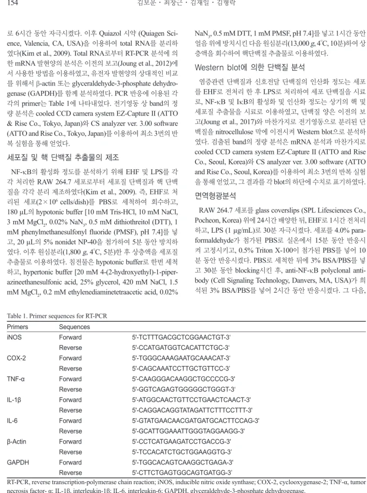
LPS로 유도되는 NF-κB의 활성화에 대한 EHF의 억 제 효과
염증촉진성
cytokines, iNOS
및COX-2
의발현은전사인자 인NF-κB
에의해조절된다. LPS
로자극된RAW 264.7
세포를 이용하여NF-κB
의활성화에대한EHF
의효과를분석하였다.
먼저면역형광법으로염색을하고confocal microscopy
로분 석한결과를Fig. 4A
에나타내었다.
자극이가해지지않은상태 에서NF-κB/p65 subunit (
녹색)
는주로세포질에분포하는것 이관찰되었다. LPS
로처리된세포의경우녹색인NF-κB p65
의대부분은핵에분포하는것으로나타났고,
이는NF-κB
가 활성화되어핵으로이동했음을보여주는결과이다.
그러나세 포에EHF (50 µg/mL)
를전처리한경우NF-κB/p65
는핵주변 의세포질에대부분분포하는것으로관찰되었다.
이러한결과 는EHF
에의해NF-κB
의활성화가현저하게억제되고있음을 보여주고있다(Fig. 4A). NF-κB
의활성화에영향을미치는단 백질을Western blot
으로분석한결과를Fig. 4B
에나타내었다. LPS
에의해과도하게인산화된IKK-β
와IκBα
는EHF
처리에 의해인산화정도(p-IKK-β
및p-IκBα)
가농도의존적으로감 소함을보여주었다. LPS
에의해핵으로이동된NF-κB p65
또 한EHF
처리에의해감소됨을보여주고있다.
다음은LPS
로 자극된대식세포주에있어NF-κB
의promoter activity
에대한EHF
의효과를분석하였다(Fig. 4C).
이를위해RAW 264.7
세 포에NF-B promoter
를가진luciferase construct
를일시적으 Fig. 2. Effect of EHF on LPS-stimulated inducible nitric oxide syn- thase (iNOS) and cyclooxygenase-2 (COX-2) protein and mRNA expressions in RAW 264.7 cells. Cells pretreated with various con- centrations of EHF for 1 h were stimulated with or without LPS (1 μg/mL) for 16 h. Whole proteins were separated with SDS-PAGE.The expression of iNOS, COX-2, and β-actin was analyzed by Western blot using corresponding antibodies. Cells were incubated with various concentrations of EHF for 1 h, and then stimulated with LPS (1 μg/mL) for 6 h. Total RNA was prepared for RT-PCR.
The results presented are representatives of three independent ex- periments.
(A)
(B)
(C)
대황 헥산 분획물의 항염증 효과
157
로
transfection
하고,
이세포를다양한농도의EHF
로2
시간전 처리하고이어서LPS
로6
시간동안자극하였다. Fig. 4C
에나 타내었듯이luciferase
활성은LPS
자극에의해현저하게증가 하였으며,
이는LPS
자극에의해활성화된NF-κB
가NF-κB
의promoter
를가진luciferase
의발현을크게증가시켰음을의 미한다.
이에대한EHF
의억제효과는10 µg/mL
의낮은농도뿐만아니라
50 µg/mL
농도에이르기까지유의적으로감소하는것으로나타났다
.
이들결과는대식세포에서LPS
자극에의 해유도되는NF-κB
의활성화가EHF
에의해효과적으로억제 되고있음을보여주고있고,
이는EHF
에의한상기의염증성cytokines
및iNOS
의발현억제는부분적으로NF-κB
활성화 경로에의해조절되고있음을의미하고있다.
LPS로 유도되는 MAPKs의 인산화에 대한 EHF의 억 제 효과
EHF
에의한NF-κB
신호경로에어떠한신호단백질들이관여하는지를확인하기위하여
MAPKs (ERK, JNK, p38 MAPK)
와Akt
의인산화를Western blot
으로분석하였다. Fig. 5
에나타 내었듯이, LPS
로유도된세포에서EHF
처리에의하여ERK, JNK, p38 MAPK
및Akt
의인산화가농도의존적으로감소하였다
.
이러한결과는EHF
가IKK-β
의인산화는물론NF-κB
의 상위단백질의인산화를조절함으로써염증억제효과를나타낸 다는것을의미한다.
부종에 대한 EHF 효과
생쥐의귀에
PMA
를처리하여귀부종을유도한결과6
시간 후PMA
에의해귀부종의무게가현저히증가하였다(Fig. 6).
Indo
는비스테로이드성항염증제(nonsteroidal anti-inflamma- tory drug, NSAID)
로써부종의치료효능을평가하기위하여 양성대조약물로사용하였다. Fig. 6
에나타난바와같이부종 은EHF
처리에의해감소하는경향이나타났고,
특히1 mg
의EHF
처리에의해유의적인차이가확인되었다(P<0.05).
이러 한결과에서EHF
의부종억제효과를확인할수있었다.
고 찰
NO
는NOS
에 의해L-arginine
으로부터 생성되며,
세균의endotoxin
또는염증성cytokines
에iNOS
가급격하게유도되 어NO
생성량이급증한다(Guha and Mackman, 2001).
병리적 인조건하에서iNOS
에의한NO
의현저한증가는다른염증성 Fig. 3. Effect of EHF on the production of pro-inflammatory cytokines in LPS-stimulated RAW 264.7 cells. Cells pretreated with various concentrations of EHF were stimulated with or without LPS (1 μg/mL) for 24 h. TNF-α (A), IL-1β (B), and IL-6 (C) in the culture media were measured by ELISA. (D) Cells were incubated with various concentrations of EHF for 1 h, and then stimulated with LPS (1 μg/mL) for 6 h. Total RNA was prepared for RT-PCR. The results presented are representatives of three independent experiments. Data represent mean±SD of three independent experiments. #P<0.001 compared to non-treated group. **P<0.01 compared to LPS-only group.(C)
(A) (B)
(C) (D)
김보운
ㆍ
최창근ㆍ
김재일ㆍ
김형락158
매개체들과함께과도한염증을유발하게되고조직의손상을 유발하는것으로알려져있어염증성손상의주요매개체이다
(Pan et al., 2011). PGE
2는COX-2
의작용에의해arachidonic acid
로부터생성된다(Sil and Ghosh, 2016).
체내의염증작용 이 증가함으로써생성되는PGE
2는통증,
염증,
체온상승,
혈 소판응집등의작용을한다(Park et al., 2006).
따라서iNOS
와
COX-2
의발현을억제하거나활성을억제함으로써NO
와PGE
2의생성을억제할수있는화합물은항염증물질로이용Fig. 4. Inhibitory effect of EHF on the activation of NF-κB in LPS- stimulated RAW 264.7 cells. A, celluar distribution of p65 subunit of NF-κB (green) protein was analyzed by immunofluorescence staining with confocal microscopy. Cells were pretreated with EHF (50 μg/mL) for 1 h, followed by LPS stimulation for 30 min. DAPI (blue) was used for nuclear staining; B, nuclear localization of NF- κB and the regulation of IκB-α phosphorylation was analyzed by Western blot. Cells pretreated with EHF for 1 h were stimulated with LPS for 30 min; C, effect of EHF on NF-κB promoter activity.
Cells were transfected with 1 μg of NF-κB promoter-containing luciferase DNA for 40 h. Transfected cells pretreated with EHF for 1 h were stimulated with LPS for additional 6 h. The results pre- sented are representatives of three independent experiments; AU, arbitrary unit. #P<0.001 compared to non-treated group. **P<0.01 compared to LPS-only group.
(A)
(B)
(C)
(A)
(B)
(C)
Fig. 6. Effect of EHF on ear edema induced by phorbol 12-my- ristate 13-actate (PMA) in mice. EHF or indomethacin (Indo) were topically administered on inner surface of the right ears of mice 1 h before application of PMA. 6 h after application of PMA, ear edema and the inhibition percentage (IP) of the ear edema by the treatment were calculated. #P<0.001 compared to non-treated group. **P<0.05 compared to PMA-only treated group.
Fig. 5. Effect of EHF on the phosphorylations of MAPKs and Akt in LPS-stimulated RAW 264.7 cells. Cells were incubated with various concentrations of EHF for 1 h, and then stimulated with LPS (1 μg/mL) for 30 min. Whole cell lysates were prepared and analyzed by Western blot for phosphorylated proteins of Akt, JNK, p38 MAPK or β-actin using corresponding antibodies. The results presented are representatives of three independent experiments.
(B)
(C)
될수있다
.
염증촉진성
cytokines
들은체내에서다양한면역및염증반 응을조절하는역할을한다.
세균의LPS
에의해자극된대식 세포는TNF-α
를생성하고분비된TNF-α
는IL-1
와IL-6
의생 성을유도함으로써염증반응을증폭시키게된다(Minnich and Moldawer, 2004). LPS
에의해유도된TNF-α
는염증반응의개 시를촉진하며지속적인생성은만성염증을유발하며결국에 는패혈성쇼크,
염증등의다양한생리적과정에관여하고있다(Scheller et al., 2011). IL-1β
는대식세포에서생성되는주요염 증촉진성cytokine
으로,
세균감염에대한염증응답의개시및 강화에중요한cytokine
이다(Li and Verma, 2002). IL-6
도대식 세포에서생성되는중요한염증성cytokine
으로서급성면역반 응에작용한다(Bonizzi and Karin, 2004).
본연구에서관찰된 결과는EHF
가LPS-
자극에의해유도되는IL-1β, IL-6, TNF-
의생성을억제시키는것으로나타났고,
이는EHF
가LPS-
자극 에의한염증성응답의초기단계를억제하고있음을의미한다.
NF-κB
의활성화를억제하는단백질인IκBα
는IKKβ
의인산화에의하여
IκBα
가인산화됨으로써유비퀴틴화되어프로테아좀에의해분해된다
(Nguyen et al., 2003).
따라서IκB-α
로부터유리된
NF-κB
는핵으로이동하여염증성유전자들의발현에기여하게된다
.
또한NF-κB
활성화경로는세포내다양 한신호전달에관여하는MAPKs
나Akt
와같은protein kinase
들에의해조절된다(Liu et al., 2017; Tian et al., 2017).
많은연 구들에서p38, JNK, ERK
와같은MAPKs
가설치류의대식세 포에서LPS
에인한NF-κB
의활성화에중요한역할을한다고 밝히고있다(Guha and Mackman, 2001; Joung et al., 2017).
그리고
Akt
또한NF-κB
의활성화를조절함으로써여러염증 성유전자의발현을조절하는것으로알려져있다(Joung et al., 2015).
본연구에서LPS
자극에의해유도되는NF-κB
의활성 화에대한EHF
의억제효과는MAPKs
및Akt
와같은kinase
의 인산화과정과관련되어있음을나타낸다.
앞서설명한것처럼모자반류에서분리된
sargaquinoic acid (Gwon et al., 2015; Joung et al., 2015), fucosterol (Jung et al., 2013; Gwon et al., 2017), sargachromenol (Gwon et al., 2017)
과같은화합물들의항염증활성에대해활발한연구가진행되 었다.
본실험에사용된EHF
를HPLC
로분석한결과sargahy- droquinoic acid, sargaquinoic acid
및sargachromenol
은발견 되지않았다.
따라서이들항염증활성을나타내는유효성분을 분리동정하기위한후속적인연구가필요하다.
이상의결과에서
, LPS
로자극한RAW 264.7
세포에있어NO
와같은염증성매개체뿐만아니라염증촉진성cytokines
의생 성및발현이EHF
에의해억제된다는것을증명하였다.
이러 한EHF
의억제효과는IκB
의분해를저해함으로써NF-κB
활 성화경로를억제시키는것으로나타났다.
또한EHF
는NF-κB
활성화의상위신호전달경로인MAPKs
및Akt
에도영향을미 치는것으로확인되었다.
여러만성염증성질환에과도한염증성매개체들의발현및생성이중요한병인적역할을하고있 음을고려했을때
,
본연구결과는EHF
가항염증효능을가진 기능성식품의소재로이용될수있다는가능성을제시하고있 다.
향후EHF
의항염증효과를나타내는유효화합물의분리.동 정및활성분석에대한연구가진행되어야할것으로판단된다.
사 사
이논문은부경대학교자율창의학술연구비
(2019
년)
에의하 여연구되었음.
References
Bonizzi G and Karin M. 2004. The two NF-kappaB activa- tion pathways and their role in innate and adaptive immu- nity. Trends Immunol 25, 280-288. https://doi.org/10.1016/j.
it.2004.03.008.
Choi JS, Han YR, Byeon JS, Choung SY, Sohn HS and Jung HA. 2015a. Protective effect of fucosterol isolated from the edible brown algae, Ecklonia stolonifera and Eisenia
bicyclis, on tert-butyl hydroperoxide- and tacrine-induced
HepG2 cell injury. J Pharm Pharmacol 67, 1170-1178.https://doi.org/10.1111/jphp.12404.
Choi JS, Haulader S, Karki S, Jung HJ, Kim HR and Jung HA.
2015b. Acetyl- and butyryl-cholinesterase inhibitory activi- ties of the edible brown alga Eisenia bicyclis. Arch Pharm Res 38, 1477-1487. https://doi.org/10.1007/s12272-014- 0515-1.
D'Acquisto F, Iuvone T, Rombola L, Sautebin L, Di Rosa M and Carnuccio R. 1997. Involvement of NF-kappaB in the regulation of cyclooxygenase-2 protein expression in LPS- stimulated J774 macrophages. FEBS Lett 418, 175-178.
https://doi.org/10.1016/s0014-5793(97)01377-x.
Elliott PJ, Zollner TM and Boehncke WH. 2003. Proteasome inhibition: a new anti-inflammatory strategy. J Mol Med 81, 235-245. http://dx.doi.org/10.1007/s00109-003-0422-2.
Eom SH, Lee DS, Jung YJ, Park JH, Choi JI, Yim MJ, Jeon JM, Kim HW, Son KT, Je JY, Lee MS and Kim YM. 2014.
The mechanism of antibacterial activity of phlorofucofu- roeckol-A against methicillin-resistant Staphylococcus au-
reus. Appl Microbiol Biotechnol 98, 9795-9804. https://doi.
org/10.1007/s00253-014-6041-8.
Guha M and Mackman N. 2001. LPS induction of gene expres- sion in human monocytes. Cell Signal 13, 85-94. https://doi.
org/10.1016/s0898-6568(00)00149-2.
Gwon WG, Joung EJ, Kwon MS, Lim SJ, Utsuki T and Kim HR. 2017. Sargachromenol protects against vascular in- flammation by preventing TNF-alpha-induced monocyte adhesion to primary endothelial cells via inhibition of NF- kappaB activation. Int Immunopharmacol 42, 81-89. https://
doi.org/10.1016/j.intimp.2016.11.014.