딱지꽃 (Potentilla chinensis) 추출물의 항염증 효과
강창호, 한상현, 소재성*
Anti-Inflammatory Effect of Chloroform Extract from Potentilla chinensis
Chang-Ho Kang, Sang-Hyun Han, and Jae-Seong So*
접수: 2013년 1월 14일 / 게재승인: 2013년 2월 24일
© 2013 The Korean Society for Biotechnology and Bioengineering
Abstract: In this study, we investigated the anti-inflamma- tion effect of Potentilla chinensis (PC) on Raw264.7 mac- rophage cells. Ethanol extract of PC decreased the production of nitric oxide (NO) in LPS-stimulated RAW264.7 cells. Eth- anol extract was fractioned by n-hexane, chloroform, ethyl acetate, n-butanol, water and each fraction was tested for inhibitory effects on inflammation. Among the sequential sol- vent fractions, PC chloroform extracts (50, 100, 300, and 500 µg/mL) significantly suppressed LPS-stimulated production of NO. During the entire experimental period, 200 and 300 µg/mL of PC chloroform extracts had no cytotoxicity. LPS- induced NO and prostaglandin E2 (PGE2) production were inhibited by PC chloroform extracts up to 50% and 90% of these productions, respectively. PC chloroform extracts reduced the expression of iNOS and COX-2 gene. These results suggest that PC chloroform extracts exhibit strong effects of anti-inflammation and can be a potential candidate in the treatment of acute and chronic inflammatory diseases.
Key words: Potentilla chinensis, Nitric oxide, Prostaglandin E2, iNOS, COX-2
1. 서론
면역체계는 외부로부터 침입하는 발병체로부터 신체를 지 켜내는 중요한 역할을 하는데, 염증반응 (inflammation)은 외 부의 물리적인 상해 또는 미생물의 체내 감염으로부터 신체 를 보호하기 위한 가장 중요한 비 특이적 방어작용이다. 염 증은 체내의 자극에 의해 변화된 손상 부위를 재생하려는 기 전이며, 지속적인 염증반응은 점막 손상을 촉진시켜 결과적 으로 통증, 부종, 발적, 발열 등을 일으켜 기능장애를 유발한 다 [1]. NO는 생체 내에서 Nitric Oxide synthase (NOS)라는 효소의 촉매 작용을 통해 L-arginin으로부터 생성되는 반응 성이 강한 자유 라디칼이다. NO는 생리적인 현상인 혈압조 절과 신경전달 매개체로 작용하며, 혈액응고 면역기능 등의 역할을 하는 것으로 알려져 있다 [2,3].
NO의 합성 효소인 NOS는 정상 생리적 조건에서 존재하는 constitutive형 (cNOS)과 면역반응에 의해 일어나는 inducible 형 (iNOS)으로 나뉘며, cNOS는 다시 혈관에 작용을 하는 endothelial형 (eNOS)과 신경에서 주된 작용을 하는 neuronal 형 (nNOS)으로 나뉘게 된다 [4]. 유도성 iNOS는 세포 내 칼 슘 농도와 외부에서 주입된 calmodulin의 자극과는 무관하게 활성화된 세포에서만 활성을 보이며 다양한 세포에서 유도 되어 병리생태학에서 중요한 역할을 한다 [5,6]. NOS의 과다 발현은 뇌출혈, 뇌성마비, 뇌졸중 등의 뇌 손상이나 알츠하 이머병 및 파킨슨병과 같은 퇴행성 뇌 신경질환의 신경독성 과 밀접한 관련이 있다고 알려져 있다 [7]. NO는 염증반응의 매 개체로 수초가 지나면 nitrite (NO2), nitrate (NO3)등의 안정한 화 합물로 존재하게 되며, 이 과정에서 형성되는 Peroxynitriteanion (ONOO-)이 염증을 일으키게 되며[8,9], 세포 내 독소작용을 인하대학교 생물공학과
Department of Biological Engineering, Inha University, Incheon 402-751, Korea
Phone: +82-32-860-8666, Fax: +82-32-872-4046 E-mail: sjaeseon@inha.ac.kr
연구논문
야기하여 조직의 손상을 일으키는 것으로 알려져 있다. 또한 NO는 2형 동위효소 (COX-2)를 활성화시켜 염증성 물질인 Prostaglandin E2 (PGE2)의 생성을 증가시킨다. Prostaglandin 은 cyclooxygenase (COX)에 의해 생성되며 염증성 cytokine 은 연골세포 및 활액세포에서 COX-2의 발현을 증가시키면 서 염증 및 통증반응을 유발한다.
딱지꽃 (Potentilla chinensis)은 장미과의 여러해살이풀이 다. 높이 30-60 cm까지 자라며 잎은 깃 모양으로 깊이 갈라 진 버들잎 모양이다. 꽃은 노란색이고 가지 끝에 모여 피며 우리나라 각지의 산비탈, 길가, 밭 부근, 산림의 풀밭, 들판 특히 개울가의 모래땅에서 잘 자란다 [10]. 전초는 각혈과 설 사, 대장염 치료에 쓰며 염증을 약화시키기 위해 감기, 위장 질병, 관절류머티즘, 통풍, 기관지천식, 기침 등에 쓴다 [11].
딱지꽃의 주요성분으로 전초와 뿌리에서 Ascorbic acid, 줄기 에서 tannin, oil, quercetin, kaempferol, delphinidin, cyanidin 등이 분리되었고, p-coumaric acid, feru acid, ellagic acid 등이 있다 [12]. 약리실험에서 아메바 원충을 죽이는 작용과 항균 작용이 있는 것으로 밝혀졌으나, 아직까지 딱지꽃에 대한 성 분연구는 미비한 실정이다.
본 연구는 항염증 효과를 나타내는 기능성 식품 및 의약품 소재 개발을 위한 기초 연구로서 딱지꽃의 NO와 PGE2 합성 억제효과를 규명하기 위해 용매 분획별 NO와 PGE2에 대한 저해 활성을 조사하였으며, 최종적으로 선정된 딱지꽃의 chloroform 분획 추출물에 대한 iNOS와 COX-2의 발현 억제 효과를 확인하였다.
2. 재료 및 방법 2.1. 시료 및 시약
본 실험에 사용한 시약은 Sulfonic acid, phosphoric acid, N-1- napthylethylenediamine dihydrochloride, Lipopolysaccharides (LPS), N-monomethyl-L-arginine (L-NMMA), 3-(4,5- dimethyl- thiazol-2-yl)-2,5-diphenyltetrazolium bromide (MTT), Fetal bovine serum (FBS), penicillin-streptomycin, TRIzol reagent, dimethy sulfoxide (DMSO)등은 Sigma (St. Louis, MO, USA) 제품을 이용하였다. Dulbeco"s Modified Eagle"s Medium (DMEM)은 Gibco (Rckville, MD, USA) 제품을 구입하여 사 용하였고, PGE2 assay kit는 R&D system (Minneapolis, MN, USA) 제품을, Maxim RT-PCR kit는 Quiagen 제품을 사용하 였다. 그 외의 시약과 용매는 일급 또는 특급을 사용하였다.
사용 기기로는 흡광도 측정을 위하여 microplate reader (Bio- rad Model 550, CA, USA)를 사용하였으며, DNA의 상대적 인 발현 비율을 측정하기 위하여 densitometer (Bio-Rad Lab., CA, USA)를 이용하였다.
2.2. 시료 추출
본 실험에 사용한 딱지꽃은 서울경동시장 약재도매 상가에 서 원산지를 확인한 후 구입하였으며, 건조 및 마쇄하여 사
용하였다. 시료의 추출은 시료 100 g에 에탄올 (95% EtOH, 시약용 1급)을 가하여 실온에서 24시간 동안 방치하고 여과 하여, 다시 잔사에 에탄올을 가하여 침지한 후 여과하는 과 정을 3회 반복하여 실행하였다. 얻은 여액은 45oC 수욕상에 서 진공회전 농축기 (EYELA, Tokyo, Japan)로 감압 농축한 후 건조하여 냉장 보관하며 에탄올 추출물 시료로 사용하였 다. 또한 에탄올 추출물에서 증류수로 현탁시킨 후 n-hexane, chloroform, ethyl acetate, n-butanol, water순으로 용매 분획을 하여 실험에 사용하였다.
2.3. 세포 배양
RAW264.7 대식세포는 한국세포주은행 (KCLB, Korea Cell Line Bank, Seoul, Korea)에서 분양받아 사용하였다. 세포는 10% FBS, 1% penicillin-streptomycin을 포함하는 DMEM 배 지에서 37℃, 5% CO2 조건에서 배양하였다.
2.4. MTT assay에 의한 세포 생존율 측정
세포에 대한 독성 측정은 MTT 환원방법을 이용하여 측정하 였다 [13]. RAW264.7 세포를 96-well plates에 2 × 10⁵ cells/
mL 농도로 분주하고 시료를 농도별로 세포에 처리하여 24 시간 동안 배양한다. MTT 용액을 20 µL 가하고 37oC, 5%
CO2 incubator에서 4시간 동안 반응시킨 후, 세포에 DMSO 200 µL를 넣어 섞어준 다음 microplate reader를 이용하여 546 nm에서 흡광도의 변화를 측정하여 대조군에 대한 세포 생존율을 백분율로 표시하였다.
2.5. NO와 PGE2 생성량 측정
시료의 NO 생성 저해 활성은 Nitrite assay를 사용하여 측정 하였다 [14]. NO합성의 indicator로 사용되는 NO2농도는 Griess reagent (1% Sulfonic acid, 0.1% N-1-napthylethyl- ene- diamine dihydrochloride, 2.5% phosphoric acid)을 이용하여 배양배지에서 측정하였다. RAW264.7 세포를 96-well cell culture plate에 well당 2 × 10⁵ cells/mL가 되도록 분주한 후, LPS (1 µg/mL)와 추출물을 각 농도에 맞게 처리하여 20시간 동안 배양하였다. 세포배양액 상층액 100 µL를 동량의 Griess reagent와 혼합하여 실온에서 10분간 배양하여 546 nm에서 microplate reader로 측정하였다. 모든 실험에서 fresh culture medium을 blank로 사용되었다. PGE2의 생성량을 측정하기 위해 nitrite를 측정하였던 동일한 배지로 PGE2 assay를 사용 하여 enzyme-linked immune sorbent assay (ELISA)를 이용하 여 제조사에서 제시한 방법으로 실험하였다.
2.6. 염증 유발 효소 발현 측정
Reverse transcription-polymerase chain reaction (RT-PCR)을 사용하여 딱지꽃 chloroform 추출물의 농도별 mRNA 상에서 의 발현 정도를 확인하였다. 다음과 같은 조건으로 RT-PCR 을 실시하였다. cDNA 합성 45oC, 30분 denaturation 94oC, 5 분 denaturation 94oC, 5분 annealing 50~55oC, 1분 elongation 72oC, 2분을 25 cycles한 다음, final elongation step을 72oC에
서 5분간 수행하였다. RT-PCR의 생성물은 1% agarose gel을 사용하여 100v에서 30분간 전기영동하여 UV에서 관찰하였 다 각 primer의 염기서열은 다음과 같다. GAPDH, sense 5’- TGAAGGTCGGTGTGAACGGATTTGGC-3’ anti-sense 5’-C ATGTAGGCCATGAGGTCCACC- AC-3’; iNOS, sense 5’-CC CTTCCGAAGTTTCTGGCAGC-3’ anti-sense 5’-GG CTGTC AGA-GCCTCGTGGCTT-3’; COX-2, sense 5’-GGAGAGAC TATCAAGATAGT-3’ anti-sense 5’-ATGGTCAGTAGACTTTT ACA-3’.
2.7. 통계처리
모든 실험은 3회 반복하여 측정하였고, 그 결과는 평균±표 준편차로 나타냈으며 통계적 분석은 SPSS 10.0 프로그램을 이용하여 각 처리구 간의 유의성(p < 0.05, 0.01)검증을 위해 분산분석(analysis of variance, ANOVA) 후 tukey test로 다중 비교를 실시하였다.
3. 결과 및 고찰 3.1. 수율
딱지꽃의 용매별 수율을 분석한 결과, 에탄올 추출물은 4.34% (w/w)를 나타냈으며, n-hexane, chloroform, ethyl acetate, n-butanol로 용매 분획한 결과 각각 2.34, 1.28, 0.57, 0.59%
(w/w)의 수율을 보였다.
3.2. NO와 PGE2 생성 억제효과
NO는 NO 합성효소에 의해 L-arginine으로부터 생성되는 무 기 유리체로 면역반응, 세포독성, 신경전달계 및 혈관이완 등 여러 생물학적인 과정에 관여하며 농도에 따라 세포기능 유지에 중요한 작용을 하기도 하고 세포독성을 일으키기도 한다 [15]. 본 연구에서는 딱지꽃 추출물이 NO와 PGE2의 발 현에 미치는 영향을 조사하기 위해 마우스의 대식세포인
RAW264.7에 LPS의 단독처리 또는 용매 분획별 추출농도별 로 동시에 처리하여 NO 저해 활성을 조사하였다 (Fig. 1).
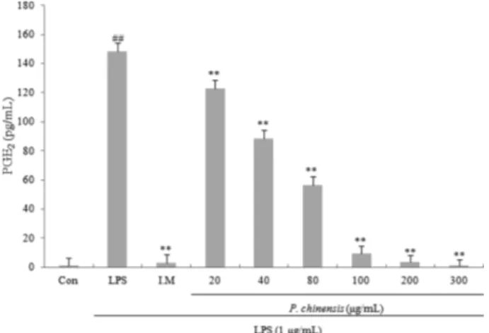
LPS를 처리한 RAW264.7 대식세포에서 생성된 NO의 함 량이 14.42 µM 수준으로 증가하였으며, 딱지꽃의 각 용매 분 획별 추출물을 농도별 (50, 100, 300, 500 µg/mL)로 처리하였 을 때 chloroform 분획물에서 다른 분획물보다 높은 NO 생성 저해 활성을 보였다 (p < 0.05). 300 µg/mL의 농도까지 세포 생존력에서 독성을 보이지 않는 것을 확인할 수 있었으며 (Fig. 2), 최종 시료로 chloroform 분획물을 선정하였다. 따라 서 chloroform 분획물의 처리농도를 독성을 보이지 않은 20, 40, 80, 100, 200, 300 µg/mL로 하여 NO와 PGE2의 생성 억제 효과 실험을 진행하였다. Fig. 3을 보면, 100 µg/mL이상의 농 도에서 NO합성 저해제인 L-NMMA와 유사한 효과를 나타 냄을 확인할 수 있었으며, PGE2 저해활성에서는 80 µg/mL 에서 60%, 100 µg/mL이상의 농도에서 90% 정도의 저해효 과를 나타냈다 (Fig. 4). 위 결과는 딱지꽃의 chloroform 분획 물이 독성을 나타내지 않으면서 LPS에 의해 생성 및 분비가
Fig. 1. Inhibition of NO production of PC extracts in LPS-induced RAW264.7 macrophage cells. Data represent the means±SD with three separate experiments. One-way ANOVA was used for comparisons of multiple group means followed by t-test (significant as compared to control. *p < 0.05, **p < 0.01).
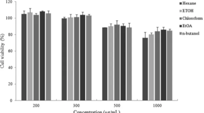
Fig. 2. Cytotoxicity effects of solvent fractionates from PC ethanolic extract in RAW264.7 macrophage cells. Data represent the means±SD with three separate experiments.
Fig. 3. Inhibitory effect of CHCl3 fractionates on nitrite production from LPS-induced in RAW264.7 macrophage cells. Con: LPS not induced group, LPS: LPS induced group, L-NMMA is an inhibitor of NO synthase. Data represent the means±SD with three separate experiments. One-way ANOVA was used for comparisons of multiple group means followed by t-test (significant as compared to control. *p < 0.05, **p < 0.01).
증가된 NO와 PGE2를 효과적으로 억제시킨다는 것을 나타 낸다.
3.3. 염증유발 효소 발현 측정
딱지꽃의 chloroform 분획물의 NO와 PGE2의 합성 저해효과 가 유전자의 조절에 의한 것인지 확인해보기 위하여 두 효소 의 발현 억제효과를 RT-PCR을 통해 확인해 보았다. iNOS는 평소에는 세포 내에 존재하지 않으나 일단 유도되면 장시간 동안 다량의 NO를 생성하며, 생성된 NO는 병리적인 혈관확 장, 세포독성, 조직손상 등과 같은 생체에 유해한 작용을 나 타낸다. 그리고 염증상태에서 iNOS에 의해 생성된 NO는 혈 관 투과성, 부종 등의 염증반응을 촉진시킬 뿐만 아니라 염 증매개체의 생합성을 촉진하여 염증을 심화시키는 것으로 알려져 있다 [16,17]. 다수의 염증 억제 약물들의 작용기전은
prostagladin 합성 억제를 나타내며 이는 COX-2의 생성 및 활성저해에 의한 것이다 [18]. 따라서 COX-2에 의한 prostagladin의 합성은 염증반응을 매개하는 것으로 여겨 진다. 염증발현 유도물질인 LPS 처리군과 비교해 보았을 때 iNOS의 경우 100 µg/mL이상의 농도에서 높은 발현 억제효 과를 나타냄을 확인할 수 있었으며, COX-2의 경우 50 µg/
mL이상의 농도에서 높은 발현 억제효과를 나타냄을 확인할 수 있었다. 이를 통해 딱지꽃의 chloroform 분획물을 처리한 양쪽 group 모두 농도에 따라 iNOS와 COX-2 유전자의 발현 이 억제되는 것을 확인할 수 있었다 (Fig. 5). 추후 딱지꽃의 chloroform 분획부분의 물질을 구조적으로 밝히고, 항염증 활성을 나타내는 기전을 규명하는 연구가 추가적으로 수행 되어야 한다고 사료된다.
4. 결론
항염증 효과가 있는 기능성 식품 및 의약품 소재의 개발을 위하여 천연 식물 자원으로부터 NO와 PGE2합성을 저해하 는 물질을 찾기 위해 항염증 효과가 있다고 알려져 있는 딱 지꽃에서 69%의 높은 NO 저해활성이 있다는 것을 확인하였 다. 딱지꽃 에탄올 추출물에서 n-hexane, chloroform, ethyl acetate, n-butanol, water 순으로 용매 분획을 실시한 후 NO 생성 저해 활성을 측정한 결과, chloroform 분획물에서 가장 높은 저해 활성을 보여 최종 분리 시료로 선정하였으며, PGE2에 대한 합성 저해 효과를 확인한 결과 100 µg/mL 이상 의 농도에서 PGE2합성 저해제인 indomethacin과 유사한 효 과를 보였다. NO와 PGE2의 합성저해효과가 유전자의 조절 에 의해서인지 알아보기 위해 iNOS와 COX-2 gene의 발현 조절효과를 RT-PCR을 통하여 확인해본 결과 두 유전자의 발현을 억제함을 확인할 수 있었다. 이를 통해 딱지꽃 추출 물이 독성과 부작용이 적은 염증 치료제로 활용될 수 있는 가능성이 높음을 제시한다.
감사
본 논문은 인하대학교의 지원에 의해 이루어진 연구이며 지 원에 감사드립니다.
REFERENCES
1. Jew, S. S., O. N. Bae, and J. H. Chung (2003) Anti-inflammatory inflammatory effects of asiaticoside on inducible nitric oxide syn- thase and cyclooxygenase-2 in RAW264.7 cell line. J. Toxicol.
Pub Health 19: 33.
2. Lee, D. E., J. R. Lee, T. W. Kim, Y. K. Kwon, S. H. Byun, S. W.
Shin, S. I. Suh, T. K. Kwon, J. S. Byun, and S.C. Kim (2005) Inhi- bition of lipopolysaccharide-inducible nitric oxide synthase, TNF- Fig. 4. Inhibitory effect of CHCl3 fractionates on PGE2 production
from LPS-induced in RAW264.7 macrophage cells. Con: LPS not induced group, LPS: LPS induced group, I.M: indomethacin is an inhibitor of PGE2 synthase. The data represent the means±SD of three separate experiments (significant as compared to control.
*p < 0.05).
Fig. 5. Inhibitory effects of the LPS-induced iNOS and COX-2 mRNA expression of CHCl3 fractionates. C: LPS not induced group, LPS: LPS induced group. Data represent the means±SD with three separate experiments. One-way ANOVA was used for comparisons of multiple group means followed by t-test (significant as compared to control. *p < 0.05, **p < 0.01).
α, IL-1β‚ and COX-2 expression by flower and whole plant of Lonicera japonica. Korean J. Oriental Physiology & Pathology 19: 481.
3. Cheon, Y. P., M. L. Mollah, C. H. Park, J. H. Hong, G. D. Lee, J.
C. Song, and K. S. Kim (2009) Inhibition effects of water extract of Bulnesia sarmienti on inflammatory responce in LPS-induced RAW 264.7cell line. J. Life Science 19: 479.
4. Tsao, L. T., C. Y. Lee, L. J. Huang, S. C. Kuo, and J. P. Wang (2002) Inhibition of lipopolysaccharide-stimulated nitric oxide production in RAW 264.7 macrophages by a synthetic carbazole, LCY-2-CHO. Biochem. Pharmacol. 63: 1961-1968.
5. Vodovotz, Y., D. Russell, Q. W. Xie, C. Bogdan, and C. Nathan (1995) Vesicle membrane association of nitric oxide synthase in primary mouse macrophage. J. Immunol. 154: 2914-2925.
6. Lowenstein, C. J., E. W. Alley, P. Raval, A. M. Snowman, S. H.
Synder, S. W. Russell, and W. J. Murphy (1993) Macrophage nitric oxide synthase gene: two upstream regions mediate induction by interferon gamma and lipopolysaccharide. Proc. Natl. Acad. Sci.
USA 90: 9730-9734.
7. Chabrier, P. E. and M. Auguest (1999) Nitric oxide synthases: tar- gets for therapeutic strategies in neurological diseases. Cell. Mol.
Life Sci. 55: 1029-1035.
8. Teresa, D. W., S. Tamir, H. Ji, J. S. Wishnok, and S. R. Tannen- baum (1995) Nitric Oxide Induces Oxidative Damage in Addition to Deamination in Macrophage DNA. Chem. Res. Toxicol. 8: 473- 477.
9. Szab, C., B. Zingarelli, M. O’Connor, and A. L. Salzman (1996) DNA strand breakage, activation of poly (ADP-ribose) syn- thetase, and cellular energy depletion are involved in the cytotox- icity of macrophages and smooth muscle cells exposed to peroxynitrite. Proc. Natl. Acad. Sci. USA 93: 1753-1758.
10. Chung, T. H. (1955) Korean flora. pp 315. Shizisa, Seoul, Korea.
11. Zhen, L., T. Zhang, L. Ye, and Y. Jia (2004) Protective effects of the ethanolic extract from potentilla chinensis on mice acute hepatic injury induced by carbon tetrachloride. Liaoning Journal of Traditional Chinese Medicine 31: 422-423.
12. Tomczyk, M. and K. P. Latte (2009) Potentilla-A review of its phytochemical and pharmacological profile. J. Ethnopharm. 122:
184-204.
13. Je, J. Y., P. J. Park, E. K. Kim, and C. B. Ahn (2009) Antioxidant and angiotensin I converting enzyme inhibitory activity of Bambu- sae caulis in liquamen. Food. Chem. 113: 932-935.
14. Reissig, J. L., J. L. Strominger, and L. F. Leloir (1955) A modified colorimetric method for the estimation of N-acetylamino sugars. J.
Biol. Chem. 217: 959-966.
15. Kim, J. Y., K. S. Jung, and H. G. Jeong (2004) Suppressive effects of the kahweol and cafestol on cyclooxygenase-2 expression in macrophages. FEBS. Lett. 569: 321-326.
16. Santos-Gomes, P. C., R. M. Seabra, P. B. Andrade, and M.
Fernandes-Ferreira (2003) Determination of phenolic antioxidant compounds produced by calli and cell suspensions of sage(Salvia officinalis L.). J. Plant. Physiol. 160: 1025-1032.
17. Tesuka, Y., S. Irikawa, T. Kaneko, A.H. Banskota, T. Nagaoka, Q.
Xiong, K. Hase, and S. Kadota (2001) Screening of Chinese her- val drug extracts for inhibitory activity on nitric oxide produc- tion and identification of an active compound of Zanthoxylum bugeanum. J. Ethnopharmacol. 77: 209-217.
18. Weisz A., I. Cicatiello, and H. Esumi (1996) Regulation of the mouse inducible-type nitric oxide synthase gene promoter by inter- feron, bacterial lipopolysaccharide, and NG-monomethyl-L- argin- ine. J. Biol.Chem. 316: 209-215.