384
서 론체내의염증반응은외부로부터침입한병원성물질이나조 직의손상에대한방어작용으로나타나는데
,
이는정상적인조 직의구조와기능을회복하기위해필수적으로일어나는반응 이다.
정상적인염증반응은시간이지남에따라염증촉진성매 개체(pro-inflammatory mediators)
의생성은감소되고,
항염증 성매개체(anti-inflammatory mediators)
는증가됨으로써스스 로염증반응이제한되는조절과정을가지고있다(Lawrence et al., 2002).
그러나이러한염증반응조절과정에이상이생기거 나최초염증반응유발원인이완전히제거되지못하였을경우 염증촉진성매개체들은과잉으로지속적으로생성되게되고,
이경우만성적인염증상태가유지됨으로써조직손상을유발 하는데작용을할수있다(Kaplanski et al., 2003).
동맥경화,
염증성관절염및암
,
그리고노화및알쯔하이머병(Alzheimer’s disease, AD)
를포함하는퇴행성신경질환과같은질환들의발 병에이러한만성적인염증반응이관련되어있는것으로알려 져있다(Libby, 2006; Packard and Libby, 2008; Schwab and McGeer, 2008; Solinas et al., 2010).
체내의면역반응에관여하는세포중하나인대식세포
(mac- rophages)
는이러한염증반응에중요한역할을하고있다.
대식 세포는interferon-γ (IFN-γ), interleukin (IL)-1β, IL-6, tumor necrosis factor-α (TNF-α)
와같은 염증촉진성cytokines,
그 리고 세균 세포막성분인lipopolysaccharides (LPS)
등의 자 극에노출됨으로써활성화된다(Xie et al., 1993).
활성화된대 식세포는염증촉진성cytokines
이외에inducible nitric oxide synthase (iNOS)
및cyclooxygenase-2 (COX-2)
와같은효소 의발현을통해nitric oxide (NO)
및prostaglandin E
2(PGE
2)
Article history;
Received 26 July 2013; Accepted 2 August 2013
*Corresponding author: Tel: +82. 51. 629. 5849 Fax: +82. 51. 629. 5843 E-mail address: [email protected]
Kor J Fish Aquat Sci 46(4) 384-392, August 2013 http://dx.doi.org/10.5657/KFAS.2013.0384 pISSN:0374-8111, eISSN:2287-8815
ⓒ The Korean Society of Fishereis and Aquatic Science. All rights reserved
LPS로 유도된 RAW 264.7 대식세포에 대한 미역( Undaria pinnatifida) Ethyl Acetate 분획물의 항염증 효과
부경대학교 식품영양학과
최민우ㆍ김재일
*Anti-Inflammatory Effect of Ethyl Acetate Fraction Isolated from Unda- ria pinnatifida on Lipopolysaccharides-Stimulated RAW 264.7 Cells
An ethanolic extract of Undaria pinnatifida was fractionated using several solvents. Of the fractions, the ethyl acetate fraction had the greatest inhibitory effect on lipopolysaccharide (LPS)-induced nitric oxide (NO) production in RAW 264.7 macrophage cells.Using this fraction ( U . pinnatifida ethyl acetate extract, UPE), we investigated the molecular mechanism underlying its inhibitory effect on LPS-stimulated RAW 264.7 cells. Pretreatment of the cells with up to 100 µg/mL UPE significantly inhibited NO production and inducible nitric oxide synthase (iNOS) expression, in a dose-dependent manner. Similarly, UPE treatment markedly reduced the production of pro-inflammatory cytokines, such as interleukin (IL)-1, IL-6 and tumor necrosis factor-α (TNF-α), while it strongly suppressed the nuclear trans- location of nuclear factor-kappa B (NF-κB) by preventing proteolytic degradation of inhibitor of nuclear factor κB (IκB)-α. Moreover, UPE treatment significantly reduced the phosphorylation of phosphatidylinositol 3-kinase (PI3K)/Akt and mitogen-activated protein kinase (MAPK) in LPS-stimulated cells. These results indicate that UPE contains anti-inflammatory compounds and suggest that it might be used as a functional food material that assists in prevention of inflammatory diseases.
Key words: Undaria pinnatifida , Anti-inflammatory effect, Pro-inflammatory cytokines, iNOS, NF-κB
Min-Woo Choi and Jae-Il Kim*
Department of Food Science and Nutrition, Pukyong National University, Busan 608-737, Korea
미역 에틸아세테이트 추출물의 항염증 효과
385
와같은다양한염증매개체들을생성하게되고
(Nathan, 1992;
Zhang and Ghosh, 2000),
이들매개체들의과도하고도지속적 인생성은다양한만성염증성질환의발병에기여하고있는것 으로알려져있다.
대식세포에있어염증성
cytokines
및COX-2, iNOS
와같은 매개체들의발현은전사수준에서주요전사인자인nuclear fac- tor kappa-B (NF-κB)
에의해조절된다(Baeuerle and Henkel, 1994). NF-κB
는dimer
를이루는두개의subunit
로이루어져 있고포유류에서가장일반적인형태는p50/65
의heterodimer
이다.
자극이없는상태에서NF-κB
는inhibitor of kappa B
(IκB)
와결합한상태로불활성형태로세포질에격리되어있다
(D’Acquisto et al., 1997; Makarov, 2001).
그러나LPS
와같 은자극이주어지는경우IκB
는IκB kinase
에의해인산화되면서떨어져나와분해되고
,
유리된NF-κB
는핵으로이동하여다양한염증성매개체와같은표적유전자의발현을유도하 게된다
(Chen et al., 1995).
이러한과정에있어IκB kinase
의 활성화에는extracellular signal-regulated kinase (ERK), c-Jun N-terminal kinase (JNK), p38 kinase
를 포함하는mitogen- activated protein kinases (MAPKs)
그리고Akt
와같은kinase
에의해조절되는것으로알려져있다(Marks-Konczalik et al., 1998; Zhang and Ghosh, 2000).
바다에널리자생하는해조류는비타민과무기질이다량함 유되어있으며식이섬유가풍부하다
.
그중갈조류는엽록소와 갈색을띄는크산토필계카로티노이드의일종인fucoxanthin
과fucoidan, alginic acid, laminarin
등의다당류, fucosterol,
polyphenol
과같은다양한생리활성물질들을가지고있는것으로보고되었다
(Hosokawa et al., 2004; Kim et al., 2012; Kim et al., 2013; Lee et al., 2003; Lee et al., 2004; Maeda et al., 2006; Park et al., 2010; Sachindra et al., 2007; Yan et al., 1999;
Yoo et al., 2012; Zhang et al., 2013).
갈조류의하나인미역(Undaria pinnatifida)
은우리나라,
일본,
중국과같은동아시아 지역의나라에서식용으로이용되고있다.
전통적으로한국의 여성들은산후조리기간에미역국을먹었는데,
이는출산후에 회복을돕고피를맑게하는작용을한다고믿었기때문이었다(Khan et al., 2008).
갈조류에서분리한fucoxanthin
이나fucoi- dan, fucosterol
과같은화합물의생리활성은다양한연구를통 해많이보고되어있지만,
미역에탄올추출물로부터얻은분획 물을이용한연구결과는아직보고된바가없다.
최근본연구 실에서수행한예비실험에서미역의ethanol (95%, v/v)
추출물 로부터유기용매(hexane, ethyl acetate, butanol)
를이용하여각 분획물들을분리하였고그활성을분석한결과, ethyl acetate
분 획물에서항염증효과가있음을일부확인하였다(Table 1).
본 연구에서는이러한미역의ethyl acetate
추출물(U. pinnatifida ethyl acetate extract, UPE)
의항염증효과및관련분자적기전 을LPS
로자극한RAW 264.7 macrophage cell
을이용하여분 석함으로써,
다양한염증성질환의발병을예방또는지연시킬수있는식품재료로서의이용가능성을검토하였다
.
재료 및 방법
추출과 분획
부산기장에서채취한미역의건조분말
2 kg
을환류냉각기가부착된집기병에담고
95% ethanol (EtOH, v/v) 4 L
를넣어가 열,
추출하고(50°C, 3
시간)
추출액을여과하여rotary vacuum evaporator
를사용하여농축하였다.
이를3
회반복하여총178 g
의EtOH extract
를얻었다.
이를H
2O:EtOH (9:1, v/v)
의혼합용매로녹인후동량의
n-hexane
을넣어분액깔대기에평형화시켜상층액의
n-hexane
가용부를모아sodium sulfate anhy- drous
로처리한다음여과,
농축과정을거쳐n-hexane fraction
을얻었다.
이를동일한방법으로ethyl acetate (EtOAc)
로추출 하여EtOAc extract (UPE) 12.5 g
을얻었다.
세포 배양 및 처리
RAW 164.7 macrophage cells (ATCC, Rockville, MD, USA)
는10% fetal bovine serum (FBS)
와penicillin (100 units/ml), streptomycin sulfate (100 µg/mL)
을첨가한Dulbecco’s modi- fied Eagle’s medium (DMEM)
을사용하였고, 5% CO
2, 37°C
배양기에서배양하였다. Cell culture plate
에RAW 264.7 cell
이70-80%
정도채워지면phosphate-buffered saline (PBS)
로 한번씻어낸후,
계대배양하였다. UPE
는100% dimethyl sulf- oxide (DMSO)
에녹여사용하였고,
배양세포처리전에배지에 희석하여처리하였다.
세포 독성 시험
RAW 264.7 cell
을96-well plate
에5×10
5cells/well
로분주 하고37°C
에서24
시간동안배양하였다.
이후에UPE
가0, 25,
Table 1.Inhibitory activity of various solvent fractions of Undaria pinnatifida ethanolic extract on nitric oxide (NO) generation in RAW 264.7 cellsFractions Weight
(yield) Inhibitory activity on NO generation* (IC50, µg/mL)**
n-Hexane 7.1 g
(0.36%) 121.06 ± 1.68
Ethyl acetate 12.5 g
(0.63%) 71.44 ± 4.35
n-Butanol 13.0 g
(0.65%) > 200
*RAW 264.7 macrophage cellspretreated with various solvent frac- tions for 1 h was incubated with 1 μg/mL of LPS for 24 h. NO generation as nitrite concentration in the media was measured by Griess test as described in the Materials and Methods section.
**IC50: The half maximal inhibitory concentration.
50, 100, 200 µg/mL
농도로희석된DMEM
배지로교체하여1
시간배양하였고,
이후에LPS (1 µg/mL)
를함유한DMEM
배지에다시24
시간 배양하였다.
이후CellTiter96
ⓇAqueous 3-(4,5-dimethylthiazol-2-yl)-5-(3-carboxymethoxyphenyl)- 2-(4-sulfophenyl)-2H-tetrazolium (MTS)
시험키트(Promega, Madison, WI, USA)
를사용하여제조사의 방법에따라세포 생존율을분석하였다. MTS
용액은FBS-free DMEM
에5%
(v/v)
의 농도로섞어100 µL
씩 처리하였다. 1
시간 후에mi- croplate reader (Glomax Multi Detection System, Promega, Madison, WI, USA)
를이용하여490 nm
의파장에서흡광도 를측정하였다.
NO 및 염증성 Cytokines 생성량의 측정
분리한각분획물들의항염증효과비교를위해
RAW 264.7
세포에서의NO
생성에대한억제효과를분석하였다.
세포를 분획물들로1
시간동안전처리한다음LPS (1 µg/mL)
로24
시간동안자극하고,
그배지를원심분리(2,000×g, 4°C, 10
분)
하여회수하였다. NO
의농도는배지(100 µL)
와Griess
시약(0.1% naphthylethylene diamine dihydrochloride + 1% sul- fanilamide + 5% phosphoric acid)
을동일한비율로반응시켜microplate reader
로540 nm
의파장에서흡광도를측정하였다(Kim et al., 2009).
그결과(Table 1), NO
생성에대한저해효 능(IC
50, ug/mL)
이가장뛰어난EtOAc
추출물(UPE)
을이후연 구에사용하였다.
NO
생성억제에대한농도별UPE
의효과는상기와동일한방법으로분석하였다
.
배지중의IL-1β, IL-6, TNF-α
의양은enzyme-linked immunosorbent assay kit (ELISA, R&D Sys- tems, Minneapolis, MN, USA)
를이용하여제조사의방법에 따라측정하였다.
iNOS mRNA 발현양의 분석
RAW 264.7
세포(1×10
6cells/well)
를0, 25, 50, 100 µg/mL
의농도로UPE
를1
시간동안처리한후, LPS 1 µg/mL
의농 도로6
시간동안자극시켰다.
이후Quiazol
시약(Quiagen Sci- ence, Valencia, CA, USA)
을 이용하여total RNA
를 분리하 였다(Kim et al., 2009). Total RNA
로부터reverse transcrip- tion-polymerase chain reaction (RT-PCR)
분석에의한iNOS mRNA
발현양의분석은이전의보고(Kim et al., 2009)
에서사 용한방법을이용하였고,
유전자발현양의상대적인비교를위 해서housekeeping gene
인glyceraldehye-3-phosphate dehy- drogenase (GAPDH)
를 함께분석하였다. PCR
반응에 이용 된각각의primer
는다음과같다: iNOS sense, 5’-GCC TTC AAC ACC AAG GTT GTC TGC A-3’; iNOS antisense, 5’- GTC ATT GTA CTC TGA CTC TGA GGG CTG ACA C-3’;
GAPDH sense, 5’-GAC CCC TTC ATT GAC CTC AA-3’;
GAPDH antisense, 5’-CTT CTC CAT GGT GGT GAA GA-
3’.
전기영동상band
의정량분석은cooled CCD camera sys- tem EZ-Capture Ⅱ (ATTO & Rise Co., Tokyo, Japan)
과CS analyzer ver. 3.00 software (ATTO)
를이용하여최소3
번의반 복실험을통해얻었다.
세포질 및 핵 단백질 추출물의 제조
NF-κB
의활성화정도를분석하기위해UPE
및LPS
를각각처리한
RAW 264.7
세포로부터세포질및핵단백질추출물을각각분리제조하였다
(Kim et al., 2009).
간략하게,
처리 한 세포(2×10
6cells/dish)
를PBS
로세척하여회수하고, 180 µL
의hypotonic buffer [10 mM Tris-HCl, 10 mM NaCl, 3 mM MgCl
2, 0.02% NaN
3, 0.5 mM dithiothreitol (DTT), 1mM phenylmethanesulfonyl fluoride (PMSF), pH 7.4]
를넣고, 20 µL
의5% nonidet NP-40
을첨가하여5
분동안반응시켰다.
이 후원심분리(1,800×g, 4°C, 5
분)
한후상층액을세포질추출 물로 이용하였다.
침전물은hypotonic buffer
로 한번세척하 고, hypertonic buffer [20 mM 4-(2-hydroxyethyl)-1-piper- azineethanesulfonic acid, 25% glycerol, 420 mM NaCl, 1.5 mM MgCl
2, 0.2 mM ethylenediaminetetraacetic acid, 0.02%
NaN
3, 0.5 mM DTT, 1 mM PMSF, pH 7.4]
를넣고1
시간동안 얼음위에서반응시킨다음원심분리(13,000×g, 4°C, 10
분)
하 여상층액을회수하여핵단백질추출물로이용하였다. Western Blot 분석에 의한 단백질 양 분석
iNOS
단백질의발현양, MAPK
및Akt
의양과인산화정도 는세포를UPE
및LPS
로처리한이후whole cell lysate
를제 조하여시료로, NF-κB
및IκB
의활성화및인산화정도는상 기의핵및세포질추출물을시료로이용하였고,
단백질의양은 이전의보고(Kim et al., 2009)
와마찬가지로sodium dodecyl
sulfate-polyacrylamide gel electrophoresis (SDS-PAGE)
이후nitrocellulose membrane
에전이시켜Western blot
방법으로조 사하였다.
검출된band
의정량분석은mRNA
분석과마찬가지 로cooled CCD camera system EZ-Capture Ⅱ
와CS analyzer
ver. 3.00 software
를이용하여최소3
번의반복실험을통해얻 었고,
그결과를각blot
의하단에수치로표기하였다.
그리고,
Western blot
사용된각각의1
차항체들은다음과같다: iNOS
(sc-650), β-actin (sc-47778), phospho-Akt (sc-4060), Akt (sc-
1618), phospho-ERK (sc-7883), ERK (sc-94), phospho-JNK
(sc-6254), JNK (sc-7345), NF-κB/p65 subunit (sc-8008),
Poly(ADP-ribose) polymerase (PARP, sc-7150)
는Santa
Cruz Biotechnology (Santa Cruz, CA, USA)
에서구입하였고,
phospho-IκB-α (4814), IκB-α (9246), phospho-p38 (4511),
p38 (9212)
는Cell Signaling Technology (Danvers, MD,
USA)
에서각각 구입하였다. Horseradish peroxidase (HRP)
가conjugate
되어있는각각의2
차항체들[rabbit anti-goat IgG
(LF-SA5004), goat anti-mouse IgG (LF-SA5001), goat anti-
미역 에틸아세테이트 추출물의 항염증 효과
387
rabbit IgG (LF-SA5002)]
은AbFrontier (Seoul, Korea)
에서구 입하였고, Enhanced chemiluminescence (ECL) detection kit
은GE Healthcare Bio-Science (Piscataway, NJ, USA)
를사용 하였다. β-actin
과PARP
는각각세포질과핵의control
단백질 로서분석에포함시켰다.
면역형광분석법
RAW 264.7
세포를glass coverslips (SPL Lifesciences Co., Gyeonggi-do, Korea)
위에24
시간배양한뒤, UPE
로1
시간전 처리하고, LPS (1 µg/mL)
로30
분자극시켰다.
세포를4.0%
paraformaldehyde
가첨가된PBS
로실온에서15
분동안반응 시켜고정시키고, 0.5% Triton X-100
이첨가된PBS
를넣어10
분동안반응시켰다. PBS
로세척한뒤에3% BSA/PBS
를넣고30
분동안blocking
시킨후, anti-NF-κB polyclonal antibody
가 희석된3% BSA/PBS
를넣어2
시간동안반응시켰다.
그다음, Alexa Fluor
Ⓡ488-conjugated secondary antibody (Invitrogen, Carlsbad, CA, USA)
가희석된3% BSA/PBS
를넣고1
시간 동안반응시킨뒤, 2 µg/mL
의4,6-diamidino-2-phenylindole (DAPI)
로핵을염색하고LSM700 laser scanning confocal mi- croscope (Carl Zeiss, Oberkochen, Germany)
로관찰하였다. NF-κB Promoter/Luciferase assay
RAW 264.7
세포(2×10
5cells/well)
가 들어있는24-well plate
의각well
에1 µg
의pNF-κB firefly luciferase DNA
와20 ng
의pRL-TK renilla luciferase DNA
를lipofectamine/plus reagent Invitrogen, Carlsbad, CA, USA)
와함께처리하여40
시간동안transfection
시켰다.
그다음, UPE
를1
시간전처리하 고, LPS (1 µg/mL)
로6
시간자극시켰다.
이후PBS
로세척하 고100 µL
의lysis buffer (0.5 mM HEPES, pH 7.8, 1% Triton N-101, 1 mM CaCl
2, and 1 mM MgCl
2)
로lysate
를만들고, luciferase assay kit
를사용하여firefly luciferase activity
와re- nilla luciferase activity
를측정하였다. Renilla luciferase
의발 현은지속적으로일어나는반면, firefly luciferase
는NF-κB
에 의해서만발현이되므로세포수에의한오차를보정할수있다. 통계 처리
본연구의모든실험은세번이상반복하였으며
,
얻어진결과들을평균값과표준편차
(mean±SD)
를계산하여나타내었다
.
실험군간의유의성검증은Student’s t-test
로검증하였다.
결과 및 고찰
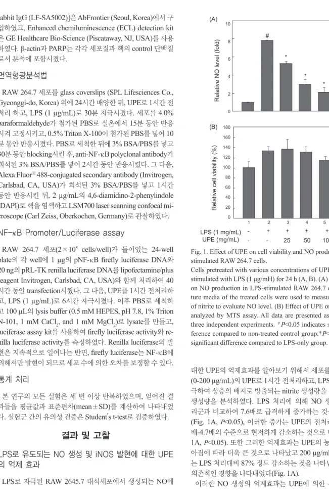
LPS로 유도되는 NO 생성 및 iNOS 발현에 대한 UPE 의 억제 효과
LPS
로자극된RAW 2645.7
대식세포에서생성되는NO
에대한
UPE
의억제효과를알아보기위해서세포를다양한농도(0-200 µg/mL)
의UPE
로1
시간전처리하고, LPS
로24
시간자 극하여상층의배지로방출되는nitrite
생성량을측정하여NO
생성량을 분석하였다. LPS
처리에 의해NO
생성량이 무처 리군과비교하여7.6
배로급격하게증가하는것을알수있고(Fig. 1A,
P<0.05),
이러한증가는UPE
의전처리에의해1.1
배-4.7
배의수준으로현저하게감소하는것으로나타났다(Fig.
1A,
P<0.05).
또한그러한억제효과는UPE
의농도가점차높아짐에따라더욱큰것으로나타났고
200 µg/mL
의농도에서는
LPS
처리대비87%
정도감소하는것을나타냄으로써농도 의존적인경향을나타내었다(Fig. 1A).
이러한
NO
생성의 억제효과는UPE
에 의한세포독성으로#
*
*
*
*
10 8 6 4 2 0
180 160 140 120 100 80 60 40 20
0 1 2 3 4 5 6
- + + + + + - - 25 50 100 200 LPS (1 mg/mL)
UPE (mg/mL)
Relative cell viability (%)
(B)
Relative NO level (fold)
(A)
- + + + + - - - 25 50 100 100 LPS (1 µg/mL)
UPE (µg/mL)
iNOS β-Actin
iNOS GAPDH
(A)
(B)
0.12±0.02 1.00*±0.00 0.55*±0.01 0.21*±0.01 0.09*±0.02 0.04±0.01
0.01±0.01 1.00*±0.00 0.49*±0.03 0.22*±0.03 0.13*±0.04 0.02±0.01
Western blot
RT - PCR
800
600
400
200
0
IL-1β (pg/mL) IL-6 (ng/mL) TNF-α (ng/mL)
(B) (A)
(C)
LPS (1 mg/mL) UPS (mg/mL)
100 80 60 40 20
0 140 120 100 80 60 40 20 0
- + + + + - - - 25 50 100 100
#
*
*
*
*
*
#
#
Fig. 1. Effect of UPE on cell viability and NO production in LPS- stimulated RAW 264.7 cells.
Cells pretreated with various concentrations of UPE for 1 h were stimulated with LPS (1 μg/mH) for 24 h (A, B). (A) Effect of UPE on NO production in LPS-stimulated RAW 264.7 cells. The cul- ture media of the treated cells were used to measure the amount of nitrite to evaluate NO level. (B) Effect of UPE on cell viability analyzed by MTS assay. All data are presented as mean±SD of three independent experiments. #P<0.05 indicates significant dif- ference compared to non-treated control group.*P<0.05 indicates significant difference compared to LPS-only group.
최민우
ㆍ
김재일388
RAW 264.7
세포가사멸되는것에의해서도나타날수있으므로
,
이러한가능성을배제하고자동일한UPE
처리조건하에서 세포생존율의변화를MTS assay
로분석하였다. Fig. 1B
에나 타내었듯이세포의생존율은UPE
처리에의해뚜렷한변화가 없었다.
이러한세포독성실험결과에서UPE
처리에의한NO
생성의억제효과는세포독성에의한것이아니라는것을확인 할수있었다.
다음은
NO
를 생성하는 효소인iNOS
의 발현에대한UPE
의효과를알아보고자하였다.
전술과같이RAW 264.7
세포 에UPE
를0-100 µg/mL
의농도로전처리한이후LPS
로자극 하였고,
이후total RNA
및cell lysate
를분리하여각각iNOS
의유전자및단백질을발현수준을분석하였다.
그림2B
에나 타내었듯이iNOS mRNA
발현양은LPS
처리에의해현저하 게높게유도되는것을알수있었고(P<0.05),
이는UPE
처리 에의해농도의존적으로감소하는경향을나타내었다(P<0.05).
iNOS
단백질의발현양도mRNA
와유사한경향으로UPE
처 리에의해현저하게감소하는것으로나타났지만,
그억제효과 는저농도인25 µg/mL
의UPE
처리부터뚜렷하게나타남으로 써전사수준에서효과적으로 조절되고있음을나타낸다(Fig.
2A, P<0.05).
NO
는NOS
에의해L-arginine
으로부터생성된다. iNOS
는 세균의endotoxin
및염증성cytokines
에의해강하게유도된다(Guha and Mackman, 2001).
병리학적인조건하에서iNOS
에의한
NO
의현저한증가는다른염증성매개체들과함께과도 한염증을유발하게되고조직의손상을유발하는것으로알려 져있어염증성손상의주요매개체이다(Nathan, 1992; Pan et al., 2011).
따라서이러한NO
의생성과iNOS
의발현및활성 을억제할수있는화합물은항염증물질로이용될수있을것이#
*
*
*
*
10 8 6 4 2 0
180 160 140 120 100 80 60 40 20
0 1 2 3 4 5 6
- + + + + + - - 25 50 100 200 LPS (1 mg/mL)
UPE (mg/mL)
Relative cell viability (%)
(B)
Relative NO level (fold)
(A)
- + + + + - - - 25 50 100 100 LPS (1 µg/mL)
UPE (µg/mL)
iNOS β-Actin
iNOS GAPDH
(A)
(B)
0.12±0.02 1.00*±0.00 0.55*±0.01 0.21*±0.01 0.09*±0.02 0.04±0.01
0.01±0.01 1.00*±0.00 0.49*±0.03 0.22*±0.03 0.13*±0.04 0.02±0.01
Western blot
RT - PCR
800
600
400
200
0
IL-1β (pg/mL) IL-6 (ng/mL) TNF-α (ng/mL)
(B) (A)
(C)
LPS (1 mg/mL) UPS (mg/mL)
100 80 60 40 20
0 140 120 100 80 60 40 20 0
- + + + + - - - 25 50 100 100
#
*
*
*
*
*
#
#
*
*
*
*
6 4 2 0
180 160 140 120 100 80 60 40 20
0 1 2 3 4 5 6
- + + + + + - - 25 50 100 200 LPS (1 mg/mL)
UPE (mg/mL)
Relative cell viability (%)
(B)
Relative NO level (fold)
- + + + + - - - 25 50 100 100 LPS (1 µg/mL)
UPE (µg/mL)
iNOS β-Actin
iNOS GAPDH
(A)
(B)
0.12±0.02 1.00*±0.00 0.55*±0.01 0.21*±0.01 0.09*±0.02 0.04±0.01
0.01±0.01 1.00*±0.00 0.49*±0.03 0.22*±0.03 0.13*±0.04 0.02±0.01
Western blot
RT - PCR
800
600
400
200
0
IL-1β (pg/mL) IL-6 (ng/mL) TNF-α (ng/mL)
(B) (A)
(C)
LPS (1 mg/mL) UPS (mg/mL)
100 80 60 40 20
0 140 120 100 80 60 40 20 0
- + + + + - - - 25 50 100 100
#
*
*
*
*
*
#
#
Fig. 2. Effect of UPE on LPS-induced iNOS protein and mRNAexpression in RAW 264.7 cells.
(A) Western blot analysis of iNOS protein expression. Cells pre- treated with various concentrations of UPE for 1h were stimulated with or without LPS (1 μg/mL) for 16 h. (B) RT-PCR analysis of iNOS mRNA expression. Cells were incubated with various con- centrations of UPE for 1 h, and then stimulated with LPS (1 μg/
mL) for 6 h. mRNA levels of iNOS, GAPDH were determined by RT-PCR analysis using respective gene-specific primers. The results presented are representatives of three independent experi- ments. Quantitative data shown underneath of each blot represent mean±SD of three independent experiments.#P<0.05 indicates sig- nificant difference compared to non-treated control group.*P<0.05 indicates significant difference compared to LPS-only group.
Fig. 3. Effect of UPE on production of pro-inflammatory cytokines in LPS-stimulated RAW 264.7 cells.
Cells pretreated with various concentrations of UPE were stimu- lated with or without LPS (1 μg/mL) for 24 h. IL-1β (A), IL-6 (B), and TNF-α (C) in the culture media were measured by ELI- SA. Data represent mean±SD of three independent experiments.
#P<0.05 indicates significant difference compared to non-treated control group.*P<0.05 indicates significant difference compared to LPS-only group.
미역 에틸아세테이트 추출물의 항염증 효과
389
다
.
본결과에서UPE
에의한NO
생성의억제는iNOS
의발현 이낮아지는것에의한것임을나타낸다.
LPS로 유도되는 염증성 Cytokines의 생성에 대한 UPE의 억제효과
LPS
로자극된RAW 2645.7
세포에서생성되는염증촉진성cytokines
의생성에대한UPE
의효과를ELISA
방법으로분석 하였다. LPS
자극에의해IL-1β, IL-6, TNF-α
와같은cytokines
의생성량은크게증가하는것으로나타났고(Fig. 3A, B, and C,
P<0.05),
이러한증가는UPE
처리에의해현저하게감소하 는것으로관찰되었다(Fig. 3A-C). IL-6
및TNF-α
의경우100 µg/mL
의UPE
농도에서만각각21%
와63%
의뚜렷한감소(
P<0.05)
를나타낸반면, IL-1β
의경우처리한모든농도에서 농도의존적으로감소하는경향을나타내었다(
P<0.05).
이들의염증촉진성
cytokines
들은체내에서다양한면역및 염증반응을조절하는역할을한다.
세균의LPS
에의해자극된 대식되는TNF-α
를생성하고분비된TNF-α
및LPS
는IL-1β
와IL-6
의생성을유도하게된다(Beutler and Ceramin, 1989).
TNF-α
는패혈성쇼크,
염증,
세포상해성등의다양한생리학적 과정에관여하고있다(Dinarello, 1999). IL-1β
는대식세포에서 생성되는주요염증촉진성cytokine
으로서,
세균감염에대한 염증성응답의개시및강화에중요한cytokine
이다(Lebovic et al., 2000). IL-6
도대식세포에서생성되는중요한염증촉진성cytokine
으로서급성면역응답에작용한다(Yoshimura, 2006).
본연구에서관찰된결과는
UPE
가LPS-
자극에의해유도되는IL-1β, IL-6, TNF-α
의생성을억제시키는것으로나타났고,
이 는UPE
가LPS-
자극에의한염증성응답의초기단계를억제하 고있음을의미한다.
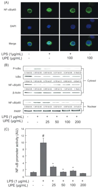
LPS로 유도되는 NF-κB의 활성화에 대한 UPE의 억 제 효과
앞에서 서술하였듯이여러 염증촉진성
cytokines
및iNOS
의발현은전사수준에주요전사인자인NF-κB
에의해조절된 다(Baeuerle and Henkel, 1994).
따라서LPS
로자극한RAW 264.7
세포에서NF-κB
의활성화변화에이에대한UPE
의처 리효과를분석하였다.
먼저면역형광법으로염색을하고con- focal microscopy
로분석한결과를Fig. 4A
에나타내었다.
아무 런자극이가해지지않은상태에서NF-κB/p65 subunit (
녹색)
는DAPI
로염색된핵(
청색)
주변에대부분분포하는것이관찰 되지만, LPS
로자극한경우녹색인NF-κB p65
의대부분은청색인핵과함께분포하는것으로나타났고이는
NF-κB
가활성화되어핵으로이동했음을보여주는결과이다
.
이러한세포에UPE (100 µg/mL)
를전처리한경우NF-κB p65
는다시핵주 변의세포질에대부분분포하는것으로관찰되었고,
이는UPE
에의해
NF-κB
의활성화가현저하게억제되고있음을보여주는결과이다
(Fig. 4A).
Fig. 4. Inhibitory effect of UPE on the degradation of IκB-α and the activation of NF-κB in LPS- stimulated RAW 264.7 cells.
(A) Cellular distribution of p65 subunit of NF-κB (green) protein analyzed by immunofluorescence staining with confocal micros- copy. Cells were pretreated with UPE (100 μg/mL) for 1 h, fol- lowed by LPS stimulation for 30 min. DAPI (blue) was used for nuclear staining. (B) Nuclear localization of NF-κB and the regula- tion of IκB-αphosphorylation analyzed by Western blot. Cells pre- treated with UPE for 1 h were stimulated with LPS for 30 min. (C) Effect of UPE on NF-κB promoter activity. Cells were transfected with 1 μg of NF-κB promoter-containing luciferase DNA for 40 h.
Transfected cells pretreated with UPE for 1 h were stimulated with LPS for additional 6 h. The results presented are representatives of three independent experiments. Quantitative data shown under- neath of the blotsin (B) represent mean±SD of three independent experiments.#P<0.05 indicates significant difference compared to non-treated control group.*P<0.05 indicates significant difference compared to LPS-only group.
NF-κB/p65
DAPI
Merge
LPS (1μg/mL)
UPE (μg/mL) - + + - - - 100 100
LPS (1 μg/mL) UPE (μg/mL)
14 12 10 8 6 4 2 0
NF- κB promoter activity (AU)
- + + + + + - - 25 50 100 200
#
* *
*
p-Akt Akt p-p38
p38 p-ERK
ERK p-JNK
JNK
β-Actin
LPS (1 μg/mL)
UPE (μg/mL) - + + + + - - - 25 50 100 100
0.18±0.03 1.00*±0.00 0.21*±0.01 0.20*±0.02 0.23*±0.07 0.19±0.04
0.15±0.02 1.00*±0.00 0.35*±0.02 0.33*±0.02 0.31*±0.01 0.17±0.02
0.82±0.01 1.00*±0.00 0.99*±0.05 0.83*±0.03 0.82*±0.03 0.78±0.02
0.12±0.03 1.00*±0.00 0.31*±0.02 0.29*±0.03 0.29*±0.04 0.17±0.03
- + + + + + - - 25 50 100 200
P-IκBα IκBα NF-κB/p65 β-Actin
NF-κB/p65 PARP
Cytosol
LPS (1 μg/mL) UPE (μg/mL)
Nuclear
0.13±0.02 1.00*±0.00 0.50*±0.03 0.31*±0.01 0.34*±0.06 0.16±0.02
0.50±0.03 1.00*±0.00 0.75*±0.03 0.52*±0.01 0.50*±0.03 0.41±0.01 1.93±0.19 1.00*±0.00 0.75*±0.07 0.78*±0.11 0.89*±0.01 0.83±0.10
1.94±0.08 1.00*±0.00 0.69*±0.02 1.08*±0.12 1.20*±0.03 1.18±0.31