Yeungnam Univ J Med 2017;34(1):106-110
비전형적인 가와사키 병 환자에서 발생한 좌주간지 거대 동맥류를 동반한 급성심근경색
김민욱, 김현수, 이명동, 정현숙, 윤성보, 김영우 홍익병원 내과
Acute myocardial infarction with a giant left main aneurysm in atypical Kawasaki disease
Min Wook Kim, Hyun Soo Kim, Myung Dong Lee, Hyun Sook Jung, Seong-Bo Yoon, Young Woo Kim
Department of Internal Medicine, Hongik Hospital, Seoul, Korea
Kawasaki disease (KD) is an acute vasculitis of small and medium sized arteries. Even many years after onset, aneurysms and stenosis in coronary arteries may lead to an acute myocardial infarction, which is described as atypical or missed KD in childhood. KD is an underlying disease of young adults with acute myocardial infarction. We report on a rare case involving a total occlusion in the proximal left anterior descending coronary artery combined with a giant left main aneurysm in a young adult patient with acute myocardial infarction ascribed to antecedent KD that is undefined but almost certain.
Keywords: Kawasaki disease; Myocardial infarction; Coronary aneurysm
Copyright ©2017 Yeungnam University College of Medicine
This is an Open Access article distributed under the terms of the Creative Commons Attribution Non-Commercial License (http://creative- commons.org/licenses/by-nc/4.0/) which permits unrestricted non-commercial use, distribution, and reproduction in any medium, provided the original work is properly cited.
Received: March 30, 2016, Revised: May 27, 2016 Accepted: June 8, 2016
Corresponding Author: Seong-Bo Yoon, Department of Internal Medicine, Hongik Hospital, 225 Mokdong-ro, Yangchun-gu, Seoul 07937, Korea
Tel: +82-2-2600-0436, Fax: +82-2-2600-4605 E-mail: kerveros@chol.com
서 론
가와사키 병은 주로 영유아 및 청소년에게 발병하는 원인 미상의 급성 다기관 침범 혈관염이다. 급성 가와사키 병을 치료하지 않은 환자의 15-25%에서 관상동맥류 또는 혈관 확장증을 가지며, 장기간 추적 관찰 동안 모든 환자의 약 5%에서 허혈성 심질환이 발생한다[1,2]. 또한 가와사키 병은 혈관 연축, 혈전 발현, 관상동맥박리 및 관상동맥변형 등이 잘 동반되며, 젊은 성인에서 심근경색을 발생시킬 수 있다 [3,4]. 그러나 가와사키 병의 후유증으로 인한 성인 환자에서 급성심근경색의 진단 및 치료에 대한 경험은 매우 적으며,
유년기에 진단을 놓친 가와사키 병의 후유증이 수년 후에 급성심근경색 및 급사로 나타날 수도 있다. 저자들은 38세 남자 환자에서 가와사키 병의 후유증으로 인한 것으로 추정 되는 좌주간지 거대 동맥류를 동반한 급성 심근경색을 진단 하고 좌전하행지 스텐트 삽입술로 치료하여, 문헌고찰과 함 께 이를 보고한다.
증 례
38세 남자 환자가 수면 중에 흉통이 한 시간 이상 지속되 어 내원하였다. 환자는 특별한 과거력, 가족력, 음주력 및 흡연력은 없었다. 내원 당시 창백한 얼굴과 식은땀을 흘리고 있었으며, 활력징후는 혈압 100/70 mmHg, 맥박 64회/분, 호 흡수 24회/분이었고, 체온은 정상이었다. 말초 혈액검사에서 백혈구 13,300/mm3, 헤모글로빈 14.9 g/dL, 혈소판 245,000/
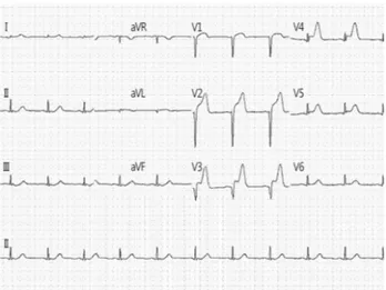
mm3, 크레아틴인산활성효소 70.0 U/L, creatine kinase-MB 0.5 ng/mL, troponin-I 0.017ng/mL였다. 심전도에서 전벽 유
Fig. 1. Electrocardiogram in the emergency room showed defi- nite ST segment elevation and Q wave in the anterior leads (V
2-5) and ST segment depression in leads III, aVF.
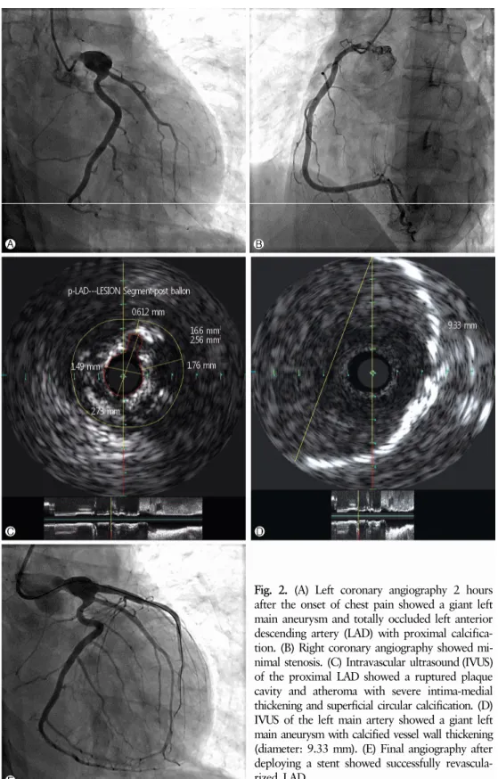
도전극(V2-5)에서 Q파를 동반한 ST분절 상승과 III, aVF 유도 전극에서 ST분절 하강을 보였다(Fig. 1). 경흉부 심초음파 검사에서 각 심방과 심실의 크기는 정상이었으나 좌심실 구 혈률은 31%로 낮았고, 좌심실의 기저부에서 첨부까지 전중 격과 전측벽 부위의 무운동 소견을 보였다. 임상 증상, 심전 도와 경흉부 심초음파 소견으로 근위 좌전하행지와 관련된 급성경색 소견으로 진단하였으며, aspirin 300 mg과 clopido- grel 600 mg을 경구로 투여한 후 morphine과 heparin을 정맥 으로 투여하였다. 응급 관상동맥조영술을 시행한 결과 우관 상동맥은 정상이었으나 좌주간지 거대 동맥류와 근위부의 석회화를 동반한 좌전하행지의 완전 폐쇄를 확인할 수 있었 다(Fig. 2A, 2B).
응급 관상동맥 중재술을 시행하기로 결정하였으며, 6-Fr Judkin 유도 카테터(Cook, Bloomington, IN, USA)를 이용하 였으며 0.014 inch choice PT 유도철선(Cook, Bloomington) 으로 병변 통과가 안되어 0.014-inch Gaia First (Asahi, Seto, Japan)으로 병변을 통과시킨 후 혈전흡입카테터(Thrombus- ter II, Kaneka, Osaka, Japan)를 이용하여 혈전흡입을 시행하 였다. 동맥 원위부부터 좌전하행동맥 근위부의 혈관 내강 협착에 대하여 2.0, 2.5 및 3.0 mm 직경의 풍선 확장술을 순차 적으로 진행한 후 intravascular ultrasound (IVUS)의 좌전하 행지로 진입이 가능했다. IVUS에서 표재성 석회 침착을 가진 심한 내막-중막 비후를 동반한 죽상경화반이 좌전하행지 근 위부에만 국한된 것을 관찰하였고(Fig. 2C), 좌전하행지 기 시부에서 시작하여 좌주간지 입구까지 약 9 mm 직경의 심한 석회 침착을 가지며, 불규칙하게 두꺼워진 벽을 가진 거대 동
맥류를 확인하였다(Fig. 2D). 병변 부위에 3.5×2.8 mm Xience Xpedition (Abbott vascular, Santa Clara, CA, USA) 스텐트를 삽입하였다. 추적 조영술 결과 좌전하행지의 혈류가 성공적 으로 회복되었고(Fig. 2E), 관상동맥 중재술 후에 증상은 거 의 소실되었다. 입원 기간 중 다른 혼합 결체조직병 및 혈관 염 등을 감별하기 위하여 혈청검사 및 자가면역항체검사를 시행하였으나 모두 음성이었으며, 다른 주요 혈관의 동맥류 를 찾기 위하여 조영증강컴퓨터단층혈관촬영(contrast en- hanced 320-slice multi-detector computed tomography an- giography)을 시행하였으나 양측 신장동맥을 포함한 다른 주 요 동맥에서 동맥류 및 협착 등의 특이 소견은 관찰되지 않았 다. 이에 저자들은 좌주간지 거대 동맥류를 동반한 급성 심근 경색이 비전형적인 가와사키 병의 후유증으로 인해 발생한 것으로 추정하였다. 현재 환자는 외래에서 흉통 없이 경과 관찰 중으로 12개월 후에 추적 관상동맥조영술을 시행할 예 정이다.
고 찰
본 증례는 다른 고식적인 위험인자 없이 좌주간지 거대 동맥류를 가진 비교적 젊은 환자에서 발생한 급성심근경색 에 대한 것으로 가와사키 병 외에 선천성 관상 동맥류, 죽상 동맥경화성 질환, 결체조직 질환, 혈관염 등을 임상양상과 관상동맥조영술을 통하여 감별하여야 한다[5].
선천성 관상동맥류는 매우 드물며, 다른 선천성 심기형과 동반되어 나타나기 때문에 성인기 이전에 증상이 나타나며, 다발성으로 나타나는 것이 특징이다[6]. 죽상동맥경화성 질 환은 대부분 위험인자를 가지며, 동맥류가 여러 혈관에 퍼져 서 나타나고 석회화 동반도 일부 동맥류에서만 보인다. 그러 나 가와사키 병에서 석회화 동반은 대개 기존 동맥류 부위에 만 국한되어 나타나며, 대개 소혈관 분지와 말초 관상 동맥들 은 침범하지 않는다고 한다[7]. 결체조직 질환 및 혈관염의 경우 대부분 특징적인 표지자가 검출되거나 신장 혈관 및 다른 혈관의 동반된 동맥류를 가진 경우가 많은데 본 증례에 서는 이러한 이상 소견을 발견할 수 없었다. 이와 같이 유년 기에 가와사키 병을 진단 받은 적이 없다고 해서 가와사키 병을 배제할 수 있는 것은 아니며 관상동맥조영술을 통하여 가와사키 병을 추정하게 되는 경우도 있다[8].
가와사키 병은 소혈관 및 중혈관을 침범하는 혈관염이며, 관상동맥을 침범하여 동맥류의 발현 및 혈관폐쇄가 합병될 때는 치명적이다[9]. 성인에서 가와사키 병 관련 관상동맥
Fig. 2. (A) Left coronary angiography 2 hours after the onset of chest pain showed a giant left main aneurysm and totally occluded left anterior descending artery (LAD) with proximal calcifica- tion. (B) Right coronary angiography showed mi- nimal stenosis. (C) Intravascular ultrasound (IVUS) of the proximal LAD showed a ruptured plaque cavity and atheroma with severe intima-medial thickening and superficial circular calcification. (D) IVUS of the left main artery showed a giant left main aneurysm with calcified vessel wall thickening (diameter: 9.33 mm). (E) Final angiography after deploying a stent showed successfully revascula- rized LAD.
후유증으로 생긴 급성 관상동맥증후군에 대한 정보는 보다 흔한 죽상동맥경화성 질환에 의한 급성 관상동맥증후군과 비교하여 매우 부족한 상황이다.
동맥류는 시간이 지남에 따라 퇴행하게 되더라도 약 20%
에서는 임상적으로 중요한 혈관 협착을 유발한다. 거대 동맥 류를 가진 환자의 약 50%에서는 동맥 협착 또는 완전 폐쇄로
진행되며, 이 환자들의 약 2/3에서 심근경색이 발생하는 것 으로 알려져 있다[7,10]. 가와사키 병에 의해 발생한 동맥류 는 아동기에 급성 발병 후에 이차적인 혈관벽 비후화와 함께 성인기까지 오랫동안 지속되는 것으로 생각된다. 또한 관상 동맥 석회화 정도는 관상동맥 혈관벽 내막층 비후화의 진행 정도를 반영하며, 급성기 동안 직경 6mm 이상으로 커진 관상 동맥류는 10년이 지난 후에는 흔히 석회화가 진행되는 것으 로 알려져 있다[11].
가와사키 병으로 인한 관상동맥류의 급성 폐쇄는 급성 또 는 아급성기 동안 주로 발생하는 혈전 형성과 함께 관상동맥 혈류의 정체에 기인하는 것으로 생각된다. 또한 장기간 지속 되는 가와사키 병에서 관상동맥 폐쇄는 석회화를 동반한 동 맥류의 입구 또는 출구 부위의 혈관 내막층의 비후화 진행에 의한 것으로 추정되나 아직 명확하지는 않다[12].
가와사키 병의 관상동맥 후유증으로 인한 심근경색이 의 심되는 환자의 관상동맥조영술 중에 실시하는 IVUS는 관상 동맥 혈관벽의 형태분석, 혈전 분포의 정확한 측정, 그리고 중재 시술 기구 선택에 도움을 준다[13]. 본 증례는 과거에 가와사키 병의 진단 병력은 없었지만 관상동맥조영술에서 석회화를 동반한 좌주간지의 거대 동맥류가 확인되었고, IVUS에서 파악된 병변도 좌주간지 및 좌전하행지의 근위부 에 국한된 표재성 석회화를 동반한 심한 내막-중막비후를 보 여서 급성 심근경색이 가와사키 병의 후유증으로 인한 것임 을 추정하였다. Kato 등[5], Takahashi [8], 그리고 Newburger 등[14]의 연구에서도 가와사키 병의 진단 기준을 충족하지 못한 관상동맥류를 동반한 환자들이 선행된 비전형적인 가 와사키 병을 가지고 있었음을 관상동맥조영술, IVUS 등의 검사를 통하여 진단할 수 있음을 보여 주었다.
가와사키 병의 후유증으로 발병한 급성 관상동맥증후군 치료로서 관상동맥중재술은 풍선 혈관성형술(balloon angio- plasty), 회전 연마술(rotational ablation), 스텐트 삽입술(stent placement)을 포함하나, 이에 대한 연구는 상대적으로 부족하 다. 일반적으로 발병 2년 후부터는 동맥혈관 벽의 조밀한 섬유화 및 석회화가 진행되어 풍선 혈관성형술만으로는 성공 하기 어려우며, 높은 압력의 풍선 혈관성형술의 시행이 새로 운 동맥류를 유발하기도 한다. 이런 이유로 10기압 미만의 풍선 혈관성형술이 권고되고 회전 연마술 및 스텐트 삽입술 의 추가 시술이 필요하다. 스텐트 삽입술은 거대 동맥류 및 석회화가 동반된 나이가 든 환자에서 유용한 것으로 알려져 있으며, 회전 연마술과 병행하여 시행한 경우 80% 이상의 성공률을 보였다[14].
본 증례는 젊은 남자 환자에서 선행된 가와사키 병의 후유 증으로 인한 것으로 추정되는 좌주간지 거대 관상동맥류를 동반한 급성 심근경색을 진단하고 좌전하행지 스텐트 삽입 술로 치료한 경우이다. 특히 관상동맥조영술 중에 실시한 혈관 내 초음파 검사를 통해 유년기에 진단을 놓친 것으로 의심되는 비전형적인 가와사키 병의 존재를 추정해 볼 수 있었다. 비전형적인 가와사키 병의 후유증으로 인한 관상동 맥류를 동반한 심근경색 발생의 증례는 매우 드물지만, 급성 관상동맥증후군의 위험인자 및 기저질환으로서 고려해 볼 가치가 있으며 이것에 대한 적절한 진료 지침 및 치료 방법을 결정하기 위해서 더 많은 연구 및 추적관찰이 필요할 것으로 생각된다.
CONFLICT OF INTEREST
No potential conflict of interest relevant to this article was reported.
REFERENCES