Flavonoid의 HT-29 대장암세포 증식 억제 효과
조 영․최미용 한국방송통신대학교 가정학과
Inhibitory Effects of Flavonoids on Growth of HT-29 Human Colon Cancer Cells
Young Cho and Mi-Yong Choi
Department of Home Economics, Korea National Open University
ABSTRACT This study was performed to elucidate the anti-proliferative and apoptotic mechanism of flavonoids in HT-29 human colon cancer cells. We investigated the anti-proliferative activity of flavonoids in HT-29 human colon cancer cells via cell viability assay (MTT assay), caspase-3 activity, RT-PCR, and western blotting. We cultured HT-29 cells in the presence of various flavonoids (apigenin, rutin, naringenin, and myricetin) at a concentration of 100 μM. In the MTT assay, naringenin showed the strongest effect on cell viability in HT-29 colon cancer cells.
Caspase-3 activity, a marker of apoptosis, significantly increased upon naringenin treatment. For RT-PCR, myricetin significantly increased Bax protein levels, naringenin increased p53 protein levels, and rutin reduced expression of the anti-apoptotic protein Bcl-2. Western blotting of HT-29 colon cancer cells showed that myricetin increased cleaved caspase-3 protein levels, naringenin significantly increased poly (ADP-ribose) polymerase protein levels, and rutin increased E-cadherin protein levels. These results indicate that flavonoid exerts anticancer effects on human colon HT-29 cells through a caspase-dependent apoptotic pathway.
Key words: colon cancer, flavonoid, HT-29 human, RT-PCR, western blot
Received 13 October 2014; Accepted 29 January 2015
Corresponding author: Young Cho, Department of Home Econom- ics, Korea National Open University, Seoul 110-791, Korea E-mail: [email protected], Phone: +82-2-3668-4640
서 론
대장암은 소화기계 암으로 미국 등 서구사회에서 발병률 과 사망률이 높은 것으로 알려졌으나 우리나라에서도 식습 관이 서구화됨에 따라 급증하고 있으며(1,2), 노령인구의 증 가와 대장암 진단 기술의 발달로 인하여 대장암의 발병도 증가하는 추세에 있다(3). 또한 우리나라 사망원인 1위인 암 중에서도 그 발생에 있어 가장 급격하게 증가하고 있는 암 중에 하나이다(4). 대장암은 서구 남녀 모두에게 높은 발 병률을 갖고 있으며 외과적 수술로 치료할 수 있음에도 불구 하고 50%의 생존율을 갖는 악성종양 중의 하나로 수술 후 암 전이의 예방에 큰 관심이 집중되고 있다(1-5). 대장암의 주요 원인으로는 과도한 동물성 지방, 당분, 알코올 섭취와 섬유소, 항산화 비타민, 채소나 과일의 섭취 부족 등이 있다 (6). 대장암의 치료에는 일반적으로 수술 이외에 화학요법, 방사선요법 등이 행해지고 있으나, 이는 면역기능저하, 유전 자 손상 및 탈모와 같은 여러 부작용을 유발시킬 수 있다고 알려져 있어 효과적인 치료에 많은 문제점이 제시되고 있는 실정이다(7-9). 또한 암 치료를 위해 현재 사용되고 있는 항암제는 암세포뿐만 아니라 정상세포에도 영향을 미쳐 독
성 및 다양한 부작용을 야기하고 있다(10). 이로 인해 안전 성이 높고 항암 효과가 높은 천연자원 소재의 항암제에 관한 개발이 요구되어 현재 안전성이 높은 생리활성물질 탐색, 이를 이용한 기능성 식품 및 의약품 개발 등에 관한 연구가 활발히 진행되고 있다(11).
Flavonoid는 식물에 광범위하게 분포되어 있어 대부분의 모든 녹색 식물에서 발견되는 물질로, 식물에 의해서 광합성 되는 탄소의 약 2%가 flavonoid와 관련된 화합물로 변환되 며 그 구조적 특이성 때문에 생체 내에서 다양한 약리작용이 있음이 보고되었다(12,13). 특히 항산화, 항균, 항혈전, 항염 증, 심혈관계, 순환기계 질환 예방 및 항암 등에 효과가 있는 것으로 밝혀져 있다(14,15).
Apigenin은 일반적인 방향족 화합물의 하나로서 fla- vonoid 중 flavones에 속하며 파슬리, 양파, 오렌지, 차, 카 모마일, 밀 그리고 몇몇 조미료 등 많은 열매와 식물에 분포 하고 있으며, 독성이 없는 것으로 알려져 있다(16). 그리고 apigenin은 염증과 산화적 스트레스를 억제하고 전립선암, 유방암, 결장직장암, 폐암 등의 발병 위험성을 높이는 요인 으로 알려진 인슐린 유사 성장 호르몬-1(IGF-1)의 수치를 감소시키며, 또한 항산화와 항암 효과가 있고 interleukin-1 β와 TNF-α를 억제하여 항염증 작용을 한다고 밝혀져 있다 (17-20).
Rutin은 polyphenolic flavonoid로 과일, 야채, 차, 와인 등에 포함된 물질이고 항산화, 항혈전, 항염증 효과 등의 약
물학적 가치를 인정받고 있으며, 혈당 강하 효과, 지방분해 효과, 심근세포의 보호 효과, 간세포 보호 효과 등이 소수의 연구에서 보고된 바 있다(21-23).
Naringenin은 flavonoid의 일종인 flavanone 무배당체 로서 벤젠고리를 2개 가지고 있으며 포도와 오렌지, 유자 등의 감귤류에 많이 들어 있고 항산화제, 항암작용을 한다고 보고되어 있으며, 최근 연구에서 위암과 췌장암, 간암세포의 악성세포의 성장을 중지시키는 작용과 발암물질에 의해 손 상되어진 세포를 보호하는 항암작용을 한다고 알려져 있다 (24-26).
Myricetin는 flavonoid 중 flavonols에 속하고 포도, 과 일, 채소, 허브 등에 있으며 월넛에 다량 함유되어 있다. 또한 항염, 항암, 항바이러스, 항균 및 혈소판 응집 예방 역할을 한다(27-31).
Apoptosis라 불리는 세포사멸은 세포가 정상적인 상태에 서 또는 병리학적인 요인에 의하여 스스로 죽음의 길을 선택 하는 것을 말하는 것으로 계획된 세포의 죽음(programmed cell death)을 의미한다. 이는 정상세포의 기능 유지와 발달 에 매우 중요한 역할을 하고, 세포의 다양한 신호전달 경로 에 의해 조절되므로 세포사멸 기전에 결함이 생기면 세포가 비정상적으로 증식하여 암을 비롯한 여러 질병을 초래할 수 있다. 세포사멸은 세포 내외적인 세포사멸 신호에 의해 시작 되어 세포사멸을 조절하는 단백질인 Bcl-2 family 단백질, caspases 등의 다양한 조절 인자에 의해 조절된다. 대장 상 피는 지속적으로 세포교체(cell turnover)가 이루어지는 곳 으로 세포사멸이 적절하게 이루어지지 않을 경우 대장암이 발생하기 때문에 대장 상피의 기능을 유지하기 위해서 적절 한 세포사멸은 필수적이다(32,33). 그러므로 apoptosis를 유도하는 각 성분의 apoptosis 유도 기전 연구는 apoptosis 유도를 통해 항암활성을 나타내는 항암제 개발에 매우 중요 하다(1). 따라서 암세포의 사멸효과가 apoptosis의 유도에 의한 것인지 확인하기 위해서 apoptosis 기전에 관여하는 많은 유전자들의 발현을 RT-PCR, western blot, 면역세포 화학염색법 등 분자생물학적인 검사기법으로 확인하게 되 는데 이들 유전자 중에서 p53, Bcl-2, Bax, caspase-3 등 은 apoptosis 기전에서 매우 중요하다(3).
본 연구에서는 인간에서 유래한 대장암 세포주인 HT-29 세포를 사용하여 여러 종류의 flavonoid를 처리하고 암세포 증식 억제 효과와 세포사멸에 관여하는 유전자들의 발현을 확인하여, 대장암 치료제로 독성과 부작용이 적은 천연물 소재인 flavonoid를 이용한 항암제 개발 연구에 기초 자료를 제공하고자 한다.
재료 및 방법
실험재료
실험에 사용된 HT-29 세포는 ATCC(American Type Culture Collection, Manassas, VA, USA)에서 구입하였
다. 세포배양에 사용한 Dulbeeco's modified Eagle's me- dium(DMEM)은 10% FBS(fetal bovine serum)와 100 unit/mL의 penicillin 및 100 ng/mL의 streptomycin과 함 께 37°C, 5% CO2 세포 배양기에서 배양하여 실험에 이용하 였다.
시약
Caspase-3 assay kit은 Caspase-3/CCP32 Colorimetric Assay Kit(BioVision, Milpitas, CA, USA)을 구입하였다.
그리고 apigenin, myricetin, naringenin, rutin의 시약, 3- (4,5-dimethylthiazol-2-yl)-2,5-diphenyltetrazolium bromide(MTT), anti-β-actin 항체와 본 연구에 사용한 일 반적인 시약은 Sigma-Aldrich Co.(St. Louis, MO, USA)에 서 구입하였다. Bcl-2, Bax, p53 항체는 Santa Cruz Bio- technology Inc.(Santa Cruz, CA, USA)에서 구입하였고, cleaved caspase-3, E-cadherin, PARP에 대한 항체는 Cell Signaling(Beverly, MA, USA)에서 구입하여 사용하 였다. Horse radish peroxidase-linked anti-rabbit IgG와 horse radish peroxidase-linked anti-mouse IgG는 Amersham International(Amersham, Buckinghamshire, UK)에서 구입하여 사용하였다.
세포배양
HT-29 세포는 DMEM 배지에 10% FBS, 100 units/mL penicillin과 100 μg/mL streptomycin을 첨가한 세포 배양 액을 사용하여 37°C 습윤한 CO2 배양기(5% CO2/95% air) 에서 배양하였다. 세포가 배양 접시에 80% 정도 포화되면 phosphate-buffered saline(PBS, pH 7.4)으로 세포의 단 층을 씻어 낸 후 0.25% trypsin-2.65 mM EDTA로 처리하 여 계대 배양하였고 배지는 2일마다 교환하였다.
MTT assay
MTT assay는 12 well plate에서 암세포를 1×10⁶ cells/
mL씩 분주하여 48시간 배양 후 시료가 함유된 배지를 제거 한 뒤 MTT를 pH 7.4의 phosphate-buffered saline(PBS) 에 5 mg/mL로 용해시키고, 각 well에 apigenin(0, 50, 100, 200 μΜ), rutin(0, 50, 100, 200 μΜ), naringenin(0, 50, 100, 200 μΜ), myricetin(0, 50, 100, 200 μΜ)을 분주하여 37°C에서 5% CO2가 유지되는 배양기에서 3시간 더 배양하 였다. 배양 종료 후 상등액을 제거하고 각 well에 1 mL의 DMSO(dimethyl sulfoxide)를 첨가하여 생성된 formazan 결정을 용해시켜 다시 96 well plate에 100 μL씩 분주한 후 microplate reader(Versa Max, Molecular Devices, Sunnyvale, CA, USA)에서 540 nm로 흡광도를 측정하였 다.
Cell viability (%)=
Absorbance of sample
Absorbance of control ×100
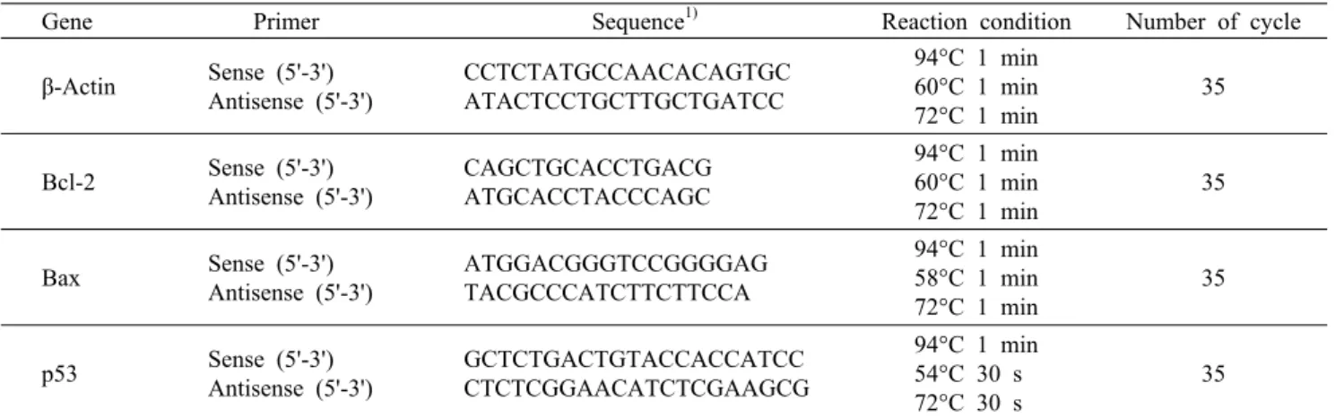
Table 1. Primers and reaction conditions for RT-PCR
Gene Primer Sequence1) Reaction condition Number of cycle β-Actin Sense (5'-3')
Antisense (5'-3')
CCTCTATGCCAACACAGTGC ATACTCCTGCTTGCTGATCC
94°C 1 min 60°C 1 min 72°C 1 min
35
Bcl-2 Sense (5'-3') Antisense (5'-3')
CAGCTGCACCTGACG ATGCACCTACCCAGC
94°C 1 min 60°C 1 min 72°C 1 min
35
Bax Sense (5'-3') Antisense (5'-3')
ATGGACGGGTCCGGGGAG TACGCCCATCTTCTTCCA
94°C 1 min 58°C 1 min 72°C 1 min
35
p53 Sense (5'-3') Antisense (5'-3')
GCTCTGACTGTACCACCATCC CTCTCGGAACATCTCGAAGCG
94°C 1 min 54°C 30 s 72°C 30 s
35
1)A: adenine, C: cytosine, G: guanine, T: thymine.
Caspase(cyseinyl aspartate-specific proteinases)-3 activity assay
Caspase-3 activity 측정에 사용된 Caspase-3 assay kit은 Caspase-3/CCP32 Colorimetric Assay kit(Bio- Vision, Inc., Mountain View, CA, USA)을 사용하였다.
HT-29 세포를 1×10⁶ cells/mL가 되도록 희석하고 60 mm dish에 분주하여 37°C, 5% CO2 incubator에서 24시간 배 양한 후 HT-29 세포가 80% 증식하면 apigenin(100 μΜ), rutin(100 μΜ), naringenin(100 μΜ), myricerin(100 μΜ) 을 처리하여 24시간 반응시켰다. 반응이 종료된 세포를 회 수한 후 lysis buffer로 세포를 회수하여 50 μΜ DEVD-ρΝ Α, caspase-3 substrate가 포함되어 있는 10 mM DTT를 혼합하여 37°C에서 1시간 동안 반응시킨 다음 ρΝΑ의 en- zyme-catalyzed 방출을 405 nm의 파장에서 ELISA plate reader(Versa Max, Molecular Devices)를 이용하여 흡광 도를 측정하였다.
mRNA 발현 실험(RT-PCR)
RNA 분리: HT-29 세포를 1×10⁶ cells/mL가 되도록 희석하고 60 mm dish에 분주하여 37°C, 5% CO2 in- cubator에서 24시간 배양한 후 HT-29 세포가 80% 증식하 면 apigenin(100 μΜ), rutin(100 μΜ), naringenin(100 μΜ), myricetin(100 μΜ)을 처리하여 24시간 반응시켰다. 총 RNA의 추출은 Trizol(Invitrogen Co., Carlsbad, CA, USA) 을 이용하여 제조사 지시 방법대로 시행하였다. PBS로 세척 한 세포에 Trizol을 1 mL 첨가하고, 마이크로 피펫을 이용 하여 균질화시켰다. 그 후 여기에 0.2 mL의 chloroform을 첨가하여 혼합한 후 10분간 원심분리(10,000×
g
, 4°C) 하여 상층액을 취해 동량의 isopropanol을 첨가한 다음 실온에 10분간 둔 후 10분간 원심분리(10,000×g
, 4°C) 하였고, 침 전된 RNA를 70% ethanol로 세척해 공기 중에서 말렸다.이것을 RNase-free water에 녹여 Genequant pro(Amer- sham Pharmacia Biotech, Inc., Piscataway, NJ, USA)를 이용하여 260 nm에서 흡광도를 측정하고 RNA 양을 정량하
였다.
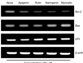
mRNA 발현(RT-PCR): 각 실험군의 총 RNA를 주형으 로 AccuPower® RT PreMix(Bioneer, Daejeon, Korea) kit을 이용하여 reverse transcription을 시행하였다. Re- verse transcription을 통하여 얻은 cDNA(complementary DNA)를 주형으로 하여 각각의 유전자에 대한 PCR을 2x Taq Pre-Mix2(SolGent, Daejeon, Korea) kit을 이용하여 수행하였다. 준비된 실험군과 primer, 2x Taq Pre-Mix를 넣고 Table 1과 같이 cycle을 실행하였다. PCR product는 2% agarose gel 상에서 전기영동 한 후 ethidium bromide (EtBr)로 염색하여 확인하였다. 각 mRNA에 대한 RT-PCR 산물의 측정된 양은 일반적으로 모든 세포에서 일정하게 발 현되는 β-actin에 대한 산물의 양으로 측정하여 발현 정도 를 비교하였다. 각 밴드는 사진을 찍어 Image J program (National Institutes of Health, Bethesda, MD, USA)을 이용하여 밀도를 측정하였다. 이번 실험에서는 RT-PCR 방 법으로 apotosis와 관련된 marker인 Bcl-2, Bax, p53의 mRNA 발현 정도를 밴드로 알아보자고 하였다.
단백질 발현 실험(western blot analysis)
HT-29 세포를 1×10⁶ cells/mL가 되도록 희석하고 60 mm dish에 분주하여 37°C, 5% CO2 incubator에서 24시간 배양한 후 HT-29 세포가 80% 증식하면 apigenin(100 μΜ), rutin(100 μΜ), naringenin(100 μΜ), myricetin(100 μΜ) 을 처리하여 24시간 반응시켰다. 차가운 PBS를 이용하여 세포를 씻어내고, 차가운 lysis buffer(20 mM Tris-HCl<
pH 7.5>, 150 mM NaCl, 1 mM Na2EDTA, 1 mM EGTA, 1% NP-40, 1% sodium deoxycholate, 2.5 mM sodium pyrophosphate, 1 mM β-glycerophophate, 1 mM Na3VO4, 1 μg/mL leupeptinTriton, 0.1 M PMSF)를 300 μL씩 넣어 shaking 한 후, ice에서 20분간 반응시킨 다음 4°C, 14,000 rpm에서 15분간 원심분리(Eppendorf, Hamburg, Ger- many) 한 후 상층액을 모아 시료로 사용하였다. Bio-rad protein assay kit(BioRad, Richmond, CA, USA)으로 단백
a b d c
a b c c
a b d c
a b d c
0 20 40 60 80 100 120
None Apigen
in 50 uM
Apigenin 100uM Apigenin 2
00uM None Rutin 50uM
Rutin 10 0uM Rutin 200uM
None Naringenin 50
uM
Naringen in 10
0uM
Naringen in 200uM
None Myricetin 50
uM
Myricetin 100uM Myricetin 200uM
Cell Viability (%) .
Fig. 1. Effects of flavonoid on HT-29 cell viability by MTT
assay. Cell viability at the indicated concentration (0, 50, 100, 200 μΜ) of flavonoid (apigenin, rutin, naringenin and myricetin) in HT-29 colon cancer cells at 48 h as assessed by MTT assay.The values are mean±SD of three independent experiments and the control levels were set at 100. Means with different letters (a-d) above the bars are significantly different at P<0.05.
질을 정량하고, 4~20% gradient sodium dodecyl sulfate polyacrylamide gel electrophoresis(SDS PAGE)에서 단 백질을 분리한 후, PVDF membrane으로 실온에서 1시간 transfer 한 뒤에 5% skim milk/TBST(20 mmol Tris- HCl, 137 mmol NaCl, 0.1% Tween 20, pH 7.4)로 실온에 서 1시간 incubation 한 후 알아보고자 하는 단백질의 anti- body를 사용하여 incubation 시켰다. TBST로 3회 세척 후 다시 anti-mouse 1 g horseradish peroxidase/TBST (Amersham)로 incubation 시켰고, TBST로 3회 세척 후 ECL detection kit(Amersham Life Science, Cleveland, USA)으로 처리해서 X-Omat film(Agfa, Gent, Belgium)으 로 현상하여 각 밴드는 Image J program(National Insti- tutes of Health)을 이용하여 밀도를 측정한 후 분자량을 비교하여 분석하였다. 이때 표준비교군으로서 일반적으로 모든 세포에서 일정하게 발현되는 β-actin을 측정하여 단백 질 발현 정도를 비교하였다. 이번 실험에서는 western blotting 방법으로 apotosis와 관련된 cleaved caspase-3, E-cadherin, PARP 단백질의 발현 정도를 밴드로 확인하였 다.
통계처리
실험에 관련된 통계처리는 SPSS(Statistical Package for Social Science, version 19.0, SPSS Inc., Chicago, IL, USA)를 이용하였고, 분산분석(ANOVA)으로
P
<0.05 수준에서 Duncan의 다중범위검증법(Duncan's multiple range test)에 의해 시료 간의 유의차를 검증하였다.결과 및 고찰
MTT assay
MTT assay는 살아있는 세포의 양을 측정하는 표준 비색 분석법(standard colorimetric assay)이다. 즉 살아 있는 세포에 MTT[3-(4,5-dimethylthiazol-2-yl)-2,5-diphenyl- tetrazolium bromide]를 처리하게 되면, MTT가 mito- chondria에 있는 reductase에 의해 환원되어 formazan이 라 불리는 crystal이 형성되는 원리를 이용한 것이다. 따라 서 어떤 물질이 세포독성을 가진 물질일 경우 이 물질 처리 에 의해 세포를 죽이게 되면 살아 있는 세포에만 formazan 이 형성되므로 formazan 형성 정도로 세포 생존율을 알 수 있는 것이다(34).
Flavonoid인 apigenin, rutin, naringenin, myricetin이 HT-29 세포의 증식에 미치는 영향을 확인하기 위하여 MTT assay를 이용하여 농도에 따른 대장암 세포 증식 억제 정도 를 측정하였다. Flavonoid를 각각 0, 50, 100, 200 μΜ 농도 로 48시간 동안 처리하였을 때, apigenin은 100, 91.95, 62.71, 41.95%, rutin은 100, 94.17, 75.78, 64.13%, nar- ingenin은 100, 93.43, 72.24, 54.54%, myricetin은 100, 93.53, 77.61, 65.17%로 나타나 농도가 증가할수록 유의적
으로 대장암 세포 증식 억제 효과가 높아지는 것을 확인했 다. 따라서 이상의 실험 결과를 바탕으로 이번 실험에서는 최소한의 용량으로 가장 효율적으로 대장암 세포 증식 억제 효과가 있는 농도인 100 μΜ을 선정하여 추후 실험에서 fla- vonoid의 apotosis에 대해 미치는 영향을 알아보았다(Fig.
1).
Caspase-3 activity
Caspase는 세포사멸이 유도될 때 활성화되는 시스틴 단 백질 분해 효소로 pro-caspase로 합성되어 있다가 신호 전 달 자극이 오게 되면 large subunit과 small subunit으로 절단되며, 이는 tetramer를 형성하여 활성화된다. 이렇게 활성화된 caspase는 PARP를 포함한 중요한 기질들을 분해 하여 oligonucleotide DNA fragmentation에 관여하게 된 다. Caspase의 아종인 caspase-8, -9와 같은 caspase 기 시체는 caspase-3, -7과 같은 caspase 효과체를 직접 또는 간접적으로 활성화시키기 때문에 결과적으로 caspase의 분 해 작용은 세포를 파괴시키므로 caspase-3의 경우는 세포 사멸의 초기지표가 된다(35-37).
이번 실험에서 각각의 flavonoid의 caspase-3 활성은 Colorimetric Assay kit을 사용하여 측정하였다. 결과는 대 조군에 비해 flavonoid 처리군이 유의적으로 활성을 증가시 켰고 그중 naringenin이 241.46%로 control군보다 2.4배 높은 값으로 활성이 가장 컸으며(
P
<0.001), 다음으로 api- genin은 175.61%로 control군보다 1.75배 높은 값을 나타 냈고, rutin과 myricetin은 126.83%와 129.27%로 서로 비 슷한 결과를 보여 이번 연구에서 초기 세포사멸에 가장 큰 효과가 있는 것은 naringenin으로 확인되었다(Fig. 2).HT-29에서 apoptosis 관련 mRNA 발현
중합효소 연쇄반응(polymerase chain reaction: PCR)
b d
b c
a
0 50 100 150 200 250 300
None Apigenin Rutin Naringenin Myricetin
Caspase-3 activity (%) .
Fig. 2. Effects of flavonoid on HT-29 cell viability by caspase-3
activity. Cell viability at a concentration (100 μΜ) of flavonoid (apigenin, rutin, naringenin, and myricetin) in HT-29 colon can- cer cells at 24 h as assessed by caspase-3 activity. The values are mean±SD of three independent experiments and the control levels were set at 100. Means with different letters (a-c) above the bars are significantly different at P<0.001.None Apigenin Rutin Naringenin Myricetin Bcl-2
Bax
p53
β-actin
Concentration 100 μM
Fig. 3. Dose-dependent effect of flavonoid (apigenin, rutin, nar-
ingenin, and myricetin) on the expression levels of Bcl-2, Bax, and p53 proteins in HT-29 cells. Cells were incubated for 24 h at 37°C with 100 μM of flavonoid (apigenin, rutin, naringenin, and myricetin) by RT-PCR. Photograph of chemiluminiscent de- tection of the RT-PCR.은 DNA의 원하는 부분을 증폭하는 것으로 내열성 DNA polymerase와 두 개의 합성 oligonucleotide(primer)를 이 용하여 목적 영역의 DNA를 특이적으로 증폭하는 방법이다 (38). 그러므로 PCR은 RNA의 발현 양상을 관찰하는 방법 이고, RT-PCR(reverse transcription-polymerase chain reaction)은 유전자가 발현되는 양을 밴드 두께로 확인하는 방법이다. Bcl-2 family 단백질은 mitochondria의 막 투과 성과 cytochrome-c의 방출을 제어하여 세포사멸을 조절하 는 것으로 알려져 있다. Bcl-2 family 단백질은 기능과 아미 노산 서열의 유사성에 따라 Bcl-2(B cell lymphoma-2)는 anti-apoptotic 단백질, Bax(Bcl-2 associated x)는 대표 적인 pro-apoptotic 단백질로 구분된다(6,39). Bcl-2 fam- ily는 apoptosis 진행에 영향을 주는 인자인데 Bax 단백질 은 세포의 증식을 억제해 apoptosis를 촉진하여 apoptosis 유도 시 발현이 증가하는 단백질이다(3). 그리고 Bcl-2 단 백질은 mitochondria의 외막, 소포체나 핵막에 위치해 있 고, 다른 암유발유전자와는 달리 세포증식에는 관여하지 않 고 세포의 생존을 조절하는 것으로 밝혀졌다. 또한 다양한 기전을 통해 세포사멸을 방지하며 caspase의 작용 기전에 영향을 미치는 것으로 보고되고 있으며, apoptosis를 억제 하는 인자로 apoptosis 유도 시 발현이 감소된다(40). 종양 억제 유전자의 산물인 p53은 세포 증식을 억제하며, 유전자 가 손상되었을 경우 복구하거나 유전자 손상에 의한 괴사를 유도하게 되고, Bcl-2의 불활성화를 직접 유도하여 apop- tosis를 유도하는 유전자로 암의 생성 및 예방에 중요한 역 할을 하는 것으로 알려져 있다(6).
Flavonoid가 세포사멸의 중요한 조절인자인 Bcl-2, Bax, p53의 mRNA 발현에 미치는 영향을 조사하기 위해 HT-29 세포에 RT-PCR을 실시하여 세포사멸을 억제하는 유전자 인 Bcl-2와 사멸을 유도하는 유전자인 Bax, 종양 억제유전 자의 산물인 p53을 통해 대조군인 β-actin과 비교 분석하였 다. 실험 결과 Bcl-2 단백질은 flavonoid 처리 시 유의적으
로 모두 감소하였으며, apigenin은 38.08%, rutin은 10.31
%, naringenin은 30.46%, myricetin은 40.28%로 그중 ru- tin이 가장 낮은 발현 양상을 나타냈고, Bax 단백질은 myr- icetin이 206.53%로 control군보다 2배 높게 발현되었다.
그리고 p53 단백질에서는 rutin이 115%, naringenin이 117.30%의 비슷한 발현 양상을 나타내 rutin과 naringenin 은 p53 단백질이 apoptosis를 유발하는 경향이 비슷한 것으 로 추정된다(Fig. 3, 4).
HT-29에서 apoptosis 관련 단백질 발현
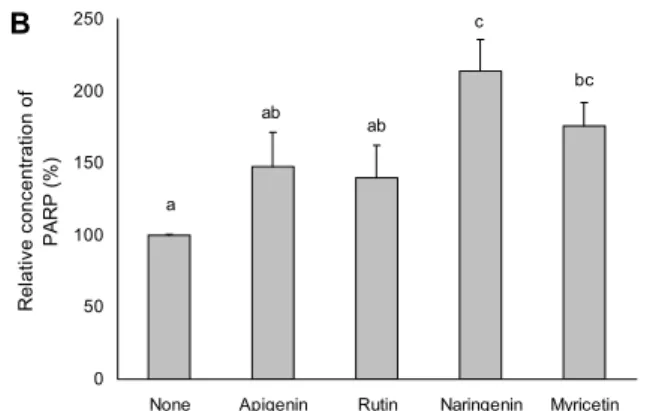
Western blotting(WB)은 단백질 확인 방법으로 여러 가 지 단백질들을 포함하고 있는 세포 또는 조직의 추출물을 전기 영동하여 그 속에 들어 있는 단백질들을 크기 별로 구 분한 다음 흡착지에 옮겨 놓고 조사하고자 하는 단백질의 항체와 결합시켜 그 단백질의 존재 유무와 크기 등을 밝히는 방법이다. 그러므로 면역 blotting이라고도 부른다. 어떤 단 백질이 일정한 세포 속에 발현되고 있는가를 확인하거나 그 발현되는 단백질의 크기 변화를 관찰하는 데 이용된다.
Caspases는 cysteine aspartic acid proteases로 cell repair 효소들을 분해시키고 세포 내 단백질의 분절을 유도 하여 세포사멸을 유도한다(32). 이 중 활성화된 caspase-3 은 apoptosis의 형태학적 특징에 관여하는 PARP 등의 단백 질을 분해하고, caspase-8, -9의 신호를 증폭시켜 최종적 으로 세포사멸을 유도한다(41). PARP(poly(ADP-ribose) polymerase)는 caspase와 상호 작용으로 핵에서 손상된 DNA를 인지하여 활성화된 후, 여러 단백질을 poly(ADP- ribose) action이라는 post-translation 과정을 통해 손상 을 복구하려고 하는 인자로 알려져 있다(42,43). 그러나 DNA 손상이 너무 심하여 PARP의 활성이 과다하게 높아지
a a
a a
b
0 20 40 60 80 100 120 140
None Apigenin Rutin Naringenin Myricetin
Bcl -2 Transcription (%) .
A
a a
a a b
0 50 100 150 200 250 300
None Apigenin Rutin Naringenin Myricetin
Bax Transcription (%) .
B
a a a
a a
0 50 100 150 200 250
None Apigenin Rutin Naringenin Myricetin
p53 Transcription (%) .
C
Fig. 4. Effects of the flavonoid (apigenin, rutin, naringenin, and
myricetin) on the protein levels of Bcl-2 (A), Bax (B), and p53 (C) in HT-29 cells. Cell lysates were analyzed by RT-PCR with and antibody against Bcl-2, Bax, p53, and β-actin. The values are mean±SD of three independent experiments and the control levels were set at 100. Means with different letters (a,b) above the bars are significantly different at P<0.05.None Apigenin Rutin Naringenin Myricetin
Cleaved Caspase-3
PARP
E-cadherin
β-actin
Concentration 100 μM
Fig. 5. Dose-dependent effect of flavonoid (apigenin, rutin, nar-
ingenin, and myricetin) on the expression levels of cleaved cas- pase-3, PARP, and E-cadherin proteins in HT-29 cells. Cells were incubated for 24 h at 37°C with 100 μM of flavonoid (apigenin, rutin, naringenin, and myricetin) by western blotting.Photograph of chemiluminiscent detection of the western blotting.
면서 급격한 에너지의 소모로 세포사멸을 유도하며 이때 caspase-3가 PARP의 DNA의 결합 부위와 활성 부위를 분 리하여 PARP의 활성을 막고 세포사멸을 일으키는 것으로 알려져 있다. PARP는 caspase-3의 주요한 표적 단백질 중 의 하나로(44), caspase-3에 의해 쪼개어져 불활성화되면 세포의 분해가 촉진되므로 cleaved PARP 수준 증가는 세 포가 apoptosis 되고 있음을 나타낸다(45). Cadherin은 유
착접합부를 구성하는 단백질 중 하나로 세포 표면에 위치하 여 세포 바깥에서 이웃의 cadherin과 동종 결합을 하고 세포 내부에서는 catenin과 결합을 하여 정상세포 간 결합에 매 우 중요한 역할을 하는 단백질이다(46,47). E(epithelial)- cadherin은 cadherin의 주된 아형의 하나로 발현 저하 시 종양세포로부터의 유리가 쉽게 이루어져 전이를 촉진시킬 수 있음이 알려져 있으며 여러 암에서 종양의 진행 상태와의 관련이 확인되었다(47,48).
본 연구에서는 HT-29 세포에서 세포사멸을 유도한 fla- vonoid가 cleaved caspase-3, PARP와 E-cadherin의 단 백질 수준을 WB 방법으로 관찰하였다. 실험 결과 flavonoid 처리 시 cleaved caspase-3 단백질은 apigenin이 108.94%, rutin이 105.83%, naringenin은 111.49%, myricetin은 112.74%로 나타나 myricetin이 가장 높은 수치를 나타내 세포사멸을 유도하는 것으로 나타났고, PARP 단백질에서 naringenin은 213.62%로 control군보다 거의 2.1배 높게 발현되었으며, E-cadherin의 단백질 수준은 rutin이 137.99
%로 가장 높은 결과를 보였다(Fig. 5, 6).
요 약
본 연구에서는 최근 식생활의 서구화로 인해 발병률이 급증 하고 있는 대장암의 진행을 억제하거나 감소시키고 인체 대 장암 세포인 HT-29의 증식을 억제하며, 세포사멸을 유도하 는 천연소재를 알아보기 위해서 flavonoid가 HT-29 인체 대장암 세포의 apoptosis 유도 및 기전에 미치는 영향을 알
a a a
a a
0 20 40 60 80 100 120 140 160
None Apigenin Rutin Naringenin Myricetin Relative concentration of Cleaved . Caspase-3 (%) .
A
bc c
ab ab
a
0 50 100 150 200 250
None Apigenin Rutin Naringenin Myricetin Relative concentration of . PARP (%) .
B
a a
a
a a
0 20 40 60 80 100 120 140 160 180 200
None Apigenin Rutin Naringenin Myricetin Relative concentration of . E-cadherin (%) .
C
Fig. 6. Effects of the flavonoid on the protein levels of cleaved
caspase-3 (A), PARP (B), and E-cadherin (C) in HT-29 cells.Cell lysates were analyzed by western blotting with and antibody against cleaved caspase-3, PARP, E-cadherin, and β-actin. The values are mean±SD of three independent experiments and the control levels were set at 100. Means with different letters (a-c) above the bars are significantly different at P<0.05.
아보았다. MTT assay 결과 apigenin, rutin, naringenin, myricetin을 100 μΜ 농도로 처리하였을 때 62.71, 75.78, 74.24, 77.61%로 이 중 naringenin이 대장암 세포 성장에 억제 효과가 가장 높은 실험 결과를 나타내었다. Caspase- 3 activity에서는 naringenin이 241.46%로 가장 높은 활성 을 나타내었다. 이를 바탕으로 세포사멸과 관련된 유전자를 확인하고자 대장암 세포에 flavonoid인 apigenin, rutin, naringenin, myricetin에 100 μΜ 농도로 처리한 후 RT- PCR을 실시한 결과, 세포사멸의 주요한 조절인자인 Bcl-2 family 단백질 중 Bcl-2는 rutin에 의해 감소되었고 Bax는 myricetin에 의해 증가하였으며, p53은 naringenin이 높게 발현되었다. 또한 western blotting을 통해 flavonoid인 apigenin, rutin, naringenin, myricetin에 100 μΜ 농도로 처리한 결과, Bcl-2 family 단백질과 더불어 세포사멸 조절 에 중요한 역할을 하는 활성형인 cleaved caspase-3은 모 두 증가하였고, 그중 myricetin이, PARP은 naringenin, E-cadherin은 rutin이 각각 높은 발현 양상을 나타내었다.
이번 실험 결과를 통해 flavonoid가 세포사멸의 주요한 조절 인자인 Bcl-2 family 단백질의 발현이나 caspase의 활성 등을 조절하여 암세포 사멸인자인 Bcl-2의 발현은 감소시 키고 Bax, p53, PARP의 발현을 증가시키는 것을 통해 대장 암 세포의 apoptosis를 유도하였다. 또한 암세포의 전이와 관련된 E-cadherin의 발현도 조절하는 것을 관찰하였다.
이상의 연구를 통해 flavonoid가 대장암 세포의 증식을 억제 하는 효과가 있음을 확인하였으며, 세포사멸과 관련된 기전 을 규명하였다. 이를 기초자료로 일상에서 쉽게 섭취할 수
있는 식품에 많이 존재하며 비교적 독성과 부작용이 적은 flavonoid를 이용한 천연 항암제 개발 가능성을 제시하였 고, 추후 대장암의 암예방제 및 암치료제로 개발될 수 있도 록 추가 연구 수행이 필요할 것으로 사료된다.
감사의 글
본 연구는 2012년 한국방송통신대학교 학술연구비 지원에 의하여 수행된 연구 결과로 이에 감사드립니다.
REFERENCES
1. Kim EJ, Park SY, Hong JE, Shin MJ, Lim SS, Shin HK, Yoon JH. 2007. Inhibitory effect of the methanolic extract of Symphyocladia latiuscula on the growth of HT-29 human colon cancer cells. J Korean Soc Food Sci Nutr 36: 431-438.
2. Kim EJ, Park HS, Lim SS, Kim JS, Shin HK, Yoon JH.
2008. Effect of the hexane extract of Saussurea lappa on the growth of HT-29 human colon cancer cells. Korean J
Food Sci Technol 40: 207-214.
3. Kim MJ. 2012. The effect of fucoidan on the apoptosis in human colon cancer HT-29 cells. MS Thesis. Duksung Wom- en's University, Seoul, Korea. p 3, 10-14, 43.
4. Baek YG. 2013. The correlation among symptoms, anxiety, depression and quality of life in patients with colorectal can- cer undergoing chemotherapy. MS Thesis. Seoul National University, Seoul, Korea. p 1.
5. Lee SH, Park SY, Kim IS, Park OJ, Kim YM. 2012. Effect of resveratrol on migration and proliferation in HT-29 colon cancer cells. KSBB J 27: 289-294.
6. Ryu MJ, Chung HS. 2011. Effects on hot water extract of
Schizandra chinensis on colon cancer cell line. Food Eng Prog 15: 64-69.
7. Andersen C, Adamsen L, Moeller T, Midtgaard J, Quist M, Tveteraas A, Rorth M. 2006. The effect of a multidimen- sional exercise programme on symptoms and side-effects in cancer patients undergoing chemotherapy-The use of semi-structured diaries. Eur J Oncol Nurs 10: 247-262.
8. Shariati A, Haghighi S, Fayyazi S, Tabesh H, Kalboland MM. 2010. The effect of exercise on the severity of the fatigue in colorectal cancer patients who received chemo- therapy in Ahwaz. Iran J Nurs Midwifery Res 15: 145-149.
9. Kim JY, Jung EJ, Won YS, Lee JH, Shin DY, Seo KI.
2012. Cultivated Orostachys japonicus induces apoptosis in human colon cancer cells. Korean J Food Sci Technol 44:
317-323.
10. Yang JH. 2005. The effect of foot reflexology on nausea, vomiting and fatigue of breast cancer patients undergoing chemotherapy. J Korean Acad Nurs 35: 177-185.
11. Park KU, Kim JY, Seo KI. 2009. Antioxidative and cytotox- icity activities against human colon cancer cells exhibited by edible crude saponins from soybean cake. Korean J Food
Preserv 16: 754-758.
12. Wagner H. 1979. Phenolic compounds in plants of pharma- ceutical interest. In Recent Advances in Phytochemistry,
Biochemistry of Plant Phenolics. Swain T, Harborne JB, van
Sumere CF, eds. Plenum Press, New York, NY, USA. Vol 12, p 589-616.13. The Korean Nutrition Society. 2011. Phytonutrient nutrition.
Life Science, Seoul, Korea. p 196.
14. Kandaswami C, Middleton E Jr. 1994. Free radical scaveng- ing and antioxidant activity of plant flavonoids. Adv Exp
Med Biol 366: 351-376.
15. Middleton E Jr, Kandaswami C, Theoharides TC. 2000. The effects of plant flavonoids on mammalian cells: implications for inflammation, heart disease, and cancer. Pharmacol Rev 52: 673-751.
16. Duthie G, Crozier A. 2000. Plant-derived phenolic anti- oxidants. Curr Opin Clin Nutr Metab Care 3: 447-451.
17. Singh JP, Selvendiran K, Banu SM, Padmavathi R, Sakthi- sekaran D. 2004. Protective role of Apigenin on the status of lipid peroxidation and antioxidant defense against hep- atocarcinogenesis in Wistar albino rats. Phytomedicine 11:
309-314.
18. Miyoshi N, Naniwa K, Yamada T, Osawa T, Nakamura Y.
2007. Dietary flavonoid apigenin is a potential inducer of intracellular oxidative stress: the role in the interruptive apoptotic signal. Arch Biochem Biophys 466: 274-282.
19. Kowalski J, Samojedny A, Paul M, Pietsz G, Wilczok T.
2005. Effect of apigenin, kaempferol and resveratrol on the expression of interleukin-1β and tumor necrosis factor-α genes in J774.2 macrophages. Pharmacol Rep 57: 390-394.
20. Jeyabal PV, Syed MB, Venkataraman M, Sambandham JK, Sakthisekaran D. 2005. Apigenin inhibits oxidative stress- induced macromolecular damage in N-nitrosodiethylamine (NDEA)-induced hepatocellular carcinogenesis in Wistar al- bino rats. Mol Carcinog 44: 11-20.
21. Pozin VM, Skuratovskaia SG, Pocheptsova GA. 1996.
Changes in the vascular wall and ischemic damages to the myocardium in reversible episodes of heart muscle ischemia.
Fiziol Zh 42: 10-16.
22. Janbaz KH, Saeed SA, Gilani AH. 2002. Protective effect of rutin on paracetamol- and CCl4-induced hepatotoxicity in rodents. Fitoterapia 73: 557-563.
23. Kamalakkannan N, Stanely Mainzen Prince P. 2006. Rutin
improves the antioxidant status in streptozotocin-induced di- abetic rat tissues. Mol Cell Biochem 293: 211-219.
24. van Acker FA, Schouten O, Haenen GR, van der Vijgh WJ, Bast A. 2000. Flavonoids can replace α-tocopherol as an antioxidant. FEBS Lett 473: 145-148.
25. So FV, Guthrie N, Chambers AF, Carroll KK. 1997. Inhibi- tion of proliferation of estrogen receptor-positive MCF-7 human. Cancer Lett 112: 127-133.
26. Kanno S, Tomizawa A, Hiura T, Osanai Y, Shouji A, Ujibe M, Ohtake T, Kimura K, Ishikawa M. 2005. Inhibitory ef- fects of naringenin on tumor growth in human cancer cell lines and sarcoma S-180-implanted mice. Biol Pharm Bull 28: 527-530.
27. Hiermann A, Schramm HW, Laufer S. 1998. Anti-inflam- matory activity of myricetin-3-O-beta-D-glucuronide and re- lated compounds. Inflamm Res 47: 421-427.
28. Hertog MG, Hollman PC, Katan MB, Kromhout D. 1993.
Intake of potentially anticarcinogenic flavonoids and their determinants in adults in the Netherlands. Nutr Cancer 20:
21-29.
29. Knekt P, Kumpulainen J, Järvinen R, Rissanen H, Heliövaara M, Reunanen A, Hakulinen T, Aromaa A. 2002. Flavonoid intake and risk of chronic diseases. Am J Clin Nutr 76: 560- 568.
30. Ono K, Nakane H, Fukushima M, Chermann JC, Barré- Sinoussi F. 1990. Differential inhibitory effects of various flavonoids on the activities of reverse transcriptase and cel- lular DNA and RNA polymerases. Eur J Biochem 190: 469- 476.
31. Landolfi R, Mower RL, Steiner M. 1984. Modification of platelet function and arachidonic acid metabolism by bio- flavonoids. Structure-activity relations. Biochem Pharmacol 33: 1525-1530.
32. Ryu MJ, Chung HS. 2012. Effects of hot water extracts of semi-fermented tea, Sun-Hyang on human colon cancer cell line. J Kor Tea Soc 18: 87-91.
33. Debatin KM. 2004. Apoptosis pathways in cancer and can- cer therapy. Cancer Immunol Immunother 53: 153-159.
34. Kim YR. 2008. Effects of Sasa borealis leaves extract on the differentiation of adipocytes and lipid metabolism. MS
Thesis. Chonnam National University, Gwangju, Korea. p 6.
35. Park HK. 2012. Induction of apoptosis by laminarin through the regulation of IGF-IR and ErbB signaling pathways in HT-29 human colon cell. MS Thesis. Pukyong National Uni- versity, Busan, Korea. p 26.
36. Cotter TG, Glynn JM, Echeverri F, Green DR. 1992. The induction of apoptosis by chemotherapeutic agents occurs in all phases of the cell cycle. Anticancer Res 12: 773-779.
37. Nagata S. 1997. Apoptosis by death factor. Cell 88: 355-365.
38. Norman D, Isidori AM, Frajese V, Caprio M, Chew SL, Grossman AB, Clark AJ, Michael Besser G, Fabbri A. 2003.
ACTH and alpha-MSH inhibit leptin expression and secre- tion in 3T3-L1 adipocytes: model for a central-peripheral melanocortin-leptin pathway. Mol Cell Endocrinol 200: 99- 109.
39. Reed JC. 1994. Bcl-2 and regulation of programmed cell death. J Cell Biol 124: 1-6.
40. Ohtsubo M, Theodoras AM, Schumacher J, Roberts JM, Pagano M. 1995. Human cycline E, a nuclear protein essen- tial for the G1-to-S phage transition. Mol Cell Biol 15: 2612- 2624.
41. Cryns V, Yuan J. 1998. Proteases to die for. Genes Dev 12: 1551-1570.
42. de Murcia G, Ménissier de Murcia J. 1994. Poly(ADP-ri-
bose) polymerase: a molecular nick-sensor. Trends Biochem
Sci 19: 172-176.
43. Pacher P, Szabo C. 2008. Role of the peroxynitrite-poly (ADP-ribose) polymerase pathway in human disease. Am
J Pathol 173: 2-13.
44. Nichoson DW, Ali A, Thornberry NA, Vaillancourt JP, Ding CK, Gallant M, Gareau Y, Griffin PR, Labelle M, Lazebnik YA. 1995. Identification and inhibition of the ICE/CED-3 protease necessary for mammalian apoptosis. Nature 376:
37-43.
45. Oliver FJ, de la Rubia G, Rolli V, Ruiz-Ruiz MC, de Murcia G, Murcia JM. 1998. Importance of poly(ADP-ribose) poly- merase and its cleavage in apoptosis. Lesson from an un-
cleavable mutant. J Biol Chem 273: 33533-33539.
46. Ozawa M, Baribault H, Kemler R. 1989. The cytoplasmic domain of cell adhesion molecule uvomorulin associated with three independent proteins structurally relate in differ- ent species. EMBO J 8: 1711-1717.
47. Lee SM. 2007. Correlation of decreased expressions of clau- din 4 and E-cadherin proteins and the clinicopathologic fac- tors of stomach cancer. MS Thesis. Chung-Ang University, Seoul, Korea. p 1.
48. Frixen UH, Nagamine Y. 1993. Stimulation of urokinase- type plasminogen activator expression by blockage of E- cadherin-dependent cell-cell adhesion. Cancer Res 53: 3618- 3623.