Pneumatosis Intestinalis Complicated by
Pneumoperitoneum in a Patient with Asthma
Joon Young Choi, M.D., Sung Bae Cho, M.D., Hyun Ho Kim, M.D., In Hee Lee, M.D., Hea Yon Lee, M.D., Hye Seon Kang, M.D., Hwa Young Lee, M.D. and Sook Young Lee, M.D.
Division of Respiratory, Allergy and Critical Care, Department of Internal Medicine, The Catholic University of Korea College of Medicine, Seoul, Korea
Pneumatosis intestinalis (PI) is a very rare condition that is defined as the presence of gas within the subserosal or submucosal layer of the bowel. PI has been described in association with a variety of conditions including gastrointestinal tract disorders, pulmonary diseases, connective tissue disorders, organ transplantation, leukemia, and various immunodeficiency states. We report a rare case of a 74-year-old woman who complained of dyspnea during the management of acute asthma exacerbation and developed PI; but, it improved without any treatment.
Keywords: Pneumatosis Cystoides Intestinalis; Asthma; Adrenal Cortex Hormones
ment, to a septic condition that can lead to death
3.
PI associated with asthma is extremely rare and, to our knowledge, only three cases have been reported worldwide
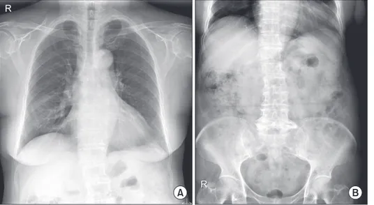
4-6. We present a case of a patient who was diagnosed with asth- ma and later developed PI, which was detected incidentally by follow-up X-ray. The patient had no symptoms and recovered without any treatment.
Case Report
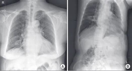
A 74-year-old Korean woman visited our hospital with com- plaints of severe cough and dyspnea for two weeks. She had a previous history of pulmonary tuberculosis but had no other respiratory diseases including asthma. Before she visited our clinic, she had used a bronchodilator inhaler prescribed previ- ously by another clinic. Her symptoms were relieved some- what after the medication, but dyspnea began to worsen the day before her visit to our hospital.
On physical examination, her breathing sounds were coarse with wheezing in both lung fields. There was no abnormal finding on chest X-ray, but luminal narrowing with wall thick- ening in the right middle lobe was noted in chest computed tomography (CT). The patient was diagnosed as acute asthma exacerbation. Treatment with a short-acting β2-agonist, an- ticholinergic nebulizer, and systemic steroid (intravenous hydrocortisone 5 mg/kg for 5 days then tapered to 2.5 mg/
kg) was started and was continued for 10 days. Under the as- Copyright © 2014
The Korean Academy of Tuberculosis and Respiratory Diseases.
All rights reserved.
Introduction
Pneumatosis intestinalis (PI) is defined as the presence of a cystic gas pocket formed within the submucosal layer or subserosal layer of the gastrointestinal tract. The primary type accounts for about 15% of the cases, and the secondary type is associated with other gastrointestinal, pulmonary, and colla- gen vascular diseases, organ transplantation, and immunode- ficiency
1,2. Most cases are asymptomatic, and fewer than 15%
of the patients show symptoms such as abdominal distension, vomiting, abdominal pain, and diarrhea. PI has diverse clinical outcomes ranging from benign, which does not require treat-
CASE REPORT
http://dx.doi.org/10.4046/trd.2014.77.5.219ISSN: 1738-3536(Print)/2005-6184(Online) • Tuberc Respir Dis 2014;77:219-222
219
Address for correspondence: Sook Young Lee, M.D.
Department of Internal Medicine, Seoul St. Mary’s Hospital, The Catholic University of Korea College of Medicine, 222 Banpo-daero, Seocho-gu, Seoul 137-701, Korea
Phone: 82-2-2258-6079, Fax: 82-2-596-2158 E-mail: [email protected]
Received: Jun. 9, 2014 Revised: Jul. 28, 2014 Accepted: Aug. 13, 2014
cc