가톨릭대학교 의과대학 내과학교실
윤재호, 여창동, 신은중, 송소향, 김치홍, 문화식, 송정섭, 박성학
Diffuse Nodular Interstitial Infiltrations with Bilateral Hilar Lymphadenopathy
Jae Ho Yoon, M.D., Chang Dong Yeo, M.D., Eun Joong Shin, M.D., So Hyang Song, M.D., Chi Hong Kim, M.D., Hwa Sik Moon, M.D., Jeong Sup Song, M.D., Sung Hak Park, M.D.
Department of Internal Medicine, College of Medicine, The Catholic University of Korea, Seoul, Korea
Lymphocytic interstitial pneumonia(LIP) is an uncommon condition in which the alveolar septa and extra-alveolar interstitial space are markedly expanded by small lymphocytes, plasma cells and histiocytes. Chest radiographs generally show nonspecific patterns with the most common pattern showing bibasilar reticular or reticulonodular infiltrates. Hilar or mediastinal lymphadenopathy and pleural effusions are usually absent. We encountered a 42-year-old female patient who was admitted to hospital because of exertional dyspnea and palpitation. The chest X-ray showed an enlarged bilateral hilar shadow and diffusely increased bronchovascular markings in both lung fields. The chest CT showed diffuse nodular infiltrations with mild septal thickening and combined patchy ground glass opacity in both lungs, and conglomerated mediastinal and bilateral hilar lymphadenopathy. A diagnosis of LIP was made from the tissue pathology taken by a thoracoscopic lung biopsy. The patient showed clinical and radiographic improvement after 3 months of treatment with prednisolone. We report a case of LIP presenting as diffuse nodular interstitial infiltrations with multiple mediastinal and bilateral hilar lymphadenopathy. (Tuberc Respir Dis 2006; 61: 294-298) Key words: Lymphocytic interstitial pneumonia, Diffuse nodular infiltrations, Bilateral hilar lymphadenopathy.
Address for correspondence : Chi Hong Kim, M.D.
Department of Internal Medicine, St. Vincent's Hospital, College of Medicine, The Catholic University of Korea, 93-6 Ji-dong, Paldal-gu, Suwon 442-723, Korea Phone: 031-249-7361, Fax : 031-253-8898 E-mail : [email protected]
Received : May. 25. 2006 Accepted : Jul. 4. 2006
증 례
환 자: 윤 O 옥, 42세 여자.
주 소: 1개월간 악화된 운동시의 호흡곤란.
현병력: 환자는 20년간 간헐적으로 운동시 호흡곤 란이 있었으나, 별다른 진단 및 치료 없이 지내다가, 내원 전일 호흡곤란을 동반한 심계항진이 10분 이상 지속되어 개인의원 방문하였으나, 이상 소견 발견하 지 못하고 증상 호전되어 귀가한 후, 다음날 다시 같 은 증상이 나타나 심질환 및 부정맥에 대한 평가를 위 해 본원 응급실로 전원되었다.
과거력: 특이 사항 없음.
사회력: 하루 4분의1갑의 흡연력이 있음.
가족력: 특이 사항 없음.
진찰 소견: 내원 당시 환자는 혈압은 100/60mmHg, 맥박은 130회/분, 호흡수는 22회/분, 체온은 36.7°C이 었다. 환자는 급성 병색을 보였으나 의식은 명료하였 고, 두경부 검사상 경정맥 확장 소견은 없었으며, 갑 상선 및 경부 림프절은 촉지되지 않았고 편도선 종대 는 없었다. 심잡음은 없었으나 빠른 심음이 청진되었 고, 양측 전 폐야에서 얕은 호흡과 함께 미세한 수포 음이 청진되었다. 복부진찰에서 특이 소견 없었으며, 사지검사에서도 곤봉지 등의 이상 소견은 없었다. 신 경학적검사에서 특이 사항은 없었다.
검사실 소견: 심전도 검사에서는 규칙적인 동성 빈 맥 이외의 이상 소견은 보이지 않았다. 산소 투여 전 시행한 동맥혈가스분석에서 pH 7.41, PaCO2 42.3 mmHg, PaO2 68.0mmHg, HCO3 27.0mmol/L, 산소포 화도 94.8%로 저산소증 및 저환기 소견을 보였고, 폐 포-동맥 산소 분압차이 {(A-a)DO2}는 29.125mmHg 로 증가해 있었다. 분당 2L의 산소 투여 후 시행한 동 맥혈가스분석에서 pH 7.40, PaCO2 41.2mmHg, PaO2
87.1mmHg, HCO3 25.0mmol/L, 산소포화도 96%로 저
A B
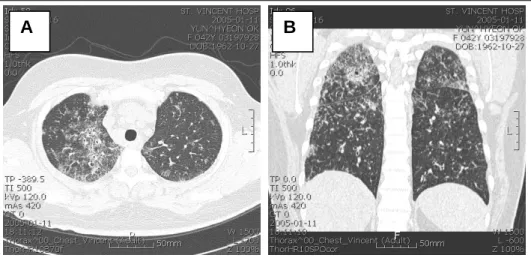
Figure 2. Chest CT shows diffuse bronchovascular bundle thickening in both parahilar regions, and a 4mm sized nodule in anterior segment of RUL(A). Diffuse nodular infiltration with mild septal thickening and combined patchy ground glass opacities in both lungs, with some tiny cystic change in both lung apices are noted(B).
Figure 1. Chest PA shows somewhat enlarged bilateral hilar shadow and diffusely increased bronchovascular markings in both lungs.
산소증이 개선되었다. 말초혈액 검사에서 백혈구 10,320/mm3(호중구 47.6%, 림프구 35.3%, 호산구 1.9%), 혈색소 10.8g/dL, 혈소판 393,000/mm3 이었고, 혈청 생화학 검사상 대부분 정상 범위에 있었으나, 총 단백 9.3g/dl, 알부민 3.4g/dl로 글로불린 비율이 증가 되어 있었다. 소변 검사는 정상이었다. 면역학 검사에 서 ferritin 64.63 ng/mL(정상6.9-282.5), IgG 4377 mg/dL(정상880-1800), IgA 893mg/dL(정상126-517), IgM 408mg/dL(정상 52-270)로 모두 증가해 있었으
며, T3 0.74ng/mL(정상0.58-1.59), T4 6.98ug/mL(정 상4.87-11.72), TSH 1.41uU/mL(정상0.35-4.94)로 정 상 소견이었다. 그밖에 CEA 0.66ng/mL, Anti-HIV 음성, AFB도말 음성이었고, 객담배양검사에서 균은 배양되지 않았다.
방사선 소견: 응급실에서 시행한 단순 흉부 X-선 에서 양측 폐문음영의 증가 소견과 함께 양 폐야에 간 질성 침윤 소견과 기관지혈관음영의 증가가 관찰되었 다(Figure 1). 흉부 CT에서는 양측 폐에 폐포소엽사 이막 비후를 동반한 미만성의 결절성 폐침윤 소견과 함께 젖빛유리음영의 동반이 관찰되었다. 양측 폐첨 부에는 부분적인 낭성 변화가 있었으며, 우폐상엽 앞 분절에서 4mm 크기의 결절이 관찰되었다(Figure 2).
종격동과 양측 폐문에는 대칭적인 림프절 종대가 관 찰되었고, 경부 및 양측 액와부에도 여러 개의 작은 림프절 종대가 확인되었다(Figure 3).
임상 경과: 입원 2병일에 시행한 24시간 심전도 검 사 결과 이상 소견 없었으며, 폐기능검사 결과 FVC 2.57L(예측치의 71%), FEV1 2.12L(예측치의 76%), FEV1/FVC 83%, DLCO 71%로 일산화탄소 확산능 감소 및 제한성 환기장애 소견을 보이고 있었다. 3병 일에 시행한 소변 및 혈청 전기 영동 검사에서 다클론 성 감마글로불린병증으로 확인되었으며, 4병일에 흉 강경을 이용한 폐 조직검사를 시행하고, 4일 후 특별 한 문제 없이 퇴원하였다. 퇴원 후 외래에서 확인한
A B
Figure 3. Chest CT shows conglomerated mediastinal and bilateral hilar lymphadenopathy(A) and several tiny lymphadenopathy in lower neck and axilla bilaterally(B).
A B C
Figure 4. Follow-up chest PA(A) shows decreased bilateral hilar enlargement and slightly cleared diffuse interstitial infiltrations in both lungs. Follow-up chest CT shows markedly improved pulmonary nodular infiltration, ground glass opacities, and mild intra and interlobular septal thickening(B) and markedly decreased size of the mediastinal and bilateral hilar lymph nodes(C).
조직검사에서 lymphocytic interstitial pneumonia로 진단되어, 0.5mg/kg 프레드니솔론 경구 복용을 시작 하였다. 이후 1개월 간격으로 프레드니솔론 투여량을 감량하였으며, 3개월째, 메틸프레드니솔론으로 바꾸 어 12mg에서 4mg까지 감량하였다. 환자가 간헐적으 로 호소하던 운동시의 호흡곤란 및 심계 항진은 호전 되었고, 스테로이드 투여 3개월 후 외래에서 시행한 흉부 X-선에서는 양측 폐문음영의 감소와 미만성 간 질성 폐침윤의 호전이 확인되었다(Figure 4A). 흉부 CT에서도 양측 폐문과 종격동에서 보이던 림프절의 크기가 감소하였으며, 젖빛유리음영의 미만성 결절과 폐포소엽사이막 비후의 호전이 확인되었다(Figure
4B).
병리 소견: 흉강경하 폐조직 검사의 광학 현미경 소견에서는 주로 폐포벽 주위의 간질조직에 림프구와 형질세포 등의 염증세포의 침윤이 확인되었다. 또한 간질의 비후와 함께 림프구와 형질세포들이 다량 침 윤하여 림프소절을 형성하였고 그 내부에는 부분적인 섬유화가 관찰되어 만성적인 lymphocytic interstitial pneumonia에 합당한 소견으로 확인되었다(Figure 5).
고 찰
림프구형 간질성 폐렴(Lymphocytic interstitial
A B
Figure 5. Small round inflammatory cells composed of lymphocytes and plasma cells infiltrate alveolar septa and extra-alveolar interstitial space forming lymphoid follicles with follicle center.(A: X100, B:X400, H&E stain)
pneumonia(LIP))은 1966년 Carrington과 Liebow 등 에 의해 처음 기술되었으며, 림프구형 침윤성 폐질환 중에서 임상적으로 가장 양성인 폐렴의 유형으로1, T 림프구와 형질세포, 조직구의 간질 침윤 및 림프구 배 중심의 조직학적 특징을 가지는 병리학적 용어이다2 . LIP는 림프구들이 간질과 폐포 내강을 단조롭게 침윤 하는 것이 특징으로, 통상형 간질성 폐렴(Usual interstitial pneumonia)과 박리형 간질성 폐렴(Des- quamative interstitial pneumonia)과 구별되며3, 정상 조직인 기도나 폐혈관, 주변 림프절, 폐 이외의 조직 침윤은 보통 동반하지 않고 주로 폐간질로만 침윤되 는 모양을 보여4, 악성 림프종과는 구별되는 특징이 있다. LIP는 외인성 알레르기 폐포염(extrinsic allergic alveolitis), 비특이형 간질성 폐렴(Nonspecific inter- stitial pneumonia), 결절형 림프구증식증(Nodular lymphoid hyperplasia), MALT(mucosa-associated lymphoid tissue) 림프종과 조직학적으로 구별이 힘 든 경우가 많아 정확한 진단을 위해 임상적, 방사선학 적으로 면밀한 검토가 필요하며, 면역조직화학적 평 가 및 유전자 검사 등을 통해 대부분 구별이 가능하다2. LIP의 원인은 명확히 알려져 있지는 않으나, 원발 성으로 발생하기도 하고 결체조직질환, 이상 단백혈 증, 악성 빈혈, 아밀로이드증 등의 여러 자가면역질환 및 선천적, 후천적인 면역 결핍 증후군과 연관되어 발 생하기도 한다5. 특히 Sjögren syndrome과의 관련성
이 높아 보고된 LIP의 적어도 25%에서 Sjögren syndrome을 동반하고 있는 것으로 알려져 있고6, SLE, HIV 또는 EBV 감염과 동반된 예가 종종 보고 되고 있으며7, 특히 저감마글로불린혈증 환자에서는 다수에서 악성 림프종화 하는 것으로 알려져 있다7. 50-75% 정도의 LIP에서 이상 단백혈증을 동반하는 데 대부분 다클론성(polyclonal)으로, 본 증례에서도 다클론성 감마글로불린병증 (Polyclonal gammopa- thy)이 확인되었으나, 일부에서는 단일클론성 감마글 로불린병증(Monoclonal gammopathy(IgG or IgM)) 을 보이는 경우도 있다8. 소수에서는 결핵, 만성소화 장애증(Celiac disease)에서 동반되거나 딜란틴 등의 약제 투여 후 발생했다는 보고가 있다9.
LIP의 방사선학적 소견은 보통 비특이적이고 다양 하며 폐 침범 정도에 따라 달라, 암종의 림프관 전이 또는 단순폐렴과 혼동되는 경우가 많다. 가장 흔하게 는 양측 폐에서 망상형, 망상결절형 폐침윤을 보이며
10,11
, 진행된 LIP에서는 림프구의 간질 침윤이 진행하 면서 낭성 변화를 일으켜 X-선에서 기관지 공기 음영 을 보이게 된다. 종격동 및 양측 폐문부의 림프절 비 후 및 흉수 소견은 비교적 드물며12, 흉수가 동반된 경 우 림프종의 가능성이 높다.
Jonkoh 등은 LIP 의 흉부 CT 소견으로 폐실질의 젖빛유리음영 결절, 폐포소엽사이막 비후, 중심소엽 결절, 낭성 변화 및 경화를 주요 소견으로 제시하였으
며13, 추적 CT 촬영에서 이전의 낭성 변화 부위는 비 가역적으로 병변이 남아있었으나, 폐실질 부위의 침 윤 소견은 가역적으로 호전을 보이는 경우가 있었다.
진행된 LIP에서는 낭성 변화 및 경화부위가 봉소상 (honeycombing appearance)으로 진행하였고, 중심소 엽결절에서 새로운 낭성 변화가 나타났다. 봉소상 폐 가 관찰되는 경우는 대체로 간질성 폐섬유화로의 진 행을 의미하며, 대체로 진행된 LIP에서 나타나는 소 견이다14.
본 증례는 흉강경하 폐생검을 통해 조직학적으로 증명된 LIP로, 흉부CT에서 부분적인 낭성 변화를 동 반한 양폐야의 광범위한 젖빛유리음영 결절과 폐포소 엽사이막 비후가 확인되었으며, 종격동과 폐문부의 림프절 종대가 동반된 드문 경우이다. 환자는 3개월간 의 글루코코티코이드 투여로 간질성 폐침윤 및 림프 선 종대가 모두 호전을 보였고 간헐적으로 나타난 운 동시의 호흡곤란 또한 소실된 상태로 추적 관찰 중이 다.
요 약
단순 흉부 X-선에서 양측성 폐문 림프절 비후를 보이는 대표적인 질환으로 유육종증, 림프종, 결핵, 폐 암, 아밀로이드증, 브루셀라증, 콕시디오이데스진균증 (Coccidioidomycosis) 등이 있다. LIP의 방사선학적 소견은 보통 비특징적이고, 초기 병변에서 양측 폐하 에 망상, 망상결절성 침윤을 보이며 병이 진행됨에 따 라 낭성 변화를 동반하고, 봉소상을 보이는 경우가 있 으나, 본 증례의 환자와 같이 종격동과 양측 폐문에 림프절 종대를 동반하는 경우에 대한 보고는 흔치 않 다. 저자들은 흉부 X-선 및 CT상 종격동과 양측 폐문 부의 림프절 종대를 동반한 간질성 결절성 폐침윤 소 견을 나타낸 환자에서 흉강경하 폐 생검을 통해 진단 된 LIP를 경험하였기에 문헌고찰과 함께 보고하는 바 이다.
참 고 문 헌
1. Heitzman ER, Markarian B, Delise CT. Lymphopro-
liferative disorders of the thorax. Semin Roentgenol 1975;10:73-81.
2. Nicholson AG. Lymphocytic interstitial pneumonia and other lymphoproliferative disorders in the lung.
Semin Respir Crit Care Med 2001;22:409-22.
3. Liebow AA, Carrington CB. Diffuse pulmonary lymphoreticular infiltrations associated with dysproteinemia. Med Clin North Am 1973;57:809-43.
4. TeirsteinAS, Rosen MJ. Lymphocytic interstitial pneu- monia. Clin Chest Med 1988;9:467-71.
5. Itescu S, Brancato LJ, Boxbaum J, Gregersen PK, Rizk CC, Croxson TS, et al. A diffuse infiltrative CD8 lymphocytosis syndrome in human immunodeficiency virus infection: ahost immune response associated with HLA-DR5. Ann Intern Med 1990;112:3-10.
6. Alkhayer M, McCann BG, Harrison BD. Lymphocytic interstitial pneumonitis in association with SjÖgren’s syndrome. Br J Dis Chest 1988;82:305-9.
7. Yum MN, Ziegler JR, Walker PD, Ridolfo AS, Brashear RE. Pseudolymphoma of the lung in a patient with systemic lupus erythematosus. Am J Med 1979;66: 72-6.
8. Montes M, Tomasi TB Jr, Noehren TH, Culver GJ.
Lymphoid interstitial pneumonia with monoclonal gammopathy. Am Rev Respir Dis 1968;98:277-80.
9. Jung HJ, Cho ER, Shim JJ, In KH, Yu SH, Kang KH, et al. A case of lymphocytic interstitial pneumonitis.
Tuberc Respir Dis 1993;40:602-9.
10. Bragg DG, Chor PJ, Murray KA, Kjeldsberg CR.
Lymphoproliferative disorders of the lung: histopa- thology, clinical manifestations, and imaging features.
AJR Am J Roentgenol 1994;163:273-81.
11. Strimlan CV, Rosenow EC 3rd, Weiland LH, Brown LR. Lymphocytic interstitial pneumonitis: review of 13 cases. Ann Intern Med 1978;88:616-21.
12. Fishman AP, Elias JA, Fishman JA, Grippi MA, Kaiser LR, Senior RM. Fishman's pulmonary diseases and disorders. 3rd ed. New York: McGraw-Hill, Inc;
1998. p.1862-3.
13. Jonkoh T, Ichikado K, Akira M, Honda O, Tomiyama N, Mihara N, et al. Lymphocytic interstitial pneu- monia: follow-up CT findings in 14 patients. J Thorac Imaging 2000;15:162-7.
14. Suh YA, Kim SI, Kim DH, Kwak JY, Lee JC, Baek HJ, et al. A case of lymphocytic interstitial pneu- monia. Tuberc Respir Dis 2001;51:390-4.