Single Center Experience of Five Diffuse Panbronchiolitis Patients Clinically Presenting as Severe Asthma
Diffuse panbronchiolitis (DPB) is a bronchiolitis affecting the whole lung fields which can be treated by macrolide. Especially East Asian patients are more susceptible to diffuse panbronchiolitis. As asthma and DPB both can cause airway obstruction, differential diagnosis is important for the 2 diseases. Here we report 5 patients with DPB clinically presenting as severe asthma in Korea, who were well treated by macrolide. Among the 5 patients, 2 could stop their asthma inhalers and the other 3 could reduce asthma medications after diagnosis and treatment of DPB. In conclusion, considering DPB as differential diagnosis for asthmatics in Asian ethnic groups is important.
Keywords: Diffuse Panbronchiolitis; Asthma; Clarithromycin Kyung Hee Park, Hye Jung Park,
Jae-Hyun Lee, and Jung-Won Park Department of Internal Medicine and Institute of Allergy, Yonsei University College of Medicine, Seoul, Korea
Received: 22 September 2014 Accepted: 27 October 2014 Address for Correspondence:
Jung-Won Park, MD
Division of Allergy and Immunology, Department of Internal Medicine, Yonsei University College of Medicine, 250 Seongsan-ro, Seodaemun-gu, Seoul 120-752, Korea Tel: +82.2-2228-1961, Fax: +82.2-393-6884 E-mail: [email protected]
http://dx.doi.org/10.3346/jkms.2015.30.6.823 • J Korean Med Sci 2015; 30: 823-828
INTRODUCTION
Asthma is an obstructive lung disease diagnosed by variable airway obstruction. However, before diagnosis, other underly- ing causes should be excluded (1). One of them is diffuse pan- bronchiolitis (DPB) which incidence is high among Asians, es- pecially Koreans and Japanese (2). Clinical manifestations of DPB are cough, dyspnea, and crackling sounds. A chest image shows bilateral centrilobular nodules. Although DPB is distinct from asthma, their clinical manifestations can overlap (3) and co-occurrence of DPB and asthma is possible in East Asian pa- tients (4).
DPB can be treated successfully with long-term, low-dose macrolide administration. The effectiveness of macrolide has been confirmed in a double-blind, placebo-controlled study (5). Macrolide is an antimicrobial and anti-inflammatory agent (6). Macrolide can alter neutrophil functions, inhibit oxidant production, and influence the production of various cytokines and chemokines, such as interleukin (IL)-1, IL-6, IL-8, IL-10, and tumor necrosis factor (7). In this report, we describe 5 Ko- rean patients with DPB who clinically presented with severe asthma but for whom computed tomography (CT) findings were suggestive of DPB; these patients showed a good response to long-term macrolide treatment.
CASE DESCRIPTION Case 1
A 71-yr-old woman referred for a 10-yr history of cough, dys- pnea, sputum, post-nasal drip on 4th June 2010. Her breathing
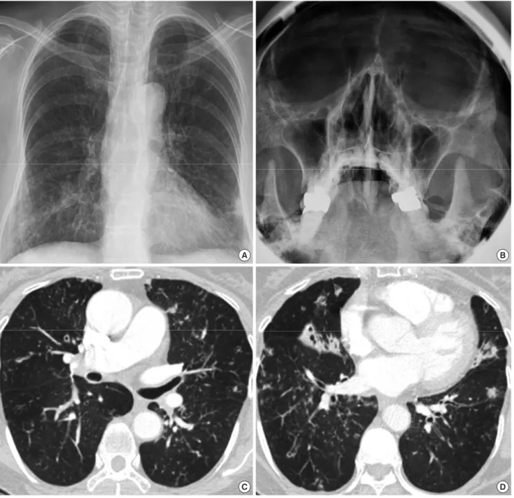
sound was clear. A chest x-ray (CXR) showed increased bron- chovascular markings in the both lower lungs and consolida- tion in the left lower lung field (LLLF) (Fig. 1A). Initial forced expiratory volume in 1 second (FEV1) was 1.21 L (64.9%) and forced vital capacity (FVC) was 1.70 L (75.5%) and FEV1/FVC was 71.1% without a bronchodilator (BD) response. The metha- choline bronchial provocation test (MBPT) was positive as a re- sult of provocative concentration (PC20) was 3.8 mg/mL. Induc- ed sputum showed mild eosinophilia (3%). During one month of asthma treatment with inhaled budesonide combined with formoterol, theophylline, and montelukast, she complained of mild fever and yellowish sputum. Thus, we performed a chest CT and water’s view x-ray. Bilateral maxillary sinusitis was ob- served (Fig. 1B). CT showed bronchial wall thickening with cen- trilobular nodules, which was suggestive of DPB (Fig. 1C, D). To differentiate from mycobacterial infection (8), acid fast bacilli (AFB) studies were done and showed negative. The patient took clarithromycin 250 mg twice a day for 9 months, after which her FEV1 improved from 64.9% to 84.2%, FVC and FEV1/FVC also improved. A follow-up examination, LLLF consolidation and sputum eosinophilia disappeared. In addition, she was able to reduce the daily amount of inhaled steroid and discontinue theophylline.
Case 2
A 52-yr old man visited for a 4-yr history of cough, dyspnea, snee- zing, rhinorrhea, and wheezing on 25th June 2010. Initial FEV1
was 1.74 L (54.4%), FVC was 2.40 L (61.3%), and FEV1/FVC was 72.5%. BD response was positive (FEV1 increased 20.6%, 270 mL). CXR showed multiple nodules in both lungs. A sinus CT
showed bilateral maxillary sinusitis. A chest CT showed a tree- in-bud appearance and air trapping in both lower lungs. Induc- ed sputum showed neutrophilia. He showed a poor response to inhaled budesonide combined with formoterol and montelu- kast. As AFB studies were negative, we administered clarithro- mycin. The patient took clarithromycin 250 mg twice a day for 9 months, after which FEV1 improved from 54.4% to 95.3%, FVC improved from 61.3% to 92.8%, and FEV1/FVC also improved.
Cough and sputum disappeared. He now takes montelukast and clarithromycin without respiratory symptoms.
Case 3
A 25-yr-old man had a 5-yr history of cough and dyspnea on 28th March 2007. Initial FEV1 was 2.51 L (62.6%), FVC was 3.65 L (77.8%), and FEV1/FVC was 68.8%. BD response was negative.
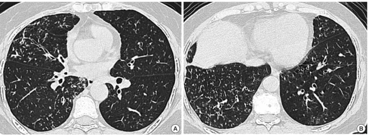
The results of a MBPT were positive (PC20 4.36 mg/mL). A CXR showed miliary nodules in both lungs, but he didn’t have sinus- itis. A chest CT showed bronchiolectasis and centrilobular nod- ules on both lungs (Fig. 2A, B). Induced sputum showed severe eosinophilia (42%). The AFB studies were negative. He took in- haled fluticasone combined with salmeterol, montelukast, and theophylline, and he intermittently used 30 mg of prednisolone
Fig. 1 Fig. 1 Fig. 1 Fig. 1
A
C
B
D Fig. 1. Images of the Case 1. Initial chest x-ray showed bilateral haziness at bilateral lung field (A) and both maxillary sinusitis (B). Chest CT showed diffuse bronchial wall thick- ening with centrilobular nodules (C, D).
(PL) for asthma attacks. After the patient took clarithromycin 250 mg twice a day for 9 months, his FEV1 improved from 62.6%
to 88.4%, and he could reduce the amount of PL. Before the pa- tient had started taking clarithromycin, he had taken 720 mg of PL per year. After he treated with clarithromycin, he was able to reduce use of oral steroids by about 80%, such that he took 150 mg per year.
Case 4
A 60-yr-old woman referred for a 5-yr history of severe asthma on 18th August 2010. Cough and dyspnea were her main symp- toms. Her initial FEV1 was 0.88 L (42.9%), FVC was 1.10 L (44.9%), and FEV1/FVC was 73.4% without a BD response. A crackling sound was heard in both lungs. A chest CT showed diffuse bron- chial wall thickening and centrilobular nodules with a tree-in- bud appearance in both lower lungs (Fig. 3A, B). Induced spu- tum showed 2% eosinophils. A MBPT could not be performed.
Water’s view showed bilateral maxillary sinusitis. She had been using inhaled budesonide combined with formoterol, a tiotro-
pium inhaler, montelukast, and theophylline for a month. After a month, she complained of continuous cough and sputum.
The crackling sound was consistently heard. The AFB studies were negative, and clarithromycin was introduced. The patient took clarithromycin 250 mg twice a day for 5.5 months with as- thma medication, after which her FEV1 improved from 0.88 L (42.9%) to 1.96 L (97.1%), and FVC improved from 44.9% to 98.8%.
Follow-up chest CT after a year showed improved centrilobular nodules in both lungs. She was able to discontinue budesonide and formoterol.
Case 5
A 69-yr-old man came with a 3-yr history of cough, sputum, and dyspnea on 11th July 2012. His initial FEV1 was 1.88 L (79.2%), FVC was 2.34 L (76.8%), and FEV1/FVC ratio was 80.3%. FEV1
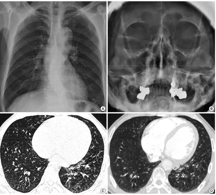
increased 13.2% (250 mL) after bronchodilator inhalation. In- duced sputum showed neutrophilia without eosinophils. CXR showed tiny nodules and bronchial wall thickening in both low- er lungs (Fig. 4A). Water’s view showed left maxillary sinusitis
Fig. 2 Fig. 2
A B
Fig. 2. Images of the Case 3. Bronchiolectasis and centrilobular nodules were observed on almost entire lung in his chest CT (A, B).
Fig. 3 Fig. 3
A B
Fig. 3. Images of the Case 4. Initial CT showed bronchial wall thickening, centrilobular nodules (A, B).
Fig. 4 Fig. 4 Fig. 4 Fig. 4
A
C
B
D Fig. 4. Images of the Case 5. Tiny nodules, bronchial wall thickening (A) and left maxillary sinusitis (B) were observed. Initial CT showed severe and diffuse centrilobular nodules (C). After treatment, multiple nodules were decreased (D).
(Fig. 4B). A chest CT showed severe and diffuse centrilobular nodules and branching linear opacities in both lungs (Fig. 4C).
The AFB studies were negative. He was prescribed fluticasone combined with salmeterol, pranlukast, myrtol, and clarithro- mycin. After the patient took clarithromycin 250 mg twice a day for 40 days, his cough and sputum decreased. A follow-up CT showed a slight decrease in bronchial wall thickening and mul- tiple nodules throughout both lungs (Fig. 4D). After 8 months of clarithromycin, he was able to use only fluticasone and sal- meterol inhaler with clarithromycin. His FEV1 increased from 79.2% to 127.0%, and FVC increased from 76.8% to 118.9%.
The study occurred in the allergy clinic of a tertiary teaching hospital in Korea. We collected 5 patients who visited as severe
asthma but treated well with DPB treatment. Institutional re- view board reviewed and approved this study (4-2014-0362).
DISCUSSION
All patients in this study were referred to our clinic with treat- ment resistant severe asthma, but our report shows that DPB should be excluded before diagnosing asthma in East Asian pa- tients. As DPB is a progressive inflammatory airway disease and can be treated by long-term administration of macrolide (9), ac- curate diagnosis is especially important. Both asthma and DPB patients complain of chronic cough, sputum, and dyspnea, and it is not easy to discriminate between the two diseases. All 5 pa-
tients had CT findings that were consistent with DPB, and all showed marked improvement in lung function and clinical sym- ptoms with long-term macrolide treatment.
In this study, all patients were treated with clarithromycin, which consists of 14-membered ring macrolides. The mecha- nism by which macrolides manage DPB may be related to their immune modifying effects. Only 14-membered ring macrolides (erythromycin, clarithromycin, and roxithromycin) and 15-mem- bered ring macrolides (azithromycin) are effective; 16-mem- bered ring macrolides are not effective. The structure of macro- lides determines their immune modifying activity (10), and it has been reported that macrolides could reduce airway mucus secretion and decrease airway neutrophil accumulation through reduced expression of adhesion molecules or cytokine expres- sion from macrophages, lymphocytes, monocytes, fibroblasts, and eosinophils (10). The treatment effects of macrolides in DPB have been reported independent of the presence or absence of Pseudomonas infection, and the treatment effect can be observ- ed below the minimum inhibitory concentration of macrolides, (10, 11) suggesting that immune modifying effects of macrolides are crucial for management of DPB.
Previously, the lower airway has been regarded as a bacteria- free area, but recent data show that bacterial colonization can occur in asthma (12, 13) and chronic obstructive pulmonary disease (COPD) (14). These bacteria may exacerbate chronic airway diseases (14). In addition to their immune-modifying effects, macrolides have anti-microbial properties that may play a role in the management of other chronic airway diseases, such as asthma, COPD (15), cystic fibrosis, and chronic rhinosinus- itis (16). For cystic fibrosis patients on long-term macrolide treat- ment, the prevalence of sputum culture positivity for Haemoph- ilus influenzae and Streptococcus pneumoniae are markedly de- creased, although positive rates of these bacteria with macro- lide resistance are much higher (17, 18). Decreased bacterial burden may contribute to the effect of long-term treatment of macrolides. Emergence of macrolide-resistant bacteria is an important concern related to the long-term administration of macrolides, but no cases have been reported in which macro- lide-resistant bacterial infection led to a life threatening episode (2). In conclusion, it is essential to consider DPB for treatment resistant asthmatics in East Asian patients. DPB can be treated well by long term use of clarithromycin.
DISCLOSURE
There are neither conflicts of interest nor financial support to declare.
AUTHOR CONTRIBUTION
Conceived and designed the study: Park JW. Wrote the paper:
Park KH, Park HJ, Lee JH, Park JW. ICMJE criteria for authorship read and met: Park KH, Park HJ, Lee JH, Park JW. Agree with man- uscript results and conclusions: all authors
ORCID
Kyung Hee Park http://orcid.org/0000-0003-3605-5364 Hye Jung Park http://orcid.org/0000-0002-1862-1003 Jae-Hyun Lee http://orcid.org/0000-0002-0760-0071 Jung-Won Park http://orcid.org/0000-0003-0249-8749 REFERENCES
1. Bateman ED, Hurd SS, Barnes PJ, Bousquet J, Drazen JM, FitzGerald M, Gibson P, Ohta K, O’Byrne P, Pedersen SE, et al. Global strategy for asth- ma management and prevention: GINA executive summary. Eur Respir J 2008; 31: 143-78.
2. Azuma A, Kudoh S. Diffuse panbronchiolitis in East Asia. Respirology 2006; 11: 249-61.
3. Tsang KW, Ooi CG, Ip MS, Lam WK, Ngan H, Chan EY, Hawkins B, Ho CS, Amitani R, Tanaka E, et al. Clinical profiles of Chinese patients with diffuse panbronchiolitis. Thorax 1998; 53: 274-80.
4. Matsuno O, Ueno K, Hayama Y, Honda H, Yamane H, Saeki Y. Deterio- ration of asthma in a patient with diffuse panbronchiolitis (DPB) after macrolide therapy. J Asthma 2010; 47: 486-8.
5. Kadota J, Mukae H, Ishii H, Nagata T, Kaida H, Tomono K, Kohno S. Long- term efficacy and safety of clarithromycin treatment in patients with dif- fuse panbronchiolitis. Respir Med 2003; 97: 844-50.
6. Poletti V, Casoni G, Chilosi M, Zompatori M. Diffuse panbronchiolitis.
Eur Respir J 2006; 28: 862-71.
7. Schultz MJ. Macrolide activities beyond their antimicrobial effects: mac- rolides in diffuse panbronchiolitis and cystic fibrosis. J Antimicrob Che- mother 2004; 54: 21-8.
8. Park HY, Suh GY, Chung MP, Kim H, Kwon OJ, Chung MJ, Kim TS, Lee KS, Koh WJ. Comparison of clinical and radiographic characteristics be- tween nodular bronchiectatic form of nontuberculous mycobacterial lung disease and diffuse panbronchiolitis. J Korean Med Sci 2009; 24:
427-32.
9. Kudoh S, Azuma A, Yamamoto M, Izumi T, Ando M. Improvement of survival in patients with diffuse panbronchiolitis treated with low-dose erythromycin. Am J Respir Crit Care Med 1998; 157: 1829-32.
10. Kanoh S, Rubin BK. Mechanisms of action and clinical application of macrolides as immunomodulatory medications. Clin Microbiol Rev 2010; 23: 590-615.
11. Fujii T, Kadota J, Kawakami K, Iida K, Shirai R, Kaseda M, Kawamoto S, Kohno S. Long term effect of erythromycin therapy in patients with chron- ic Pseudomonas aeruginosa infection. Thorax 1995; 50: 1246-52.
12. Bisgaard H, Hermansen MN, Buchvald F, Loland L, Halkjaer LB, Bonn- elykke K, Brasholt M, Heltberg A, Vissing NH, Thorsen SV, et al. Child- hood asthma after bacterial colonization of the airway in neonates. N Engl J Med 2007; 357: 1487-95.
13. Marri PR, Stern DA, Wright AL, Billheimer D, Martinez FD. Asthma-as- sociated differences in microbial composition of induced sputum. J Al- lergy Clin Immunol 2013; 131: 346-52.e1-3.
14. Sethi S, Murphy TF. Infection in the pathogenesis and course of chronic obstructive pulmonary disease. N Engl J Med 2008; 359: 2355-65.
15. Wenzel RP, Fowler AA 3rd, Edmond MB. Antibiotic prevention of acute exacerbations of COPD. N Engl J Med 2012; 367: 340-7.
16. Wallwork B, Coman W, Mackay-Sim A, Greiff L, Cervin A. A double- blind, randomized, placebo-controlled trial of macrolide in the treat- ment of chronic rhinosinusitis. Laryngoscope 2006; 116: 189-93.
17. Hansen CR, Pressler T, Hoiby N, Johansen HK. Long-term, low-dose
azithromycin treatment reduces the incidence but increases macrolide resistance in Staphylococcus aureus in Danish CF patients. J Cyst Fibros 2009; 8: 58-62.
18. Phaff SJ, Tiddens HA, Verbrugh HA, Ott A. Macrolide resistance of Staph- ylococcus aureus and Haemophilus species associated with long-term azithromycin use in cystic fibrosis. J Antimicrob Chemother 2006; 57:
741-6.