Acute lung injury, the most severe form of which is ARDS is defined as rapid alteration of the alveoli that re- sults in impairment of gas exchange. Despite improve- ment in supportive care for patients with acute lung in- jury, the mortality rate for ARDS has hovered around the 50 % for the past decade (1). Recent studies have
suggested that once a patient has survived initial injury, subsequent gas exchange problems may arise in part from an inadequately regulated healing response (2).
Although the plain radiographic and CT findings of ARDS are well known, HRCT findings in survivors of ARDS have not been fully evaluated. We recently en- countered four patients who survived from ARDS who were diagnosed according to the definition of Murray et al. (3). We describe the chest radiographic and HRCT findings in these cases, and believe they may explain the pathogenesis of fibrotic lung.
Case 1
A 30-year-old pregnant woman at gestational week 35
Acute Re s p i ra tory Distress Sy n d rome (ARDS) :
H RCT Findings in Survivo rs
1Jung Im Jung, M.D., Seog Hee Park, M.D., Jae Mun Lee, M.D., Jeong Sup Song, M.D.2, Kyo - Young Lee, M.D.3
The purpose of this report is to describe the high-resolution computed tomography ( H RCT) findings of the lung in survivors of acute respiratory distress syndrome (ARDS). Among eleven patients who survived ARDS for one ye a r, chest radiography and HRCT revealed pulmonary fibrosis in four. Causes of ARDS included pneumonia during pregnancy, near drowning, pneumonia during liver cirrhosis, and postopera- t i ve sepsis. Thoracoscopic biopsy and histopathologic correlation were available in one patient.
H RCT showed diffuse interlobular septal thickening, ground glass opacity, p a r e n c hymal distortion, and traction bronchiectasis. Fuzzy centrilobular nodules were seen in two patients and one patient had multiple, large bullae in the left hemithorax. In all patients, lesions affected the upper and anterior zones of the lung more prominently. The distribution of pulmonary fibrosis was characteristic and re- flected the pathogenesis of lung injury; fibrosis was largely due to hy p e r oxia caused by ventilator care. In one patient, histopathologic correlation showed that imaging findings were accounted for by thickening of the alveolar septum along with infiltra- tion of chronic inflammatory cells and fibrosis. Fuzzy centrilobular nodules corre- sponded with bronchiolitis.
Index words :Lung, CT Lung, fibrosis
Computed tomography (CT), high-resolution
1Department of Radiology, College of Medicine, The Catholic University of Korea
2Department of Internal Medicine, College of Medicine, The Catholic University of Korea
3Department of Pathology, College of Medicine, The Catholic University of Korea.
Received February 10, 1999 ; Accepted May 7, 1999
Address reprint requests to : Jung Im Jung, M.D., Department of Radiology, St. Mary’s Hospital, College of Medicine, The Catholic University of Korea.
#62 Youido-dong, Youndungpo-gu, Seoul 150-010, Korea.
Tel. 82-2-3779-1277 Fax. 82-2-783-5288 E-mail; [email protected]
was admitted with complaints of chronic cough and dyspnea. Arterial blood gas analysis showed pH 7.402, P C O22 5 .3 mmHg, and PO25 5 .2 mmHg. Body tempera- ture was 39.2°C. A chest radiograph revealed ill defined patchy opacity in both lungs and consolidation in the left lower lung (Fig. 1A). Two days later, radiography showed diffuse haziness and consolidation in both lungs. The baby was delivered normally and a ventila- tor was applied. Follow-up chest radiographs revealed gradual resolution of the haziness in both lungs.
Chryseobacterium meningoseptum and Buckhholderia pick- e t t i were cultured in the sputum. About a month later, persistent ground glass opacity and coarse reticular den- sity were seen on chest radiographs in the left upper, and right middle and lower lobes (Fig. 1B). A pul- monary function test performed two and a half months later revealed a severely restrictive pattern and diffu- sion defect. HRCT performed on the 98th hospital day showed diffuse ground glass opacity with interlobular
septal thickening in both the upper and mid-lung zones.
Diffuse bronchial dilatation and mild architectural dis- tortion were also noted. Involvement of the anterior as- pect of the lung predominated (Fig. 1C). Follow-up chest radiography indicated that pulmonary fibrosis remained unchanged. A summary of this case can be found in Table 1.
Case 2
A 43-year-old woman came close to drowning and re- ceived cardio-pulmonary resuscitation. A chest radi- ograph obtained at that time showed diffuse ground glass opacity and patchy consolidation in both lungs. Six days later, she was severely dyspneic and arterial blood gas analysis showed pH 7.47, PaO25 0 .1 mmHg, PCO2 4 4 .7 mmHg, and SaO28 7 .1 %. Ventilation was started.
Chest radiography again revealed persistent ground glass opacity and consolidation in both lungs though, a follow-up chest radiograph obtained three weeks later
A
C
B
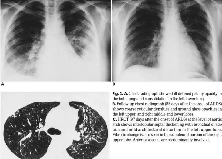
Fig. 1. A. Chest radiograph showed ill defined patchy opacity in the both lungs and consolidation in the left lower lung.
B. Follow up chest radiograph (85 days after the onset of ARDS) shows coarse reticular densities and ground glass opacities in the left upper, and right middle and lower lobes.
C. HRCT (97 days after the onset of ARDS) at the level of aortic arch shows interlobular septal thickening with bronchial dilata- tion and mild architectural distortion in the left upper lobe.
Fibrotic change is also seen in the subpleural portion of the right upper lobe. Anterior aspects are predominantly involved.
showed resolution of the consolidation. Coarse reticular density and patchy ground glass opacity were noted in both lungs. HRCT performed on the 37th hospital day showed diffuse interlobular septal thickening and ground glass opacity in both lungs, these features were more prominent in the upper and anterior lung zones than in the posterior and lower zones. Fuzzy centrilobu- lar nodules were also noted throughout the entire lungs, without zonal predominance (Fig. 2A) (Table1). In order to determine disease activity, thoracoscopic biopsy of the right middle lobe was performed on the 46th admis- sion day. Photomicroscopy showed thickening of the alveolar septum together with infiltration of chronic in- flammatory cells and mild fibrosis. Foci of atelectasis with alveolar macrophage accumulation and hemor- rhage in alveolar spaces and proliferation of type II p- neumocytes were also seen, and associated dilated bron- chioles and bronchiolitis were noted (Fig. 2B). A pul- monary function test showed moderate restrictive air- way disease and diffusion defect. Because of inflamma-
tion and fibrosis, the patient was placed on high dose s- teroid therapy. Follow-up chest radiography revealed resolution of ground glass opacity and reticular density.
Case 3
A 68-year-old woman was admitted because of Parkin- s o n i s m a n d hepatic encephalopathy and three weeks lat- er, began to complain of dyspnea. A chest radiograph showed total haziness of the left lung and arterial blood gas analysis showed pH 7.387, PaO25 4 .7 m m H g , P C O2 45.7 mmHg, and SaO284.2 %. Hypoxia improved after ventilation, and Klebsiella pneumoniae was identified in blood culture. After treatment with antibiotics, con- solidation was completely resolved, but because of weaning failure, ventilation was continued. On the 46th admission day, the patient lapsed into a coma with a high temperature. A chest radiograph revealed total haziness, with air-bronchograms in the right lung and ill defined haziness in the left (Fig. 3A). Haziness and con- solidation were extended to both lungs, but eight days
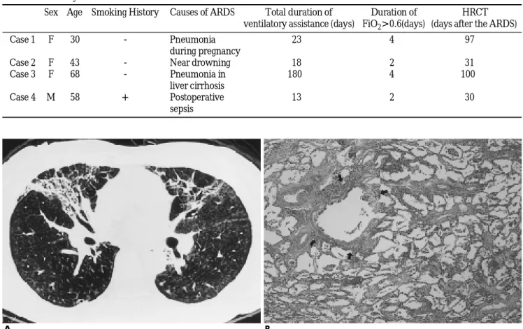
Table 1. Summary of Patients Characteristics
S e x A g e Smoking History Causes of ARDS Total duration of Duration of H R C T ventilatory assistance (days) F i O2> 0 . 6 ( d a y s ) (days after the ARDS)
Case 1 F 3 0 - Pneumonia 2 3 4 9 7
during pregnancy
Case 2 F 4 3 - Near drowning 1 8 2 3 1
Case 3 F 6 8 - Pneumonia in 1 8 0 4 1 0 0
liver cirrhosis
Case 4 M 5 8 + Postoperative 1 3 2 3 0
s e p s i s
A B
Fig. 2. A. HRCT (31 days after the onset of ARDS) shows fibrosis in both lungs. Involvement is more prominent in the upper and anterior lung zones than in the lower and posterior zones. Fuzzy centrilobular nodules are also seen throughout the entire lung fields, suggesting bronchiolitis.
B. Photomicroscopic examination (H-E stain, ×40) reveals thickening of alveolar septum with infiltration of chronic inflammatory cells and mild fibrosis. Bronchioles are dilated and bronchiolitis is associated (arrows).
later, improved markedly. Newly developed diffuse reticular density and ground glass opacity were seen in the right lung, however, and the left lung showed multi- ple radiolucent bullae at its periphery. Follow - up showed that bullae in the left lung had become larger and the mediastinum had shifted to the right side.
Reticular density with ground glass opacity of the right lung was persistent. HRCT obtained on the 146th ad- mission day demonstrated interlobular septal thicken- ing with ground glass opacity in the right lung, as well as subpleural lines and dilated bronchi. The left lung contained bullae of various sizes (Fig. 3B) (Table 1).
Case 4
A 58-year old man was transferred because of spleen laceration suffered in a traffic accident. It was found in- cidentally that GB polyp was present, and splenectomy and cholecystectomy were performed. Three days after surgery the patient complained of dyspnea and on the 4th postoperative day, chest radiography revealed ill - defined patchy opacity in both lungs. Arterial blood gas analysis showed pH 7.39, PaO245 mmHg, PCO2
40 mmHg, and SaO284 % while chest radiography showed ill-defined consolidation in both lung fields.
Extensive consolidation in both lungs persisted despite improved arterial blood gas, but consolidation later im- proved gradually. Staphylococcus aureus was identified in blood culture. Chest radiography indicated near - complete resolution of consolidation, but reticular den- sity with patchy ground glass opacity remained in both lungs. HRCT performed on the 30th postoperative day showed diffuse interlobular septal thickening and ground glass opacity in both lungs, mild parenchymal distortion, bronchiectasis, bronchioloectasis, and fuzzy centrilobular nodules. The anterior and upper lung zones were more prominently affected than the posteri- or and lower lung zones (Fig. 4). On the 32nd postopera- tive day a pulmonary function test revealed reduced dif- fusion capacity, indicating loss of functional alveolar capillary surface (Table 1).
D i s c u s s i o n
ARDS is a descriptive term that has been applied to a- Fig. 4. HRCT (30 days after the onset of ARDS) shows inter-
lobular septal thickening with ground glass opacity in the an- terior aspects of both upper lung zones.
A B
Fig. 3. A. Chest radiograph (onset of ARDS) shows total haziness with air-bronchograms in the right lung and ill - defined haziness in the left lung.
B. HRCT (100 days after the onset of ARDS) shows interlobular septal thickening with ground glass opacity in the right lung.
Subpleural lines are also seen. Bronchi are diffusely dilated (arrows). The left lung is replaced by large bullae.
cute and diffuse infiltrative lung lesions of diverse etiol- ogy accompanied by severe arterial hypoxemia.
Roentgenographic manifestations are areas of patchy, ill-defined opacity that initially extended throughout both lungs. Twenty - four hours to four days later, patchy zones of consolidation rapidly coalesce to form massive air-space consolidation in both lungs. Characteristically, all lung zones from the apex to the base and to the ex- treme periphery of the lung are involved (4, 5).
Plain radiographic findings in survivors of ARDS are variable (6-8). Elliot et al. reported survivors whose chest radiograph obtained at an interval between one and 90 weeks after the onset of respiration distress was normal (6, 7). Lakshminarayan et al. (8) described their findings in ten subjects examined at interval of 16 weeks to 42 months after the onset of distress. Chest radiographs were normal in five cases, showed bilateral basal inter- stitial infiltrates of varying severity in five others, as in our cases, and features of emphysema in one.
Although plain radiographic findings varied, function- al abnormalities in survivors of ARDS were persistent ( 6-10). Elliot et al. (6) reported persistent abnormalities of DLco as in our cases 1, 2, and 4. They also observed persistent abnormalities of oxygen transfer across the lung during exercise, a condition associated with pul- monary fibrosis. Elliot et al (10) explained that function- al impairment and pulmonary fibrosis were largely due to hyperoxia caused by ventilator care. Where there was a high fractional concentration of inspired O2
( F i O2> 0.6), severity of fibrosis correlated closely with the duration of ventilator care (10). Desi et al recently reported that in patients with acute respiratory distress syndrome, the duration of associated ventilation -in par- ticular, pressure - controlled inverse ratio ventilation is independently related to the extent of reticular pattern seen during follow up CT (11). The variety of plain radi- ograph features seen in survivors of ARDS is therefore due to the varying duration of hyperoxia during ARDS.
For diagnosis in patients with chronic diffuse infiltra- tive lung disorders, HRCT is superior to plain radiogra- phy (12), detecting more easily the subtle fibrosis seen in survivors of ARDS. HRCT findings have also been shown to correlate closely with the histologic findings of interstitial fibrosis (13). Ground glass opacity usually suggests active inflammation. Interlobular septal thick- ening is seen in various stages of fibrosis, the end stage being represented by honeycombs. Open-lung biopsy was performed in one of our patients, revealing pul- monary interstitial inflammation and early fibrosis cor-
responding to HRCT findings of ground glass opacity and interlobular septal thickening.
In survivor of ARDS the affected lung zones were characteristic ; in all four patients the anterior aspect was more prominently affected than the posterior lung zone. An anterior reticular pattern resulted from alveo- lar overdistention in “u n p r o t e c t e d”n o n c o n s o l i d a t e d lung. During the acute phase of ARDS, hyperattenuated areas of unaerated or collapsed parenchyma are typical- ly seen in dependent parts of the lung. The distribution of pulmonary fibrosis in survivors of ARDS differs from other types of pulmonary fibrosis such as idiopathic pul- monary fibrosis or interstitial lung disease with collagen vascular disease (14, 15).
In one of our patients showed extensive bullae occu- pied most of the hemithorax. Interstitial emphysema, bullae, and pneumothorax are frequently found during the ARDS support, possibly due to barotrauma (6).
In summary, HRCT findings of the lung in survivors of ARDS are interlobular septal thickening and ground glass opacity with parenchymal distortion and traction bronchiectasis. Fuzzy centrilobular nodules and bullae may be associated. Lesions are more prominent in the upper and anterior lung zones than in the posterior and lower zones.
R e f e r e n c e s
1. Foweler AA, Hamman RF, Zerbe GO, Benson KN, Hyers TM.
Adult repiratory distress syndrome; prognosis after onset. Am Rev Repir Dis 1985; 132: 472-478
2. Marinelli WA, Henke CA, Harmon KR et al. Mechanism of alveo- lar fibrosis after acute lung injury. Clin Chest Med 1990; 11:657- 6 7 2
3. Murray JF, Matthay MA, Luce JM, Flick MR. An expanded defini- tion of the adult respiratory distress syndrome. Am Rev Respir Dis 1988; 138: 720-723
4. Ostendor P, Birzle H, Vogel W, Mittermayer C. Pulmonary radi- ographic abnormalities in shock. R a d i o l o g y 1975; 115: 257-263 5. Green R. Adult respiratory distress syndrome: acute alveolar dam-
age. R a d i o l o g y 1987; 163:57-66
6. Elliot CG, Morris AH, Cengiz M. Pulmonary function and exercise gas exchange in survivors of adult respiratory distress syndrome.
Am Rev Respir Dis 1981; 123: 492-495
7. Buchser E, Leuenberger, Chiolero R, Perret CL, Freeman J.
Reduced pulmonary capillary blood volume as a long-term sequel of ARDS. C h e s t 1985; 85: 608-611
8. Lakshminarayan S, Stanford RE, Petty TL. Prognosis after recov- ery from adult respiratory distress syndrome. Am Rev Resp Dis 1976; 113: 7-16
9. Simpson DL, Goodman M, Spector SL, Petty TL. Long term follow up and bronchial reactivity testing in survivors of the adult respi- ratory distress syndrome. Am Rev Respir Dis 1978; 117: 449-454 1 0 . Elliot CG, Rasmusson BY, Crapo RO, Morris AH, Jensen RL.
Prediction of pulmonary function abnormalities after adult respi-
ratory distress syndrome (ARDS). Am Rev Respir Dis 1987; 135:
6 3 4 - 6 3 8
1 1 . Desai SR, Wells AU, Rubens MB, Evans TW, Hansell DM. Acute respiratory distress syndrome: CT abnormalities at long-term fol- low up. R a d i o l o g y 1999; 210: 29-35
1 2 . Mathieson JR, Mayo JR, Staples CA, Muller NL. Chronic diffuse infiltrative lung disease: comparison of diagnostic accuracy of CT and chest radiography. R a d i o l o g y 1989; 171: 111-116
1 3 . Nishimura K, Kitaichi M, Izumi T, Nagai S, Kanaoka M, Itoh H.
Usual interstitial pneumonia: histologic correlation with high-reso-
lution CT. R a d i o l o g y 1992; 182: 337-342
1 4 . Staples CA, Muller NL, Vedal S, Abbound R, Ostrow D, Miller RR. Usual interstitial pneumonia: correlation with CT with clini- cal, functional, and radiologic findings. R a d i o l o g y 1987; 162: 377- 3 8 1
1 5 . Lim MK, Im JG, Ahn JM, Kim JH, Lee SK, Yeon KM, Han MC.
Idiopathic pulmonary fibrosis vs. pulmonary involvement of colla- gen vascular disease: HRCT findings. J Korean Med Sci 1997; 12:
4 9 2 - 4 9 8
급성 호흡 곤란 증후군 :
생존자의 고해상 전산화 단층촬영 소견11가톨릭 대학교 방사선과학교실
2가톨릭 대학교 내과학교실
3가톨릭 대학교 병리학교실
정정임・박석희・이재문・송정섭2・이교영3
급성 호흡 곤란 증후군(Acute Respiratory Distress Syndrome)에서 생존한 사람의 고 해상도 전산화 단층촬영 ( H R C T )소견을 알아보고자 하였다. 1년간 급성 호흡 곤란 증후군에서 생존한 사람 1 1명중 단순 흉부 촬영에서 폐 섬유화가 발견된 4명을 대상으로 하였다. 급성 호흡곤란 증후군의 원인으로는 임신 중 폐렴, 물에 빠진 경우, 간 경화 와 동반된 폐렴, 그리고 수술 후 패혈증이었다. 이들의 고 해상도 전산화 단층촬영 소견과 병변의 분포 를 알아보았다. 흉강경 생검을 시행한 1명에서는 조직학적 소견과 비교하였다.
급성 호흡 곤란 증후군 생존자의 고 해상도 전산화 단층촬영 소견은 엽간의 중격 비후, 간유리 음영, 간질 변형, 그리고 견인성 기관지 확장이었다. 2명에게서 불분명한 중심 소엽성 결절이 보였고, 1명은 여러 개의 큰 기포가 한쪽 흉강을 완전히 채우고 있었다. 병변의 분포는 4명의 환자 모두 에서 주로 상엽 과 앞쪽에 저명하였다. 이 러한 분포는 급성 호흡 곤란 증후군의 폐 섬유화가 주로 인공호흡기에 의한 고산소증에 의한 것이기 때문이라 고 생각되었다. 1예에서 시행한 조직학적 소견과 연관 시켜보았을 때, 방사선학적 소견은 폐간질의 만성 염증세 포의 침착과 비후, 섬유화 때문이었다. 불분명한 중심 소엽성 결절은 세 기관지염에 의한 것이었다.
대한방사선의학회지 1 9 99;41:3 27- 3 3 2