� Address for Correspondence : Hyejin Kim, M.D.
Department of Emergency Medicine, Inje University Sanggye Paik Hospital, 1342 Dongil-ro, Nowon-gu, Seoul, Korea
Tel : 82-2-950-1119, Fax : 82-2-950-1932, E-mail : [email protected]
Submitted : April 12, 2015 Revised : May 17, 2015 Accepted : October 4, 2015 인제대학교 의과대학 상계백병원 응급의학과
제상봉, 김혜진, 류석용, 조석진, 오성찬, 강태경, 최승운
- Abstract -
The Consequence of Delayed Diagnosis of an Occult Hip Fracture
Sangbong Je, M.D., Hyejin Kim, M.D., Seokyong Ryu, M.D., Ph.D., Sukjin Cho, M.D., Sungchan Oh, M.D., Taekyung Kang, M.D., Seungwoon Choi, M.D.
Department of Emergency Medicine, Inje University Sanggye Paik Hospital, Seoul, Korea
Purpose: Occult hip fracture is not evident on radiographs and the diagnosis is often missed or delayed. This study was undertaken in order to identify the clinical characteristics and complications of patients with a delayed diagnosis of an occult hip fracture.
Methods: We retrospectively reviewed patients with occult hip fracture who had normal findings on initial radi- ographs, the diagnosis was made on additional studies between August 2006 and February 2012. Patients who were diagnosed as having occult hip fractures at the first visit were categorized as non-delayed group and those who were not diagnosed at the first visit were categorized as delayed group.
Results: Non-delayed group included 43 patients (86%). In the remaining 7 patients (delayed group), the diagnosis was delayed by a mean of 9.6 days (range 3~19 days). Patients who were diagnosed with an occult fracture on the ini- tial visit presented later than those with a delayed diagnosis (41/43 .vs. 3/7, p=0.002). Other clinical features were no difference between the two groups. Patients in the delayed diagnosis group were more likely to have fracture displace- ment (4/7 .vs. 0/43)15patients in non-delayed group (34.9%) needed operative treatment, whereas all delayed patients (100%) needed operative treatment.
Conclusion: A delayed diagnosis of occult hip fractures was associated with increased rate of displacement and oper- ation. In patients suspected of having occult hip fractures, additional studies should be recommended. [ J Trauma Inj 2015; 28: 91-97 ]
Key Words: Complications, Emergency room, Hip fractures
I.
Introduction
Hip fracture in elderly patients are quite common- ly seen in emergency department. There are reports that the patients with hip fractures contribute about 20% of patients who are admitted to the orthopedic surgery department which accumulate to substantial medical costs in result.(1,2) It is also known that hip fractures are correlated with high morbidity rate and high mortality among elderly patients.(3)
Because the integrity of hip joint is inevitable to daily activities, it is important to detect hip frac- tures accurately when suspected.
In which cases where there are hip fractures with displacements, proper history taking, physical examination and simple x-ray tests will suffice in about 90% of cases.Butthe x-ray tests of elderly patients with osteoporosis or x-ray tests of patients with non-displaced hip fractures might appear nor- mal in its initial presentation and it could lead to delay in detecting hip fractures.(4-6) It is reported that about 2% of all the hip fractures are not diag- nosed by simple x-rays.(7-9)
Also the experience of the emergency physician might vary and the stressful environment of emer- gency department might also add to difficulty in detecting hip fractures briskly.When hip fractures are not found in initial x-ray but detected later by follow up x-ray or computed tomography (CT), bone scanning, magnetic resonace imaging (MRI) or when it is detected during the operation it is named occult hip fractures.(10)
Discharging patients with potential occult hip fractures could lead to increased risk of hip dis- placement and resultant elevated probability of avascular necrosis of femoral head and its complica- tion might contribute to the higher risk in future operations. Furthermore this delay could lead to unwanted pain, higher mortality, mal-union, rise of pulmonary thromboembolism and higher mortality.
Andas more time is delayed the higher the morbidity and mortality becomes. There is a report that a delay of 2 days inanoperable case could lead to two fold increase in mortality rate.(11)
Despite the advances in modern medical science and technology, the 1 year mortality of a hip fracture
patient is reported about 15~35% and in 25~50% of the patients who survive after the first year do not recoup their pre-trauma activeness.(12-14)
Our goal was to compare the clinical characteristics of patients with an occult hip fracture diagnosed on the initial visit compared to those with a delayed diagnosis.
II.
Materials and methods
We enrolled patients who were hospitalized for the management of femoral neck fracture, intertrochanteric fracture, trochanteric fracture or acetabular frac- ture at our medical institution through a retrospec- tive analysis of the medical recordsbetween August 2006 and February 2012.
Patients who visited an emergency care center with a chief complaint of hip joint pain at the initial visit were screened.
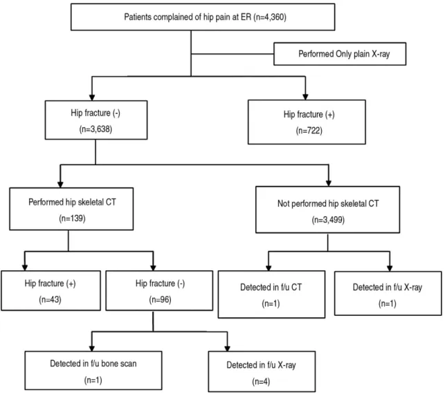
Patients who were diagnosed as having occult hip fractures at the first visit were categorized as non- delayed group. Those who were not diagnosed at the first visit were categorized as delayed group. They re-visited emergency department or outpatient clin- ic after a period of time. We excluded patients who were transferred to other hospitals and those who were diagnosed with pelvic bone fracture (Fig. 1).
We evaluated the age of patients, sex, accident mechanism, body mass index (BMI), previous frac- ture, pre-fracture mobility,late presentation, able to bear weight, pain on passive rotation, tenderness on groin area, diagnosis, displacement of fracture and treatment. We defined late presentation is presented next day after injury.
Imaging studies were interpreted by a board-cer- tified specialist in radiology.
Causes of injury were classified into a high-energy injury and a low-energy injury. A high-energy injury was defined as an injury where there is a high possi- bility that multiple organs might be damaged due to such mechanisms as falling, traffic accident and direct blow. A low-energy injury was defined as an injury which patients would sustain while falling over slippery ground in a walking or sitting position.(15)
We compared the baseline characteristics, a past history, physical examination, the displacement of hip fracture and the treatment regimens.
different groups using the independent sample t-test.
In addition, we analyzed the categorical variables using the Chi-square test or Fisher’s exact test. A p- value of <0.05 was considered statistically significant.
III.
Results
1. Baseline characteristics of the patients (Fig. 1) The total number of patients with hip fracture
In 43 patients, occult hip fracture was suspected at the first visit of patients and the diagnosis was made on CT.
In the remaining 7 patients, the diagnosis was delayed by mean of 9.6 days (range, 3~19 days).
5 patients of delayed group were diagnosed on a plain radiography at a follow-up, one did on bone scan and one did on CT.Five patients were not diag- nosed with fracture although they underwent CT at the initial visit.
Fig. 1. Flow diagram of the study participants.
Fig. 1.Hip fracture was included femur neck fracture, intertrochanter fracture, subtrachanter fracture, acetabular fracture.
Fig. 1.Hip fracture (+): patients diagnosed with hip fracture Fig. 1.Hip fracture (-): patients were not diagnosed with hip fracture Fig. 1.f/u: follow up
2. Patient characteristics of the patients with occult hip fracture (Table 1)
In a total of 50 patients who were diagnosed with occult hip fracture, the mean age was 63.9 years. 20
patients (40%) were men. 14 patients sustained a high- energy injury. 10 patients had a past history of fracture.
There were 44 patients who did not visit our med- ical institution at the time of the onset of injury.
There was one patient who had a weight-bearing ability and could perform a gait. There were 47 patients who complained of pain during the passive rotation of the hip joint. There were 39 patients who presented with inguinal tenderness. There were 22 patients who had intertrochanter fracture or trochanteric one. There were 13 patients with femoral neck fracture. There were 15 patients with acetabular fracture.
3. Comparison of the patient characteristics between the delayed group and the non-delayed group (Table 2)
There were no significant differences in the age, sex, accident mechanisms, BMI, previous fracture and pre-fracture mobility between the two groups.
In addition, there were also no significant differ- ences in the weight-bearing ability, pain on passive rotation and tenderness on groin area between the two groups. But late presentation after event were seen in 3 patients of the delayed group and 41 patients of the non-delayed group, which was a statistically significant difference (p=0.002).
Table 1. demographic characteristics of study subjects.
Variables Frequency, n
Age* (years) 63.86±20.30
Sex (female:male) 30:20
BMI* (kg/m2) 24.18±3.780
Accident mechanism
high energy injury 14
low energy injury 36
Previous fracture 10
Pre-fracture independence 02
Late presentation 44
Ability to bear weight 01
Pain on passive rotation 47
Tenderness on groin area 39
Type of fracture
Femoral neck 13
Intertrochancteric or trochancteric 22
acetabular 15
Treatment
Conservative 28
operative 22
Displacement of fracture 04
* The result are expressed as mean±standard deviation BMI: body mass index
Table 2. Baseline characteristics between the non-delayed group and delayed group.
Non-delayed group (n=43) Delayed group (n=7) p value
Mean age (years) 64.4±19.1 60.6±28.2 0.740
Sex (female:male) 25:18 5:2 0.410
BMI (kg/m2) 25.0±3.80 22.0±3.10 0.086
Accident mechanism
low energy injury 29 7 0.084
high energy injury 14 0
Previous fracture 09 1 0.571
Pre-fracture independence 02 0 0.737
Late presentation 41 3 0.002
Ability to bear weight 00 1 0.140
Pain on passive rotation 40 7 0.630
Tenderness on groin area 35 4 0.170
Type of fracture
femoral neck 08 5
inter- or trochanteric 21 1
acetabular 14 1
BMI: body mass index
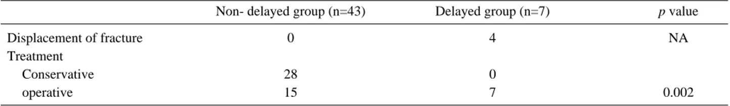
The displacement of the hip fracture was seen in 4 of seven patients of the delayed group and in no patients of the non-delayed group. 15 patients of the non-delayed group and all patients of the delayed group underwent surgical treatment, which was a statistically significant difference (p=0.002).
IV.
Discussion
The occult hip fractures are sometimes missed ini- tially and they are detected later on. Because the hip fractures might seem normal in their initial x-rays and some patients might be able to ambulate and at times the trauma vector can be minor to be noticed.(3) There was a report that occult hip frac- tures consist 2~10% of all hip fractures.(7) In the present study, the incidence of occult hip fracture was 6.5%.
To miss the diagnosis of an occult hip fracture may lead to displacement of the fracture andother- wise unnecessary surgery.(11,16) In a prospectives- tudy of 90 elderly patients with an acute hip frac- turetreated with internal fixation, Tidermark et al.
found amajordifference in outcome between undisplacedanddisplaced fractures.(11) The rate of fracture healing complicationsand reoperations in patients with displaced fractureswas high, and even in patients with uneventfullyhealed fractures, there was a substantial decrease in thequality of life.(3)
Many have reported that there were serious com- plications resulting from delayed diagnosis of the occult hip fractures.(12-14) Non-union of bones and avascular necrosis might be induced and it could lead to extension of admission days and cause
worse outcome than those without displacements.
In our study, 7 patients of total 50 patiensts who were diagnosed with occult hip fracture were not diagnosed with fractures in their initial visits to emergency department. In above stated 7 patients with occult hip fractures 4 patients had displace- ments and all 7 patients required operation. The average length of stay for non-delayed group and for delayed group was 12 days and 24.6 days each.
But, there were no serious complications in delayed occult hip fracture group.
In comparison between delayed occult hip fracture groups and non-delayed group, the time delayed in visiting emergency department(late presentation) was the only factor statistically different between two groups (p<0.002). This difference is likely due to bone remodeling which could have occurreddur- ingthe time difference and it could have lowered the probability of being detected precisely.
The patient who was late presentation after the occurrence of event in emergency department was becomed bone remodeling, then they increased the probability of being detectedfracture at the first visit.
Hence we should evaluate possible hip fracture patients with supplementary measures. There were several attempts to establish systematic approach and algorithm for potential hip fracture patients who had no definite fractures but complained of hip pains. There was a report that MRI (magnetic reso- nance imaging) examination is more accurate than any other imaging modalities.(4,6) But, in reality carrying on MRI test has its restrictions. Not all hospitals can afford costly MRI machines and even in those hospitals where they can, the radiologist
Table 3. Fracture displacement, treatment modality of the two groups.
Non- delayed group (n=43) Delayed group (n=7) p value
Displacement of fracture 0 4 NA
Treatment
Conservative 28 0
operative 15 7 0.002
NA: not applicable
might not be available after regular working hours.
Also there are absolute contraindications to MRI tests, such as patients with pacemakers or patients with metallic implants. A patient who is uncomfort- able with closed space or is in harsh pain will have difficulties due to relatively long time absorbed in the test
CT could be a second option because modern CT technology has improved significantly and the CT imaging study is comparably readily executed in emergency room settings and almost all hospitals run CT test on 24 hour bases. The multidetector CT technology has also reduced the radiation exposures such that the exposure amount is relatively small for adults and old aged patients.(19,20) The CT images now have higher resolutions and are more readily available. In recent studies they have shown that it is almost equally accurate as an MRI test and they agree that fracture can be diagnosed with very high level of certainty.(21)
Gillet al. reported the use of multislice CT scanning and suggests it has a role to play in the investigation ofoccult hip fractures. CT scanning has wide spread availability out of hours and lower cost when com- pared to MRI scanning. They showed that the propor- tion of scans in eachmodality that showed a fracture was similar (38% of CT scans, 36% of MRI scans). No MRI or CT scan missed an occult fracture which was then found when the initial X-rays were.(19)
Cabarrus et al. compared CT with MRI for in suf- ficiency fractures in the pelvis and reported 64 cases where MRI and CT could be compared sideby side.(22) Cabarrus et al. found that MRI did miss a fracture due topartial volume effects and adjacent joint effusion along with motion artefacts. This fracture was picked up by CT.
Advances in technology, suchas 64-slice scanners and sophisticated 3-dimensional reconstruction algorithms, may have made the reliability of CT comparable to that of MRI.
But, there are also reports that the CT test is prone to small impacted fractures or a fracture which is parallel with the axial plane of the CT image.(23) Kim et al stated that there was 1 patient whose hip fracture was missed initially even though they had carried out the CT test. In our study 5
patients had early CT test taken but was not diag- nosed with hip fractures. This result may have come from the fact that there are disadvantages in CT test as well.
But, We guess it is because these papers was writ- ten before their widespread usage of multislice heli- cal scanners were available.
In our study, five patients were not diagnosed with occult hip fracture although they underwent CT at the initial visit. They had severe osteoporosis or growth plate involvement.
There were several limitations in our study. First, our study was a retrospective one performed in a small cohort. Second, the accuracy of information presented is limited to the accuracy of admission details. An element of observer hetero-geneity is also present as different physicians with different levels of experience recorded clinical findings over a 6 and half year period. Third, in our study, a proto- col for referral to MRI or CT scans could not be applied in every patient with hip pain after trauma, who had normal or equivocal findings on conven- tional radiographs during the study period. Fourth, some neglected patients in whom pain was durable and fracture healed without displacement might not have been detected. Fifth, some patients could have gone to other hospitals and might have gotten diag- nosed with occult hip fractures.
A prospective study for this occult hip fracture can be executed but because of the relatively small incidence rate of occult hip fractures and of the barriers a MRI protocol intrinsically has it will be challenging to apply.
In result, the patient group who were diagnosed later on as having occult hip fracture had higher rate of hip displacement and all patients in the group received operation.
V.
Conclusion
Patient with a delayed diagnosis of occult hip fracture had a higher rate of fracture displacement and need for surgery than those diagnosed on the initial ED visit. Although CT scan detected the occult fracture in most cases, it missed five occult fractures during the initial visit.
burden of osteoporosis in Sweden. Bone 2007; 40: 1602-9.
03) Kim KC, Ha YC, Kim TY, Choi JA, Koo KH. Initially missed occult fractures of the proximal femur in elderlypatients:
implications for need of operation and their morbidity. Arch Orthop Trauma Surg 2010; 130: 915-20.
04) Lubovsky O, Liebergall M, Mattan Y, Weil Y, Mosheiff R.
Early diagnosis of occult hip fractures MRI versus CT scan.
Injury 2005; 36: 788-92.
05) Helland EB, Tollefsen I, Reksten G. Radiographic diagnosis of the occult hip fracture: experience in 16 patients. Acta Orthop Scand 2000; 71: 639-41.
06) Verbeeten KM, Hermann KL, Hasselqvist M, Lausten GS, Joergensen P, Jensen CM et al. The advantages of MRI in the detection of occult hip fractures. Eur Radiol 2005; 15: 165-9.
07) Dominguez S, Liu P, Roberts C, Mandell M, Richman PB.
Prevalence of traumatic hip and pelvic fractures in patients with suspected hip fracture and negative initial standard radi- ographs-a studyof emergency department patients. Acad Emerg Med 2005; 12: 366-9.
08) Oka M, Monu JU. Prevalence and patterns of occult hipfrac- tures and mimics revealed by MRI. AJR Am J Roentgenol 2004; 182: 283-8.
09) Perron AD, Miller MD, Brady WJ. Orthopedic pitfalls in theED: radiographically occult hip fracture. Am J Emerg Med 2002; 20: 234-7.
10) Nachtrab O, Cassar-Pullicino VN, Lalam R, Tins B, Tyrrell PN, Singh J. Role of MRI in hip fractures, including stress fractures, occult fractures, avulsion fractures. Eur J Radiol 2012; 81: 3813-23.
11) Moore MN. Orthopedic pitfalls in emergency medicine. South Med J 1988; 81: 371-8.
12) Bentley G. Treatment of nondisplaced fractures of thefemoral
14) Blickenstaff LD, Morris JM. Fatigue fracture of the femoral neck. J Bone Joint Surg Am 1966; 48: 1031-47.
15) Nodzo SR, Hohman DW, Galpin RD. Bilateral acetabular fractures in an adolescent after low-energy trauma. Pediatr Emerg Care 2012; 28: 568-9.
16) Frihagen F, Nordsletten L, Tariq R, Madesn JE. MRI diagno- sis ofoccult hip fractures. Acta Orthop 2005; 76: 524-30.
17) Zuckerman JD, Skovron ML, Koval KJ, Aharonoff G, FrankelVH. Postoperative complications and mortality associ- atedwith operative delay in older patients whohave a fracture of the hip. J Bone Joint Surg Am 1995; 77: 1551-6.
18) Sankey RA, Turner J, Lee J, Healy J, Gibbons CE. The useof MRI to detect occult fractures of the proximal femur: a study- of 102 consecutive cases over a ten-year period. J Bone Joint Surg Br 2009; 91: 1064-8.
19) GillSK, Smith J. Fox R,Chesser TJ. Investigation of Occult Hip Fractures: The Use of CT and MRI. Scientific World Journal 2013; 2013: 830319.
20) Memarsadeghi M, Breitenseher MJ, Schaefer-Prokop C, Weber M, Aldrian S, Gäbler C et al. Occult scaphoid frac- tures: comparison ofmultidetector CT and MR imaging-initial experience. Radiology 2006; 240: 169-76.
21) Collin D, Dunker D, Göthlin JH, Geijer M. Observer variation for radiography, computed tomography, and magnetic reso- nance imaging of occult hip fractures. Acta Radiol 2011; 52:
871-4.
22) Cabarrus MC, AmbekarA, Lu Y, Link TM. MRI andCT of insufficiency fractures of the pelvis and the proximalFemur.
AJR Am J Roentgenol 2008; 191: 995-1001.
23) Hayes CW, Balkissoon AR. Current concepts in imagingof the pelvis and hip. Orthop Clin North Am 1997; 28: 617-42.