서론
고령화와 진단 기술, 수술 기술의 발전으로 망막앞막 및 황반 원공의 진단 및 수술이 많이 늘고 있다[1,2]. 또한 최근 빛간섭 단층촬영술(optical coherence tomography)의 축해상도 및 스캔
속도의 발전으로 인한 새로운 영상 기술이 도입되고 있으며 최 근 빛간섭단층혈관조영술의 개발도 관혈적인 술기 없이 황반부 에서의 혈관 변화와 망막층별 구조 변화를 쉽게 관찰할 수 있게 되었다[3-5]. 빛간섭단층촬영에서 재구성하여 볼 수 있는 C-스 캔영상 또는 정면영상(en-face image)은 수술 전 또는 수술 후
내경계막벗김술에 의한 내망막패임과 관련된 요소들
Related Factors and Inner Retinal Dimples Induced by Internal Limiting Membrane Peeling
신용균1,이근우2
Yong Kyung Shin1, Geun Woo Lee2
1한림대학교 의과대학 춘천성심병원 안과학교실, 2대구가톨릭대학교 의과대학 안과학교실
1Department of Ophthalmology, Chuncheon Sacred Heart Hospital, Hallym University College of Medicine, Chuncheon, Korea
2Department of Ophthalmology, Daegu Catholic University School of Medicine, Daegu, Korea
Purpose: To describe inner retinal dimples in patients who underwent epiretinal membrane (ERM) or macular hole (MH) surgery, and to analyze factors affecting the occurrence of inner retinal dimples between ERM and MH patients.
Methods: This was a retrospective observational study. Patients who underwent ERM or MH surgery involving internal limiting mem- brane peeling from January 2018 to December 2018 were enrolled. Among 191 patients, 28 patients were identified. These patients were subdivided by ERM type (diffuse, cystoid macular edema, pseudolamellar hole type) and posterior vitreous detachment stage of MH. Two months after surgery, the number of inner retinal dimples was analyzed using en-face imaging. The location of the inner retinal dimple and intraoperative findings (pinch or retinal hemorrhage) were compared.
Results: On en-face imaging, inner retinal dimples were detected in 18 (17.3%) of 104 eyes with ERM and 10 (26.3%) of 38 eyes with MH.
The number of inner retinal dimples in the MH group was significantly greater (p = 0.049) than in the ERM group. In the ERM group, the pseudolamellar hole type was associated with more inner retinal dimples, as was higher metamorphopsia score (p = 0.023). The locations of retinal hemorrhages matched inner retinal dimples better than the locations of pinches (all p < 0.05).
Conclusions: Retinal hemorrhages, as well as the iatrogenic trauma associated with pinches, are associated with inner retinal dimples.
Inner retinal dimples were more frequently associated with MH than ERM. Among the ERM group, pseudo lamellar hole type was more observed than other types. Ophthalmologist should be aware of the risk of inner retinal dimple in cases with internal limiting membrane peeling.
Keywords: Epiretinal membrane; Inner retinal dimple; Internal limiting membrane peeling; Macular hole
Address reprint requests to Geun Woo Lee, MD
Department of Ophthalmology, Daegu Catholic University Medical Center, #33 Duryugongwon-ro 17-gil, Nam-gu, Daegu 42472, Korea
Tel: 82-53-650-4728, Fax: 82-53-650-0133 E-mail: [email protected]
Received: 2020. 4. 2.
Revised: 2020. 5. 7.
Accepted: 2020. 5. 11.
망막 표면의 모습을 높은 화질로 쉽게 파악할 수 있게 되었다 [6,7]. 망막 내경계막벗김술을 시행한 눈에서 이러한 정면영상을 통해 신경섬유층의 분리(dissociation of nerve fiver layer)를 의 미하는 것으로 알려진 내망막패임(inner retinal dimple)도 새롭 게 종종 관찰된다[8,9].
망막 내경계막 제거를 포함한 수술은 대표적으로 망막앞막과 황반원공이 있다. 망막앞막은 망막표면에 섬유세포성 증식으로 인해 막이 생겨나는 질환으로 심한 경우 시력감소나 변형시 등 의 증상을 유발하기도 한다[10]. 보통 증상을 수반하거나 구조 적 변화가 진행하는 경우에는 수술을 시행하게 되는데 이때 망 막앞막과 함께 내경계막벗김술도 많이 시행된다. 이는 내경계 막 제거가 기능적, 구조적으로 더 나은 변화를 보이는 것은 아 니나 재발률을 낮출 수 있다고 알려져 있기 때문이다[11,12]. 그 리고 황반원공은 일반적으로 65세 이상의 여성에 많이 일어나 며 병인은 명확하지 않으나 비정상적인 중심와바깥 유리체분리 에 의한 견인력에 의한 결과로 생각되고 있다[13]. 이와 관련하 여 황반원공의 경우에는 내경계막 제거와 유리체강내 가스주입 술이 치료의 표준으로 자리잡고 있다[14]. 저자들은 내경계막벗 김술 후 나타나는 내망막패임이 관찰되는 망막앞막수술 후와 황반원공수술 후에서의 두 수술 간의 내망막패임의 발생에 차 이가 나타나는지 알아보고 내망막패임 발생에 영향을 주는 인 자들에 대해 알아보고자 한다.
대상과 방법
2018년 1월부터 2018년 12월까지 특발망막앞막 및 황반원공으 로 내경계막벗김술을 시행 받았던 환자를 대상으로 후향적 관 찰 연구를 시행하였다. 이 연구는 대구가톨릭대학교병원의 임 상시험심사위원회 승인을 받았고 헬싱키선언을 준수하였다(IRB no: CR-19-146-L).
60세 이상의 수술한 환자를 대상으로 술 후 2개월 이상의 외 래방문이 이루어지고 술 전, 술 후 검사가 적절히 이루어진 경우 중 수술 후 2개월째의 망막단층촬영혈관조영술의 정면영상에서 내망막패임이 확인되는 경우를 포함하였다. 망막앞막 및 황반 원공 이외에 다른 망막 질환이 시험 안에 있거나 있었던 경우, 고도근시인 경우(굴절값 -6.0디옵터 이하 또는 안축장 26.0 mm 이상 또는 근시성 안저 변화), 첫 번째 수술 후 황반원공폐쇄에 실패한 경우, 영상의 질이 나쁜 경우는 연구에서 제외하기로 하 였다. 모든 기준에 부합하는 28명(28안)이 대상자에 포함되었다.
모든 환자들은 수술 전에 시행하는 양안 최대교정시력, 변형 시 점수(Metamorphopsia score, M-score), 구면렌즈 대응치, 산 동안저검사를 포함하는 전반적인 검사들을 시행 받았다. 변형 시 점수는 세로선과 가로선의 검사값의 평균을 이용하였고 최 대교정시력은 스넬렌시력표를 이용하여 측정한 뒤 통계를 위
해 logarithm of minimal angle of resolution (logMAR) 시력으 로 변환하였다. 그리고 수술 전과 수술 후 2개월에 정면영상을 분석에 이용하였다. 빛간섭단층촬영술(AngioVueⓇ; Optovue, Fremont, CA, USA)을 이용하여 수술 전 망막앞막을 광범위형 (diffuse type), 낭포황반부종형(cystoid macular edema type), 가 성층판원공형(pseudolamellar hole type)의 세 가지로 분류하였 고 황반원공의 경우 후유리체박리를 단계에 따라 분류하였다 (Fig. 1) [15-17].
23게이지 유리체절제술 시스템으로 한 명의 술자에 의해 모 든 수술들이 시행되었다(AssociateⓇ; Dutch Ophthalmic Research Center Inc., Zuidland, The Netherlands). 백내장수술을 받지 않 은 환자는 유리체절제술을 시행받기 전 수정체유화술 및 인 공수정체삽입술도 함께 시행하였다. 중심유리체절제술 및 후 유리체 제거 후 트리암시놀론(MaqaidⓇ; Hanmi Pharm Co., Seoul, Korea) 혹은 인도시아닌그린(DID-Indocuanine greenⓇ; DongKwang Pharm Co., Seoul, Korea)을 이용하여 망막앞막 혹은 내경계막을 가시화시켰다. 트리암시놀론(triamcinolone acetonide)은 40 mg에 4 mL 평형염액(balanced salt solution)을 혼합한 후 1 mL 주사기에 뽑아서 사용하였고 인도시아닌그린 (indocyanine green)은 25 mg에 25 mL 주사용수를 믹스한 후 1 mL 주사기로 0.5 mL를 뽑고 추가로 평형염액 0.5 mL와 혼합하 여 최종 농도가 0.5 mg/mL (0.05%)가 되도록 하였다. 인도시아닌 그린을 사용할 때는 20초간 유입용액을 정지시켜 내경계막을 염 색시킨 후 재빨리 유리체강 내의 관류 및 흡인을 시행하여 인도 시아닌그린을 제거하였다. 가시화된 망막앞막과 내경계막을 집게 (endgripping forcep)를 이용하여 핀치기술(pinch technique)로 벗 김술을 시행하였다. 황반원공 환자의 경우는 액체-공기 교환술 후의 유리체강을 10% C3F8 가스로 채우고 수술을 종료하였고 수 술 후 엎드린 자세를 최소 2주 이상 유지하였다.
A B
C
D
E
Figure 1. Epiretinal membrane type (A-C) and posterior vitreous detachment stage in macular hole (D, E). (A) Diffuse type, (B) cystoid macular edema type, (C) pseudolamellar hole type, (D) stage 2, (E) stage 3.
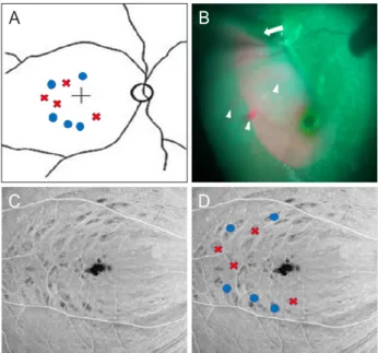
수술 후 2개월째 시험안의 optical coherence tomography angiography (OCTA)에서 보이는 표면의 정면영상을 이용하여 내망막패임의 전체 수를 파악하였고 내망막패임 존재가 헷갈리 는 경우에는 단층영상(B scan image of OCT)을 통해 재차 확인 하였다(Fig. 2). 그리고 수술 시에 녹화된 수술 영상을 통해 내 경계막벗김술 시행 시에 이용된 핀치 위치들 및 전체 횟수, 망막 출혈 위치들 및 전체 횟수를 파악하였다(Fig. 3). 정면영상에서 관찰되는 내망막패임을 핀치의 위치 및 망막출혈의 위치와 일 치하는 정도를 점수화(scoring)하였다. 즉, 핀치 위치 및 망막출 혈의 위치가 내망막패임의 위치와 완전일치하는 경우는 3점, 반 이상 일치하는 경우는 2점, 반 이하 일치하는 경우는 1점, 전혀 일치하지 않는 경우는 0점으로 점수화하였다.
두 군의 연속형 변수의 평균을 비교하는 경우에는 independent t test 또는 Mann-Whitney test를 시행하였고 비율을 비교하는 경우에는 Fisher’s exact test를 시행하였다. 추가로 망막앞막군에 서 형태 따른 비교는 Kruskal-Wallis test를 시행하였다. 이후 유 의한 경우에는 사후분석을 실시하여 차이가 나는 부분을 확인 하기로 하였다. 다른 요소와 상관관계 분석은 Spearman’s Rho test를 이용하여 분석하였다. 통계는 SPSS statistics software package version 21.0 (IBM Corp., Amonk, NY, USA)을 사용하 였고 p-value값이 0.05보다 작은 경우를 유의한 결과로 보았다.
결과
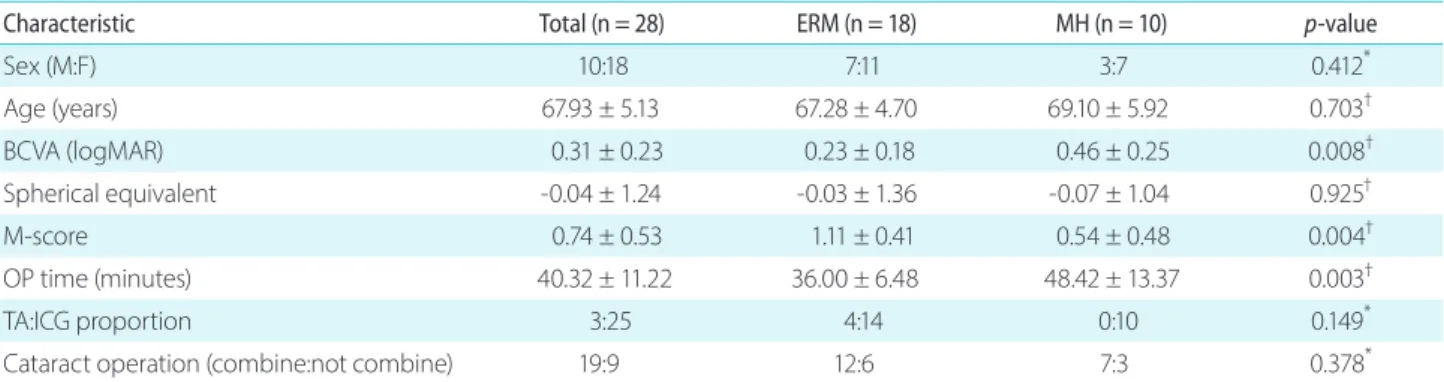
총 142명의 망막앞막 환자와 49명의 황반원공 환자의 의료기 록이 분석되었고 그중 내망막패임의 유무를 제외한 다른 기준 을 만족하는 대상안의 수는 망막앞막이 104안, 황반원공이 38안 이었다. 그중 내망막패임이 보이는 경우는 망막앞막 18안(17.3%), 황반원공 10안(26.3%)이 분석에 이용되었다(Pearson chi square test, p = 0.241). 평균 연령은 67.9세였고 망막앞막 환자(67.3세)와 황반원공 환자(69.1세) 간의 유의한 차이는 없었다. 이 외에 성별,
구면렌즈대응치, 사용한 염색약의 분율 차이, 동반 백내장수술 여부도 유의하게 차이 나지 않았다. 그러나 두 군 간에 logMAR 최대교정시력과 수술 시간, 그리고 변형시 점수에서 유의한 차이 가 보였다(Table 1, 각각 p = 0.008, p = 0.003, p = 0.004).
수술 전 시행한 OCTA의 결과를 통해 망막앞막의 형태와 황반원공의 후유리체박리 단계를 확인하였다. 망막앞막의 경 우, 광범위형은 8안, 낭포황반부종형은 6안, 가성층판원공형은 4안이었고 황반원공의 경우, 단계 2가 5명, 단계 3이 5명이었다.
수술 후 2개월째 시행한 정면영상의 결과와 수술동영상을 이 용하여 시행한 분석 결과는 Table 2에 정리하였다. 내망막패임 의 숫자는 전체 평균이 10.68개였고, 망막앞막(9.60개)과 황반원 공(11.28개) 그룹 간의 유의하게 차이가 있었다(Mann-Whitney test, p = 0.049). 이외에 전체 핀치 수와 핀치점수, 망막출혈의 수와 출혈 점수는 두 군 간에 유의하게 차이가 나지 않았다. 추 가로 시행한 전체 대상안과 각 그룹 내에서 핀치 점수와 망막출 혈 점수 간에 비교한 결과 출혈 점수가 유의하게 높은 결과값을 보이는 것을 확인하였다.
각 그룹에서 형태와 단계별로 내망막패임의 수, 핀치 수, 핀치 점수, 망막출혈 수, 망막출혈 점수의 평균값을 Table 3에 정리하 였다. 각 그룹에서 망막앞막의 형태와 후유리체박리 단계별로 내망막패임의 수에 차이가 나는지 비교 분석해보았다(Fig. 4).
우선 망막앞막에서는 형태에 따른 차이가 나타나는 것으로 나
A B
Figure 2. En-face image 2 months after surgery. (A) Inner retinal dimple is confirmed. (B) Modified image that inner retinal dimple was bordered with white line.
A
C
B
D
Figure 3. (A) Pinch (blue circle) and hemorrhage (red x) locations on the retinal image. (B) Intraoperative view of internal limiting membrane peeling. Retinal hemorrhages (white arrowheads) and pinch (white arrow) are confirmed. (C) En-face imaging of optical coherence tomography angiography. (D) Comparison of inner retinal dimples on en-face imaging with pinch locations (blue circle) and hemorrhage locations (red x).
Table 1. Baseline demographics of epiretinal membrane and macular hole groups
Characteristic Total (n = 28) ERM (n = 18) MH (n = 10) p-value
Sex (M:F) 10:18 7:11 3:7 0.412*
Age (years) 67.93 ± 5.13 67.28 ± 4.70 69.10 ± 5.92 0.703†
BCVA (logMAR) 0.31 ± 0.23 0.23 ± 0.18 0.46 ± 0.25 0.008†
Spherical equivalent -0.04 ± 1.24 -0.03 ± 1.36 -0.07 ± 1.04 0.925†
M-score 0.74 ± 0.53 1.11 ± 0.41 0.54 ± 0.48 0.004†
OP time (minutes) 40.32 ± 11.22 36.00 ± 6.48 48.42 ± 13.37 0.003†
TA:ICG proportion 3:25 4:14 0:10 0.149*
Cataract operation (combine:not combine) 19:9 12:6 7:3 0.378*
Values are presented as mean ± standard deviation or number.
ERM = epiretinal membrane; MH = macular hole; M = male; F = female; logMAR = logarithm of the minimum angle of resolution; BCVA = best-corrected visual acuity; M-score = metamorphopsia score; OP = operation; TA = triamcinolone acetate; ICG = indocyanine green.
*Fisher’s exact test; †Independent t test.
Table 2. Analysis of inner retinal dimple and intraoperative findings
Total (n = 28) ERM (n = 18) MH (n = 10) p-value*
Total number of dimple 10.68 ± 2.00 9.60 ± 1.26 11.28 ± 2.11 0.049
Total count of pinch 4.96 ± 0.65 4.48 ± 0.65 5.00 ± 0.67 0.388
Total number of hemorrhages 2.39 ± 1.40 2.33 ± 1.46 2.50 ± 1.35 0.393
Pinch score 2.43 ± 0.50 2.44 ± 0.51 2.40 ± 0.52 0.782
Hemorrhage score† 2.91 ± 0.29 2.93 ± 0.27 2.88 ± 0.35 0.426
p-value* 0.023 0.021 0.028 -
Values are presented as mean ± standard deviation.
ERM = epiretinal membrane; MH = macular hole.
*Mann-Whitney test; †Hemorrhage score means to score the degree of correlation between hemorrhage site and dimple site. If there was no hemorrhage on surgery, we excluded from score (4 in the ERM group and 2 in the MH group).
Table 3. Intraoperative findings and inner retinal dimple by ERM type and PVD stage in MH
Number of Dimple Pinch count Pinch score Number of retinal
hemorrhage Hemorrhage score* Types in ERM
Diffuse type (n = 8) 9.88 ± 0.44 4.62 ± 0.26 2.25 ± 0.16 2.50 ± 0.19 2.87 ± 0.13
Cystoid macular edema type (n = 6) 12.17 ± 1.05 4.67 ± 0.21 2.83 ± 0.17 1.83 ± 0.83 2.67 ± 0.33 Pseudolamellar hole type (n = 4) 12.75 ± 0.48 5.25 ± 0.25 2.75 ± 0.25 2.75 ± 0.95 2.67 ± 0.33
p-value† 0.041 0.271 0.101 0.608 0.663
PVD stage in MH
Stage 3 (n = 5) 10.33 ± 0.33 5.33 ± 0.33 2.67 ± 0.33 3.33 ± 0.33 2.67 ± 0.33
Stage 2 (n = 5) 10.67 ± 0.33 5.00 ± 0.58 2.67 ± 0.33 3.33 ± 0.33 2.67 ± 0.33
p-value‡ 0.941 1.000 0.549 0.650 0.495
Values are presented as mean ± standard deviation.
ERM = epiretinal membrane; PVD = posterior vitreous detachment; MH = macular hole.
*Hemorrhage score means to score the degree of correlation between hemorrhage site and dimple site. If there was no hemorrhage on surgery, we excluded from score (4 in the ERM group and 2 in the MH group); †Kruskal-Wallis test; ‡Mann-Whitney test.
타났고 사후검정에서 광범위형과 가성층판원공형 사이에서 유 의하게 차이나는 것으로 나왔다(p = 0.01). 그러나 황반원공에 서는 단계에 따른 차이는 없는 것으로 나왔다.
추가로 각 그룹 내의 내망막패임 수와 상관관계가 있는 요소 들을 분석해보았고 망막앞막에서는 변형시 점수와 내망막패임 의 수가 유의한 상관관계가 있는 걸로 나왔다(Fig. 5, p = 0.023, 상관계수 = 0.682). 그러나 황반원공에서는 변형시 점수와 내망 막패임의 수가 유의한 상관관계를 보이지 않았다(p = 0.094, 상 관계수 = 0.406).
고 찰
내경계막벗김술 시행 후 생기는 내망막패임에 대해서 2001년 Tadayoni et al. [8]이 처음 언급하였고 이는 망막신경섬유층의 분리를 의미하는 것으로 시력이나 시야에 영향을 주지 않는다 고 보고했다. 그러나 이전 연구에서는 이것이 암점과 관계가 있 을 수 있다는 보고도 있다[18]. Amouyal et al. [19]은 내망막패 임이 12개월까지는 크기와 깊이가 더 커지고 이 후 서서히 줄어 든다고 보고하였고, 다른 연구에서는 내망막패임이 추후 망막 신경섬유층이 얇아지는 것과 관계가 될 수 있으며 내망막패임 의 발생은 벗김술 시행 시에 망막표면에서 막을 잡는 것과 관련 있다고 보고했다[19-21].
본 연구에서는 망막앞막 환자와 황반원공 환자에서 내경계 막벗김술 후 내망막패임을 보이는 경우를 대상으로 두 군 간 에 어떤 차이를 보이는지 알아보았고 각 군 내에서 내망막패임 의 수와 관련된 인자들을 추가로 분석하였다. 이러한 연구는 이 전 보고가 없다.
망막앞막 환자의 17.3%, 황반원공 환자의 26.3%에서 내망막 패임이 관찰되었고 황반원공 환자군에서 유의하게 많은 내망 막패임의 수를 보였다(Mann-Whitney test, p = 0.049). 이전 연 구에서 가스충전술을 한 경우에서 내망막패임이 더 많이 관찰 된다는 보고가 있었다[22]. 이에 두 군 간의 내망막패임 발생률 이 차이가 나는 것은 유리체강을 10% C3F8 가스로 채운 황반 원공수술 과정의 차이일 수 있다. 또한 두 군 간에 수술 전 최 대교정시력과 수술 시간, 변형시 점수에서 유의한 차이가 있었 으며 망막전막 및 황반원공 자체가 다른 질환이므로 이러한 병 Figure 4. Differences between dimple number and epiretinal mem-
brane (ERM) type. DIF = diffuse type; CME = cystoid macular edema type; PLH = pseudo lamellar hole type. p = 0.047 (on Kruskal-Wallis analysis); p = 0.01 (between DIF and PLH on post hoc analysis).
14.00
12.00
10.00
8.00
Dimple number
ERM type 1. DIF, 2. CME, 3. PLH
1 2 3
Figure 5. Correlation between dimple number and M-score in the epiretinal membrane (ERM) group and macular hole (MH) group. R = Spear- man’s rho coefficient. p-value were calculated by Spearman correlation analysis.
11.00
10.50
10.00
9.50
9.00
8.50
8.00
.75 1.00 1.25 1.50 1.75
M-score in the ERM group
Dimple number
p = 0.023, r = 0.617 14.00
12.00
10.00
8.00
.0 .5 1.0 1.5 2.0
M-score in the ERM group
p = 0.094, r = 0.406
적 상황에 의한 내경계막의 유착력의 차이가 있을 수도 있을 것으로 생각된다.
우리 연구에서는 수술동영상과 정면영상을 통해 핀치 횟수 및 핀치 점수(핀치 위치와 내망막패임의 위치가 일치하는 정도), 망막출혈의 수 및 망막출혈 점수(망막출혈의 위치와 내망막패 임의 위치가 일치하는 정도)를 파악하였다. 두 그룹 간에 유의 한 차이를 보이지는 않았으나 각 그룹 내에서 핀치 점수와 망막 출혈의 점수를 비교한 결과는 유의하게 망막출혈의 점수가 높 은 것을 확인할 수 있었다. 이전 연구와 같이 내망막패임이 핀치 로 인한 의인성 외상이 주요 인자이지만 본 연구에서는 의인성 외상 이외에 망막출혈이 내망막패임과 관계될 수 있으며 본 연 구에서 오히려 더 높은 관련성을 보이는 것을 확인하였다[9,20].
이는 핀치로 인한 직접적인 외상과 내경계막벗김술로 인한 외 상이 출혈로 관찰됨으로 생각된다. 또한 모든 군에서 망막패임 의 수가 핀치 횟수 및 망막출혈의 수보다 많은 것은 의인성 외 상 이외에도 많은 인자들이 관여할 수 있다는 것을 알 수 있다.
망막앞막의 형태에 따른 내망막패임의 수의 비교는 세 군 간 에 유의한 차이를 보이는 걸로 나왔고, 특히 광범위형에 비해 가성층판원공형의 내망막패임의 수가 유의하게 큰 것으로 나왔 다. 이러한 가성층판원공형의 경우 다른 앞막형태와 다른 점을 보인다는 것을 유의하여야 한다. 황반원공의 후유리체박리 단 계에 따른 내망막패임의 수에는 차이가 없었다. 그리고 망막앞 막군에서 변형시 점수와 내망막패임의 수와 상관관계가 있는 것으로 나왔으나 황반원공군에서는 유의한 상관관계를 보이지 는 않았다. 즉, 망막앞막군에서는 술 전 변형시 점수가 심할수 록 더 많은 내망막패임을 보일 수 있다는 것이다. 이는 변형시 점수가 망막왜곡과 더 관련될 수 있을 수 있고 이는 부분적으 로 내경계막의 강한 유착을 일으켜 수술 중에 내경계막벗김으 로 인한 신경섬유층의 손상이 많기 때문인 것으로 보인다[7].
본 연구의 제한점은 첫째로 후향적으로 이루어졌으며 분석 대 상자가 수가 적었고 연구에 포함시킨 관찰 기간이 적었다는 점 이다. 즉, 이 연구를 바탕으로 추후 내망막패임이 시기능에 어 떤 영향을 주는 지에 대한 장기간의 관찰 연구 혹은 전향적 코 호트 연구가 나오면 본 연구를 이해하는 데 도움이 될 것으로 생각된다. 둘째로 본 연구에서 사용된 인도시아닌그린은 안전한 농도로 사용되었지만 이러한 염색제는 망막독성을 가지고 있으 므로 염색제의 사용 유무에 따른 연구 분석은 시행하지 못하였 다. 이러한 차이에 대한 향후 연구가 필요할 것으로 생각된다.
결론적으로 황반원공군에서 망막앞막군에 비해 내망막패임 이 더 잘 발생하고 더 많이 관찰된다. 핀치와 관계된 의인성 외 상 이외에 망막출혈도 내망막패임과 연관된다. 그리고 망막앞 막군에서 가성층판원공형이 내망막패임의 발생을 더 유발하고 술 전 변형시 점수가 클수록 내망막패임의 수가 늘어났다. 이 에 이러한 군에서는 수술로 인한 주의가 필요로 할 것으로 생 각된다. 둘째로 본 연구에서 사용된 인도시아닌그린은 안전한
농도로 사용되었지만 이러한 염색제는 망막독성을 가지고 있으 므로 염색제의 사용 유무에 따른 연구 분석은 시행하지 못하였 다. 이러한 차이에 대한 향후 연구가 필요할 것으로 생각된다.
Conflicts of Interest
The authors declare no conflicts of interest relevant to this article.
References
1. Mitchell P, Smith W, Chey T, et al. Prevalence and associations of epiretinal membranes. The Blue Mountains Eye Study, Australia.
Ophthalmology 1997;104:1033-40.
2. Kim JY, Rim TH, Kim SS. Trends of pars plana vitrectomy rates in South Korea: a nationwide cohort study. Korean J Ophthalmol 2017;31:446-51.
3. Nelis P, Alten F, Clemens CR, et al. Quantification of changes in foveal capillary architecture caused by idiopathic epiretinal membrane using OCT angiography. Graefes Arch Clin Exp Oph- thalmol 2017;255:1319-24.
4. Romano MR, Cennamo G, Schiemer S, et al. Deep and superfi- cial OCT angiography changes after macular peeling: idiopathic vs diabetic epiretinal membranes. Graefes Arch Clin Exp Oph- thalmol 2017;255:681-9.
5. Kim YJ, Kim S, Lee JY, et al. Macular capillary plexuses after epiretinal membrane surgery: an optical coherence tomogra- phy angiography study. Br J Ophthalmol 2018;102:1086-91.
6. Greven MA, Elkin Z, Nelson RW, Leng T. En Face imaging of epiretinal membranes and the retinal nerve fiber layer using swept-source optical coherence tomography. Ophthalmic Surg Lasers Imaging Retina 2016;47:730-4.
7. Hirano M, Morizane Y, Kanzaki Y, et al. En face image-based anal- ysis of retinal traction caused by epiretinal membrane and its relationship with visual functions. Retina 2020;40:1262-71.
8. Tadayoni R, Paques M, Massin P, et al. Dissociated optic nerve fiber layer appearance of the fundus after idiopathic epiretinal membrane removal. Ophthalmology 2001;108:2279-83.
9. Spaide RF. “Dissociated optic nerve fiber layer appearance” after internal limiting membrane removal is inner retinal dimpling.
Retina 2012;32:1719-26.
10. Wise GN. Clinical features of idiopathic preretinal macular fibro- sis. Schoenberg Lecture. Am J Ophthalmol 1975;79:349-7.
11. Chang WC, Lin C, Lee CH, et al. Vitrectomy with or without inter-
nal limiting membrane peeling for idiopathic epiretinal mem- brane: a meta-analysis. PLoS One 2017;12:e0179105.
12. Schechet SA, DeVience E, Thompson JT. The effect of internal limiting membrane peeling on idiopathic epiretinal membrane surgery, with a review of the literature. Retina 2017;37:873-80.
13. Bainbridge J, Herbert E, Gregor Z. Macular holes: vitreoretinal relationships and surgical approaches. Eye (Lond) 2008;22:1301- 9.
14. Bikbova G, Oshitari T, Baba T, et al. Pathogenesis and manage- ment of macular hole: review of current advances. J Ophthalmol 2019;2019:3467381.
15. Kinoshita T, Kovacs KD, Wagley S, Arroyo JG. Morphologic differences in epiretinal membranes on ocular coherence to- mography as a predictive factor for surgical outcome. Retina 2011;31:1692-8.
16. Kishi S. Impact of swept source optical coherence tomography on ophthalmology. Taiwan J Ophthalmol 2016;6:58-68.
17. Tsukahara M, Mori K, Gehlbach PL, Mori K. Posterior vitreous detachment as observed by wide-angle OCT imaging. Ophthal-
mology 2018;125:1372-83.
18. Haritoglou C, Gass CA, Schaumberger M, et al. Macular changes after peeling of the internal limiting membrane in macular hole surgery. Am J Ophthalmol 2001;132:363-8.
19. Amouyal F, Shah SU, Pan CK, et al. Morphologic features and evolution of inner retinal dimples on optical coherence to- mography after internal limiting membrane peeling. Retina 2014;34:2096-102.
20. Scupola A, Grimaldi G, Abed E, et al. Arcuate nerve fiber layer changes after internal limiting membrane peeling in idiopathic epiretinal membrane. Retina 2018;38:1777-85.
21. Mastropasqua L, Borrelli E, Carpineto P, et al. Microvascular changes after vitrectomy with internal limiting membrane peel- ing: an optical coherence tomography angiography study. Int Ophthalmol 2018;38:1465-72.
22. Park SH, Kim YJ, Lee SJ. Incidence of and risk factors for dissoci- ated optic nerve fiber layer after epiretinal membrane surgery.
Retina 2016;36:1469-73.
내경계막벗김술에 의한 내망막패임과 관련된 요소들
목적: 망막앞막 또는 황반원공수술을 받은 환자에서의 내망막패임을 기술하고, 망막앞막과 황반원공 사이의 망막패임에 영향을 미치 는 요인을 분석하고자 한다.
대상과 방법: 본 연구는 후향적 관찰 연구로 2018년 1월부터 2018년 12월까지 내경계막벗김술을 시행받은 망막앞막 및 황반원공수술 후 환자를 대상으로 하였다. 총 191명의 환자 중 기준에 부합하는 28명(28안)이 대상자에 포함되었다. 이러한 환자를 망막앞막 유형 및 후유리체막박리 단계에 따라 세분화하였다. 수술 2개월 뒤 정면영상(en-face image)을 이용하여 망막패임 수를 분석하였고 이를 수 술 중 핀치 및 망막출혈 사이와의 관계를 분석하였다.
결과: 정면 영상에서 망막앞막 104안 중 18안(17.3%)과 황반원공 38안 중 10안(26.3%)에서 망막패임이 관찰되었다. 황반원공군에 서의 망막패임 수가 망막앞막군보다 유의하게 많았다(p = 0.049). 망막앞막군에서 가성층판원공형에서 망막패임 수가 많았으며 변형 점수(M-score)가 높을수록 망막패임 수가 많았다(p = 0.023). 망막패임 위치는 핀치의 위치보다 망막출혈의 위치와 더 일치하였다.
결론: 핀치와 관련된 의인성 외상 외에 망막출혈도 망막패임과 연관이 있다. 망막패임은 황반원공에서 망막앞막보다 더 많이 관찰되었 고, 망막앞막 중에서는 가성층판원공형에서 더 많이 관찰되었다. 이러한 경우 내경계막벗김술 시행 시에 주의가 필요하다.
국문초록