Introduction
Endometrial polyps have been considered as a hyperplastic growth of the stromal and glandular parts of the endome- trium [1]. The prevalence of endometrial polyps in women is approximately 7.8%. However, it seems to be higher (9.2%) in women over the age of 30 years [2]. Most polyps are known to be benign, especially in premenopausal women.
Estimates of endometrial cancer occurrence within polyps vary across study populations as 1.1–4.9% [3]. While en- dometrial polyps are usually asymptomatic, they may lead to intermenstrual bleeding, menorrhagia, postmenopausal bleeding, infertility, and pelvic pain.
Endometrial polyps: Is the prediction of spontaneous regression possible?
Semra Yuksel, MD
1, Guray Tuna, MD
1, Hale Goksever Celik, MD
2, Suleyman Salman, MD
1Department of Obstetrics and Gynecology, 1Saglik Bilimleri University, Gaziosmanpasa Training and Research Hospital, Gaziosmanpasa/İstanbul,
2Saglik Bilimleri University, Istanbul Kanuni Sultan Suleyman Training and Research Hospital, Küçükçekmece/İstanbul, Turkey
Objective
Endometrial polyps have been considered as a hyperplastic growth of endometrial stromal and glandular tissues.
Even asymptomatic polyps in premenopausal women are usually removed as soon as they are diagnosed, although it is still unknown how often endometrial polyps disappear spontaneously. The aim of this study was to investigate the regression rate of endometrial polyps and the possible factors related to their spontaneous regression.
Methods
A total of 197 women with endometrial polyps were treated with operative hysteroscopy between January 2017 and April 2019 at our tertiary center. Of these, 123 patients who preferred conservative follow-up were enrolled in the study. Clinical and pathological data were obtained from electronic medical records.
Results
Patients with endometrial polyps were followed up for a median period of 62 days (range 30–360 days). Most women with endometrial polyps (84%) were reported to have gynecologic symptoms. Spontaneous polyp regression was observed in 28 (23%) patients who underwent surgery reevaluation. Patient age (<45 years), premenopausal period, and polyp size (<2 cm) were found to be associated with spontaneous endometrial polyp regression (P<0.05). We also observed more polyp regression in women with abnormal uterine bleeding (P=0.05). Second-look hysteroscopy showed that all postmenopausal women had persistent endometrial polyps.
Conclusion
Patient age (<45 years), premenopausal period, polyp size (<2 cm), and abnormal uterine bleeding may be associated with spontaneous endometrial polyp regression.
Keywords:
Polyps; Hysteroscopy; Intermenstrual bleeding; Adenomatous polyps
Received: 2020.08.16. Revised: 2020.10.19. Accepted: 2020.11.05.
Corresponding author: Semra Yuksel, MD
Department of Obstetrics and Gynecology, Saglik Bilimleri University, Gaziosmanpasa Training and Research Hospital, Street 885, Gaziosmanpasa/İstanbul 34255, Turkey
E-mail: [email protected] https://orcid.org/0000-0003-3773-4107
Articles published in Obstet Gynecol Sci are open-access, distributed under the terms of the Creative Commons Attribution Non-Commercial License (http://creativecommons.
org/licenses/by-nc/3.0/) which permits unrestricted non-commercial use, distribution, and reproduction in any medium, provided the original work is properly cited.
Copyright © 2021 Korean Society of Obstetrics and Gynecology https://doi.org/10.5468/ogs.20242
eISSN 2287-8580
Even asymptomatic polyps in premenopausal women are usually removed as soon as they are diagnosed, although it is still unknown how often endometrial polyps disappear spon- taneously. Hysteroscopy provides an opportunity to examine the endometrial cavity and to resect endometrial polyps in all women presenting with gynecologic symptoms suspected to have endometrial polyps. There are few publications to date regarding the natural history of polyps or the factors related to their spontaneous regression. In a study involving asymptomatic women, the rate of polyp disappearance for 1 year was reported to be 28% [4]. Another study involving postmenopausal women reported that the rate of complete spontaneous polyp regression was 6.3% in a period of 28 months [5]. Thus, some polyps can regress spontaneously, and immediate polypectomy in these women may not be necessary, avoiding associated surgical and anesthetic risks.
The ability to estimate which polyps might regress sponta- neously would allow clinicians to determine which patients require close follow-up without a need for surgical removal.
In this retrospective study, we aimed to evaluate the rate of spontaneous polyp regression in women and to investigate the possible factors related to polyp regression.
Materials and methods
Patients who underwent operative hysteroscopy and pre- ferred follow-up for endometrial polyps for various reasons were included in this retrospective study between January 2017 and April 2019.
1. Patient selection
The study group consisted of women aged between 27 and 71 years who were offered operative hysteroscopy but could not be operated in a month because of the patients’ prefer- ence or other circumstances related to patient comorbidities.
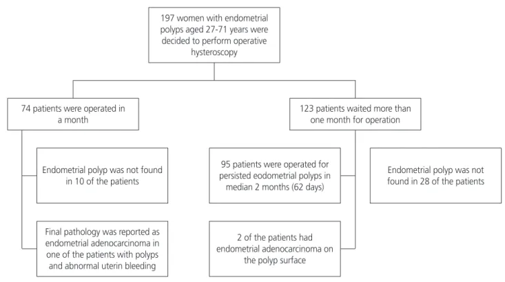
Fig. 1 shows the flowchart of patients who were assessed and followed up or excluded. Clinical and pathological data were obtained from the medical records. All clinical and de- mographic characteristics, presenting symptoms, histopatho- logical results, and intraoperative findings of the patients were recorded.
Patients were excluded if they were lost to follow-up or were operated on within a month after the diagnosis of endometrial polyp or if they had another pathology, such as submucous myoma observed in the ultrasound or office hysteroscopy or if they were taking hormonal contracep-
Fig. 1. Study flowchart.
197 women with endometrial polyps aged 27-71 years were decided to perform operative
hysteroscopy
Final pathology was reported as endometrial adenocarcinoma in one of the patients with polyps and abnormal uterin bleeding 74 patients were operated in
a month 123 patients waited more than
one month for operation
2 of the patients had endometrial adenocarcinoma on
the polyp surface Endometrial polyp was not found
in 10 of the patients
95 patients were operated for persisted eodometrial polyps in median 2 months (62 days)
Endometrial polyp was not found in 28 of the patients
tion, tamoxifen, or hormone replacement therapy. We also excluded infertile women with endometrial polyps who were not feasible for follow-up.
The first examination of the patients was performed dur- ing the follicular phase (10–14 days after last menstrual period), which was reported as the most appropriate time period to detect endometrial polyps [5]. All scans were per- formed in a standardized manner. The initial and follow- up tests were conducted by the same expert physician for each patient. The criteria for a suspected endometrial polyp on ultrasonographic examination included a hyperechoic well-defined focal mass within the endometrial cavity with a single feeding vessel on Doppler examination [6]. Patients suspected to have more than 1 polyp were also evaluated with office hysteroscopy. Saline hydrosonography (SHG) was used in women with inadequate observation during office hysteroscopy. In women with more than 1 polyp, the size of the largest polyp was used for analysis. Each polyp was mea- sured in 3 perpendicular planes, and their mean was used for size description (<1 cm, or 1–2 cm, or >2 cm). Abnormal uterine bleeding was defined as heavy menstrual bleeding (<80 mL) or bleeding for more than 7 days or the presenta- tion of bleeding between menstrual periods. Complete spon- taneous regression was noted when an endometrial polyp was not detected on follow-up ultrasound or office hysteros- copy examination.
In our clinic, we only offer follow-up of premenopausal pa- tients with asymptomatic small polyps (<1 cm), according to the 2012 Practice Guidelines for the Diagnosis and Manage- ment of Endometrial Polyps by the American Association of Gynecologic Laparoscopists (AAGL) [7]. Indications for opera- tive hysteroscopy were endometrial polyps with gynecologic symptoms (postmenopausal bleeding, abnormal uterine bleeding, pelvic pain), large endometrial polyps (>1 cm), and postmenopausal patients with endometrial polyps. After the indication for surgery, conservative follow-up was offered to premenopausal patients due to the chance of spontaneous regression and low malignancy risk. The postmenopausal group of patients with endometrial polyps who could not be operated in a month because of factors related to older age and comorbidity were also included in the study. Endometrial sampling with a Pipelle cannula was performed in patients who were older than 40 years with abnormal uterine bleed- ing due to malignancy risk.
Patients with persistent polyps underwent surgery. Using
an Olympus electrosurgery system (Olympus, Tokyo, Japan) with mannitol, the polyps were removed using a monopolar loop resectoscope. Histopathologic evaluation of the back- ground endometrium was performed in premenopausal women, menopausal women, those with large polyps (>2 cm) or irregular or thickened endometrium, and women above 40 years of age. If the rest of the endometrial cavity was atrophic, no additional sampling was performed.
2. Statistical analysis
NCSS 10 (2015; NCSS, LLC, Kaysville, UT, USA) was used for statistical analysis. Normality control was performed using the Shapiro-Wilk test. The differences in continuous variables be- tween women with regressed and non-regressed polyps were analyzed using the Mann-Whitney U test. Nominal variables were evaluated with Fisher’s exact probability tests, Yates corrected χ
2test, and Yates correction. The factors affecting regression status were determined by logistic regression anal- ysis. All variables with P<0.20 were included in the logistic regression analysis. The significance limit was set at P<0.05.
Results
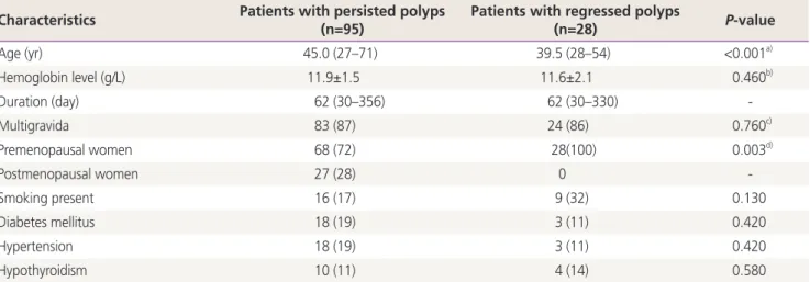
During the 2-year study period, 197 women with endome- trial polyps were candidates for hysteroscopic polypectomy in our department (Fig. 1). Of these, 74 patients underwent surgery within 1 month after diagnosis. A total of 123 pa- tients waited more than 30 days for the operation. They were followed up regularly over a median period of 62 days (range 30–360 days). The demographic characteristics and clinical properties of the study population are shown in Table 1. Of the study participants, 27 women (22%) were postmeno- pausal (more than 1 year since the last menstrual bleeding), and 96 women (78%) were premenopausal. Moreover, 64% of the women had no chronic disease other than the diagnosis of endometrial polyps. The most frequent diseases were diabetes mellitus (17%), hypertension (17%), hypo- thyroidism (11%), and coronary artery disease (4%). When comparing women with persistent or regressed polyps, no significant differences in median duration time, mean hemo- globin level, parity, smoking, and comorbidity were detected.
Women with persistent endometrial polyps were older than
women with regressed polyps (median age 45 vs. 39.5 years,
P<0.001).Most women with endometrial polyps (84%) were report- ed to have gynecologic symptoms. While 83% of premeno- pausal women had abnormal uterine bleeding (menorrhagia or metrorrhagia), 41% of postmenopausal women had post- menopausal bleeding. Endometrial polyps were detected in
81.3% (n=100) of patients by ultrasound, 62.6% (n=77) of patients by office hysteroscopy, and 1.6% (n=2) of patients by SHG. Endometrial sampling was performed in patients at risk for endometrial cancer. These are women older than 45 years with abnormal uterine bleeding, younger than 45 years
Table 2. Comparison of patients’ demographic and polyps’ morphological characteristics between polyps that persisted during the follow-up period and those that underwent complete spontaneous regression
Patient and polyp characteristics Patients with persistent polyps (n=95)
Patients with regressed
polyps (n=28) P-valuea)
Age (yr) 45 (27–71) 39.5 (28–54)
<45 23 (24) 14 (50) <0.001
≥45 72 (76) 14 (50) 0.009
Polyp number (single polyp) 40 (64) 15 (83) 0.190
Polyp number (multiple polyps) 23 (36) 3 (17) -
Polyp size by ultrasound or office hysteroscopy (cm) 0.002
<1 26 (27) 16 (57)
1–2 38 (40) 11 (39)
>2 31 (33) 1 (4)
No complaint (asymptomatic) 18 (19) 2 (7) 0.500
AUB 56 (59) 24 (86) <0.001
Adenomyomatous polyp 12 (13) 0 -
Endometrial hyperplasia 2 (1.6) -
Endometrial carcinoma 2 (1.6) -
Statistically significant P-values were displayed with bold characters.
AUB, abnormal uterine bleeding.
a)After correction for comparisons, the threshold for statistical significance is P<0.003.
Table 1. Comparison of patients with regressed and persistent polyps with regard to their demographic and clinical characteristics Characteristics Patients with persisted polyps
(n=95) Patients with regressed polyps
(n=28) P-value
Age (yr) 45.0 (27–71) 39.5 (28–54) <0.001a)
Hemoglobin level (g/L) 11.9±1.5 11.6±2.1 0.460b)
Duration (day) 62 (30–356) 62 (30–330) -
Multigravida 83 (87) 24 (86) 0.760c)
Premenopausal women 68 (72) 28(100) 0.003d)
Postmenopausal women 27 (28) 0 -
Smoking present 16 (17) 9 (32) 0.130
Diabetes mellitus 18 (19) 3 (11) 0.420
Hypertension 18 (19) 3 (11) 0.420
Hypothyroidism 10 (11) 4 (14) 0.580
Values are presented as median (interquartile range), number (%), or mean±standard deviation. Statistically significant P-values were displayed with bold characters.
a)Mann-Whitney U test; b)Independent samples t-test; c)Chi-square test; d)Fisher’s exact test.
with a history of unopposed estrogen exposure (obesity or polycystic ovary syndrome), or with postmenopausal bleed- ing. Further, 18.7% of patients had preoperative endometrial sampling, all of which were benign. A total of 42 patients (34%) had polyps smaller than 1 cm in size, 49 patients (40%) had polyps greater than 1 cm but not larger than 2 cm in size, and 32 patients (26%) had polyps larger than 2 cm at diagnosis. Additionally, 21% of patients had more than 1 polyp at diagnosis.
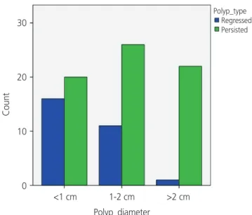
Table 2 shows the comparison of women with persistent and regressed polyps with the possible factors associated with polyp regression. Endometrial polyps regressed sponta- neously during follow-up in 28 women (23%), who were all in the premenopausal period. Regressed endometrial polyps were significantly smaller at diagnosis (<2 cm) than others, more frequent in the premenopausal group and in women younger than 45 years (P<0.05). Fig. 2 shows the regression or persistence of the polyps according to size (<1 cm, 1–2 cm,
>2 cm) in premenopausal women. Polyp regression was more frequent in patients with abnormal uterine bleeding than in asymptomatic women (P=0.05). When multiple com- parisons between variables with logistic regression were per- formed, age (>45 years) and polyp size (>2 cm) were found to be associated with endometrial polyp persistence (P<0.01 and P=0.009, respectively). The number of polyps at diagno- sis was similar between the 2 groups. In the histopathologi-
cal examination of persistent polyps, we observed a 12.6%
prevalence of adenomyomatous polyps. Clinically, the major- ity of our patients with adenomyomatous polyps presented with postmenopausal bleeding or abnormal uterine bleeding (75%).
Ninety-five women with persistent endometrial polyps underwent hysteroscopic polypectomy in the median of 2 months. We observed no complications related to the surgical procedure. Histopathological examination revealed benign endometrial polyps in 92 women (95.8%) and en- dometrial hyperplasia without atypia in 2 women (1.6%).
Endometrioid adenocarcinoma was detected at the base of the polyp in 2 patients (1.6%). Of the 2 patients with grade 1 adenocarcinoma, 1 was 42 and the other was 71 years old. Both had polyps greater than 2 cm in size. No polyp was found in 22% (n=28) of patients during hysteroscopy.
Endometrial biopsy was performed in 24 patients with endo- metrial irregularity and abnormal uterine bleeding. There was no malignancy in the pathology report in this group.
Discussion
There is little evidence in the literature regarding the spon- taneous regression rate of symptomatic endometrial polyps and related factors with the usual “see” and “treat” ap- proach [4,5]. We found that the spontaneous regression rate of endometrial polyps was 23% in our study, similar to the findings of a prospective study in asymptomatic women aged 45 to 50 years during 12 months of follow-up (27%, 8 of 31 women) [4]. DeWaay et al. [8] have previously reported a higher regression rate of endometrial polyps in a small sam- ple size study (4 of 7 women, 57.1%) after a longer follow- up period (2.4–2.7 years) than what we reported. In contrast, Wong et al. [5] found a low complete regression rate (7 of 112 women, 6.3%) without intervention or medical treat- ment during a median follow-up period of 28 months. The lower regression rate in this study can be explained by the advanced age of their study population, which included a higher rate of postmenopausal women, unlike our study (62% vs. 21%).
Our results showed that endometrial polyps smaller than 2 cm in premenopausal women and in those with abnormal uterine bleeding regress more frequently than the others.
Polyps larger than 2 cm correlate with a lower possibility
Fig. 2. The bar graph shows polyp regression and persistencerates in the premenopausal group according to the size of the en- dometrial polyps.
Count
30
20
10
0
Polyp_type Regressed Persisted
<1 cm 1-2 cm >2 cm Polyp_diameter