© 2020 The Korean Ophthalmological Society
This is an Open Access article distributed under the terms of the Creative Commons Attribution Non-Commercial License (http://creativecommons.org/licenses /by-nc/3.0/) which permits unrestricted non-commercial use, distribution, and reproduction in any medium, provided the original work is properly cited.
Original Article
The trabecular meshwork (TM) is involved in regulation of aqueous humor outflow to control intraocular pressure (IOP). It is thought that impaired drainage through the tra-
becular pathway caused by increased resistance is the pri- mary cause of increased IOP in primary open-angle glau- coma [1,2]. While most glaucoma medications approved for clinical use act either on the uveoscleral pathway and/
or aqueous humor formation, several new drugs targeting the trabecular outflow pathway have recently entered clini- cal development [3].
KATP channel openers are a structurally diverse group of drugs with a broad spectrum of potential therapeutic ap- Purpose: To investigate the pathway and effects of minoxidil on trabecular outflow in cultured human trabecular
meshwork (TM) cells.
Methods: After exposing primarily cultured TM cells to 0, 10, 50, or 100 µM minoxidil sulfate (MS), trabecular outflow was assessed by measuring TM cell monolayer permeability to carboxyfluorescein and transepithe- lial electrical resistance. To assess the pathway of permeability changes, caveolin-1, occludin, and claudin-5 levels were measured via western blot. Generation of reactive oxygen species (ROS) was measured using the dichlorofluorescein diacetate assay. To assess the involvement of nitric oxide (NO) in minoxidil-induced permeability increase, the degrees of endothelial nitric oxide synthase mRNA expression and NO production were measured with reverse transcription polymerase chain reaction and Griess assays, respectively. Perme- ability was also measured with co-exposure to 50 µM N-acetyl cysteine.
Results: MS significantly increased TM cell monolayer permeability (p < 0.05) and decreased transepithelial electrical resistance (p < 0.05). MS decreased the degree of endothelial nitric oxide synthase mRNA expres- sion but did not affect NO production. MS decreased occludin and claudin-5 levels but did not affect caveolin-1 level. MS at 100 µM increased the generation of ROS, and MS-induced permeability increase was attenuated after co-exposure to 50 µM N-acetyl cysteine.
Conclusions: Minoxidil may preferentially increase trabecular permeability via a paracellular pathway by down- regulation of tight junction proteins. This minoxidil-induced permeability through the TM may be mediated by generation of ROS.
Key Words: Minoxidil, Permeability, Reactive oxygen species, Trabecular meshwork
Received: October 9, 2019 Final revision: October 25, 2019 Accepted: November 14, 2019
Corresponding Author: Jae Woo Kim, MD, PhD. Department of Oph- thalmology, Daegu Catholic University School of Medicine, 33 Duryu- gongwon-ro 17-gil, Nam-gu, Daegu 42472, Korea. Tel: 82-53-650-4728, Fax: 82-53-627-0133, E-mail: [email protected]
Effect of Minoxidil on Trabecular Outflow via the Paracellular Pathway
Hyun Gu Kang1, Jae Woo Kim2
1Cheil Eye Hospital, Daegu, Korea
2Department of Ophthalmology, Daegu Catholic University School of Medicine, Daegu, Korea
plications. Among them, minoxidil is used topically to stimulate hair growth [4]. Minoxidil is also a direct vasodi- lator introduced for treatment of hypertension as it is capa- ble of reducing blood pressure in patients with resistant hypertension in whom therapy has failed with other multi- drug regimens [5]. Although minoxidil induced ocular hy- potension when given topically in an animal study [6], fur- ther studies focusing on IOP-lowering activity have not been reported. However, previous studies revealed that mi- noxidil induced an increase in blood-brain tumor barrier permeability [7-9].
Transport of plasma proteins and solutes across the en- dothelium involves two different routes. The first is trans- cellular via caveolae-mediated vesicular transport, and the second is paracellular through inter-endothelial junctions [10]. Paracellular permeability of the endothelial barrier is maintained by inter-endothelial junctions that are regulat- ed by expression of claudin-5 and occludin. In contrast, transcellular transport occurs via fission of plasma mem- brane macrodomains enriched with caveolin-1 (CAV-1) and caveolae from the luminal surface of the endothelium, fol- lowed by transport of caveolar vesicles to the basal surface.
Caveolae and CAV-1 mediate endocytosis and transcytosis in endothelial cells [11-13]. In the eye, CAV-1 is expressed in TM cells, and dysregulation of the expression of caveo- lin in the TM has been implicated in the pathology of pri- mary open angle glaucoma [14].
Minoxidil induces accelerated formation of transport vesi- cles in both the brain tumor capillary endothelium and tumor cells [7,15] suggesting that vesicular transport is largely re- sponsible for the enhanced permeability induced by minoxi- dil rather than the opening of endothelial tight junctions (TJ).
On the contrary, another study demonstrated that minoxidil also involves the paracellular pathway by downregulating the expression of occludin and claudin-5 [16]. Previous stud- ies have shown that minoxidil-induced permeability may be related to accelerated formation of CAV-1 protein and could be mediated by reactive oxygen species (ROS). Minoxidil also improves paracellular transport by regulating the ex- pression of TJ proteins, possibly mediated by ROS [8,9,15].
TM cells regulate trabecular outflow because they have endothelial cell-like and muscle cell-like properties [17-21].
As minoxidil acts as a vasodilator and increases permea- bility, it is possible that minoxidil increases trabecular out- flow; however, the effect and mechanism of minoxidil-in- duced permeability increase have not yet been studied in
the TM. This study was performed to determine whether minoxidil affects the permeability of TM. In addition, we studied the mechanism of minoxidil-induced permeability changes and whether this process was regulated by ROS.
Materials and Methods
Cell culture and treatment
This study followed the tenets of the Declaration of Hel- sinki and was approved by the institutional review board/
ethics committee of Daegu Catholic University Hospital (CR-18-106-L). TM cell cultures were established from enucleated human eyes obtained from an eye bank. Briefly, TM tissues were excised by dissecting a continuous strand of tissue between the line of Schwalbe and the scleral spur.
The excised TM tissues were placed in a sterile culture dish and left undisturbed at 37°C in a 5% CO2atmosphere.
After noting initial cell growth, the explants were re- moved, and the cultures were maintained with a medium containing 10% fetal bovine serum. Primarily cultured TM cells were then exposed to 0, 10, 50, or 100 µM minox- idil sulfate (MS; Sigma, St. Louis, MO, USA). In addition, TM cells were co-exposed to 50 µM N-acetyl cysteine (NAC, Sigma) to evaluate the effect of ROS on MS-in- duced permeability changes.
MTT assay for cell viability
Cell survival was determined using a rapid 3-(4, 5–di- methylthiazol-2-yl)-2, 5-diphenyltetrazolium bromide (MTT, Sigma) colorimetric assay [22,23]. For the assay, 100 μL of a MTT stock solution (5 mg MTT/mL PBS) was added to each well and incubated for 4 hours at 37°C, after which all media was removed from the well. After 0.5 mL of dimethyl sulfoxide (Sigma) was added to each well, 100 μL of solution from each well was transferred to a 96-well plate and analyzed using a multi-well scanning spectro- photometer (λ = 570 nm).
Measurement of monolayer cell permeability with car- boxyfluorescein
Permeability of the TM cell monolayer was measured using carboxyfluorescein as previously described with mi-
nor modification [24-27]. Briefly, primary cultured human TM cells were incubated in the inner chamber (insert di- ameter, 12 mm; pore size, 0.4 µm) of a 12-well plate (no.
3460, Transwell; Corning, Lowell, MA, USA) supplement- ed with 10% fetal bovine serum. After the cells reached confluence, the media was changed to 1% serum contain- ing DMEM to avoid the effects of growth factors and pro- teins. Then, the TM cells were exposed to each drug for 24 hours. After washing, 50 µM of the tracer carboxyfluores- cein (Sigma-Aldrich, St. Louis, MO, USA) was added to each well. The media was collected from the outer well to analyze fluorescence after 2 hours, and the concentration of carboxyfluorescein in the collected media was measured using a spectrofluorometer (FLUOstar Optima; BMG Labtech, Offenburg, Germany) with an excitation wave- length of 490 nm and an emission wavelength of 530 nm.
Measurement of monolayer transepithelial electrical resistance
TM cells were seeded at 2 × 10⁴ cells/well and grown until confluent in the inner well (pore size, 0.4 µm; diame- ter, 12 mm) of 12-well culture plates (Transwell, Corning) [28,29]. Four hours after each drug exposure, transepitheli- al electrical resistance (TEER) was measured with an elec- trical resistance system (epithelial voltohmmeter, EVOM2;
World Precision Instruments, Sarasota, FL, USA) accord- ing to the manufacturer’s instructions. The results were re- corded and expressed as net value (Ω cm2).
Griess assay for nitric oxide production and reverse transcription polymerase chain reaction for en dothelial nitric oxide synthase mRNA expression
To evaluate the effect of MS on production of nitric ox- ide (NO), nitrite concentrations in the media were mea- sured using the Griess reaction [30]. Briefly, media samples were collected from each well following appropriate treat- ment and reacted with modified Griess reagent (Sigma-Al- drich) by mixing equal volumes at room temperature for 15 minutes. Optical density was then measured and record- ed on a multi-well scanning spectrophotometer at 540 nm.
To evaluate the effect of MS on expression of endothelial nitric oxide synthase (eNOS) mRNA, reverse transcription polymerase chain reaction (RT-PCR) was performed as follows. After exposure to MS, total RNA was extracted
using Trizol reagent (Invitrogen, Carlsbad, CA, USA). An RNA denaturation mix consisting of isolated RNA, oligo dT primers, and nuclease-free water was denatured. RT- PCR was performed using oligonucleotide primers specific to eNOS. cDNA was synthesized by adding prime RT pre- mix, Taq Green Master Mix, and 10 pM of each forward and reverse primer (forward primer, 5’ CTG GCT TTC CCT TCC AGT TC 3’, 225 bp; reverse primer, 5’ CCT TCC AGA TTA AGG CGG AC 3’, 225 bp) were added to the synthesized cDNA. The amplification reaction was carried out for 30 cycles on a DNA Engine Cycler (Bio- Rad, Hercules, CA, USA). The amplified PCR products were analyzed using Multi-gauge software (Fujifilm, To- kyo, Japan) after electrophoresis. β-actin was used as an internal standard.
Measurement of CAV-1, occludin, and claudin-5 levels by western blotting
Cell extracts were prepared by lysing cells with RIPA buffer (Thermo Scientific, Carlsbad, CA, USA). Samples were sonicated and cleared by centrifugation (1,200 rpm) at 4°C for 10 minutes. Supernatant protein concentrations were determined using BCA protein assays (Thermo Sci- entific, Carlsbad, CA, USA). Samples containing equal amounts of protein were separated by NuPAGE 4-12% Bis- Tris gels (Invitrogen), followed by transfer of resolved pro- teins to nitrocellulose membranes using the XCell Sure- Lock electrophoresis system (Invitrogen). Nonspecific binding was blocked for 1 hour at room temperature. Blots were then probed overnight at 4°C with primary antibod- ies, followed by 1-hour incubation with secondary antibod- ies conjugated to goat anti-rabbit horseradish peroxidase (Santa Cruz Biotechnology, Dallas, TX, USA). Western blots were developed using Super Signal West Pico Chemi- luminescent substrate (Thermo Scientific). Quantification of the signals was performed using the Gel Doc XR+ sys- tem (Bio-Rad). Glyceraldehyde 3-phosphate dehydroge- nase was used as an internal standard.
Measurement of ROS production by dichlorofluoresce- in diacetate assay
Generation of ROS was measured using the dichloroflu- orescein diacetate assay [31,32]. After treatment of cells in a 96-well culture plate, the medium was removed, and
cells were subsequently washed. Cells were then treated with 5 µM dichlorofluorescein diacetate (Sigma), after which they were incubated for 30 minutes at 37°C in the dark. The change in fluorescence of oxidized dichlorofluo- rescein was measured at excitation and emission wave- lengths of 488 nm and 527 nm, respectively, using a fluo- rescence analyzer (FLUOstar OPTIMA).
Statistical analysis
All data represent the average results of at least three in- dependent experiments. Experimental differences between the results of control cultures and a single treatment group were evaluated using Student’s t-test. A p-value less than 0.05 was considered statistically significant.
Results
Cell culture
The TM cells grew into the surrounding tissue explants as a monolayer after 2 weeks of primary culture. The pres- ence of TM cells was confirmed by their characteristic morphology and characteristic growth pattern as satellite colonies distant from the tissue explants [33].
Effect of MS on the viability of TM cells
Exposure to 0, 10, 50, or 100 µM MS for 24 hours did not significantly affect TM cell survival (all p > 0.05) (Fig. 1).
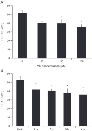
Effects of MS on TM cell monolayer permeability TEER represents the resistance to flow through the TM cell monolayer. Exposure to 10, 50, or 100 µM MS signifi- cantly decreased TEER of the TM cell monolayer (p = 0.007, 0.033, 0.004, respectively) (Fig. 2A). In addition, exposure to 10 µM MS significantly decreased TEER after 2, 3, and 4 hours (p = 0.018, 0.033, 0.013, respectively) (Fig. 2B). To evaluate the effect of MS on permeability through the para- cellular pathway, monolayer cell permeability was measured using carboxyfluorescein [10]. As a result, exposure to 10, 50, or 100 µM MS significantly increased the concentration of carboxyfluorescein in the outer well compared to non-ex- posed controls (p = 0.037, 0.038, 0.014, respectively) (Fig. 3).
% survival
MS concentration (μM) 0
120 100 80 60 40 20
10 50 100
Fig. 1. Effect of minoxidil sulfate (MS) on the survival of cul- tured human trabecular meshwork cells. MS did not affect the survival of trabecular meshwork cells compared to non-exposed controls (all p > 0.05).
A
TEER (Ω cm2)
MS concentration (μM) 0
60 50 40 30 20 10
0 10 50 100
* *
*
B
TEER (Ω cm2)
0 60 50 40 30 20 10
0 min 1 hr 2 hr 3 hr 4 hr
* *
*
Fig. 2. Effect of minoxidil sulfate (MS) on the transepithelial electrical resistance (TEER) of the trabecular meshwork cell monolayer. (A) Exposure to 10, 50, or 100 µM MS significantly decreased TEER compared with non-exposed controls (*p < 0.05).
(B) Exposure to 10 µM MS significantly decreased TEER in a time-dependent manner (*p < 0.05).
Effects of MS on NO production and eNOS mRNA expression
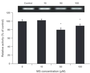
Exposure to 0, 10, 50, or 100 µM MS did not significant- ly affect nitrite concentration in the media compared to non-exposed controls (all p > 0.05) (Fig. 4). Exposure to 50 µM and 100 µM MS significantly decreased eNOS mRNA expression (p = 0.004 and 0.019, respectively) (Fig. 5).
Effects of MS on CAV-1, occludin, and claudin-5 levels Exposure to 0, 10, 50, or 100 µM MS did not affect CAV- 1 level compared to non-exposed controls (all p > 0.05)
(Fig. 6), suggesting that MS-induced permeability increase is not associated with the transcellular pathway. In con- trast, exposure to 10, 50, or 100 µM MS significantly de- creased occludin level (p = 0.045, 0.002, 0.002, respective- ly) (Fig. 7). Furthermore, exposure to 50 or 100 µM MS significantly decreased claudin-5 level (p = 0.037, 0.001, re- spectively) (Fig. 8). Taken together, these results revealed that MS increased trabecular permeability through the paracellular pathway.
Fig. 3. Effect of minoxidil sulfate (MS) on trabecular meshwork cell monolayer permeability. Exposure to 10, 50, or 100 µM MS significantly increased permeability of the trabecular meshwork (*p < 0.05). Carboxyfluorescein intensity of the outer chamber was normalized to the mean value obtained with a non-exposed control (permeability 100%).
% permeability
MS concentration (μM) 0
160 140 120
80 100
60 40 20
0 10 50 100
* *
*
Fig. 4. Effect of minoxidil sulfate (MS) on the production of ni- tric oxide. Exposure to MS did not affect the production of nitric oxide compared to non-exposed controls (all p > 0.05).
Nitrite (μM)
MS concentration (μM) 0
8 7 6
4 5
3 2 1
0 10 50 100
Fig. 5. Effect of minoxidil sulfate (MS) on the expression of endothelial nitric oxide synthase mRNA measured with reverse transcription polymerase chain reaction in trabecular meshwork cells. Exposure to 50 or 100 µM MS significantly decreased the expression of endothelial nitric oxide synthase mRNA compared to non-exposed controls (*p < 0.05).
Relative activity (% of control)
MS concentration (μM) 0
120 100 80 50 40 20
0 10 50 100
*
*
Control 10 50 100
Fig. 6. Effect of minoxidil sulfate (MS) on the level of caveolin-1 protein measured with western blot. Exposure to MS did not affect caveolin-1 level compared to non-exposed controls (all p >
0.05).
Relative activity (% of control)
MS concentration (μM) 0
120 100 80 50 40 20
0 10 50 100
Control 10 50 100
Effects of MS on ROS generation
Exposure to 100 µM MS significantly increased ROS generation after 1 and 2 hours exposure (p = 0.002, 0.001, respectively) (Fig. 9). Co-exposure to the antioxidant NAC (50 µM) significantly attenuated MS-induced permeability increase compared to exposure to 10, 50, or 100 µM mi- noxidil alone (*;p = 0.010, 0.009, 0.032, respectively) (Fig.
10).
Discussion
While the paracellular route restricts passage of solutes larger than 3 nm in radius, transcellular vesicle trafficking selectively transports macromolecules across the endothe- lium [10]. Transcellular and paracellular pathways have generally been considered independent processes regulat- ing endothelial barrier function. However, growing evi-
Fig. 7. Effect of minoxidil sulfate (MS) on the level of occludin protein measured with western blot. Exposure to 10, 50, or 100 µM MS significantly decreased occludin level (*p < 0.05).
Relative activity (% of control)
MS concentration (μM) 0
120 100 80 50 40 20
0 10 50 100
* *
Control 10 50 100
*
Fig. 8. Effect of minoxidil sulfate (MS) on the level of claudin-5 protein measured with western blot. Exposure to 50 or 100 µM MS significantly decreased claudin-5 level (*p < 0.05).
Relative activity (% of control)
MS concentration (μM) 0
120 100 80 50 40 20
0 10 50 100
*
*
Control 10 50 100
Relative fluorescence (% of control)
0 200 180 160 140 120 100 80 60 40 20
0 min 30 min 1 hr 2 hr
*
*
Fig. 9. Effect of minoxidil sulfate on generation of reactive oxy- gen species measured using the dichlorofluorescein diacetate as- say. Exposure to 100 µM minoxidil sulfate significantly increased generation of reactive oxygen species after 1 and 2 hours of expo- sure (*p < 0.05).
Fig. 10. Effect of antioxidant on the minoxidil (M) sulfate-in- duced permeability increase measured using the carboxyfluo- rescein assay. Co-exposure to 50 µM N-acetyl cysteine (NAC) significantly decreased permeability compared to exposure to M sulfate alone at each concentration (µM) (*p < 0.05). Carboxy- fluorescein intensity of the outer chamber was normalized to the mean value obtained using a non-exposed control (permeability 100%).
% permeability
160 140 120 100 80 60 40 20
0 NAC M50 M50+NAC
M M + NAC
M10 M10+NAC M100 M100+NAC
0
* *
*
dence favors the concept of interdependence between the paracellular and transcellular pathways for maintenance of tissue fluid homeostasis [10,34].
Minoxidil is used as an anti-hypertensive drug as it is known to increase blood-brain barrier permeability [6,9,17]. MS not only increases transcellular permeability by increasing CAV-1 activity, but also increases the para- cellular pathway by decreasing occludin and claudin-5 ac- tivity [7,15,16].
In the present study, we demonstrated that MS increases trabecular permeability because it decreased TEER. To clarify the route of this permeability increase, we mea- sured the levels of proteins involved in regulation of each route. We found that MS had no significant effect on the protein expression level of CAV-1, which regulates the transcellular pathway. However, MS significantly in- creased levels of the TJ proteins occludin and claudin-5, which regulate the paracellular pathway. These results in- dicate that MS increases trabecular outflow through the paracellular route rather than the transcellular route. This enhanced paracellular transport by MS is further support- ed by the finding that MS increased the permeability of carboxyfluorescein, which is transported via the paracellu- lar pathway.
Outflow through the TM occurs via both transcellular and paracellular pathways [35-37]. It is well known that in- tercellular transport accounts for only a small fraction of the aqueous humor that leaves the eye by the conventional route, and aqueous humor outflow occurs via the transcel- lular route through minute pores and giant vacuoles [38-40].
Although MS increased paracellular transport in this study, the effect of MS on the transcellular pathway cannot be ex- cluded because there is a difference between in vitro and physiological conditions, and the two pathways are closely linked and regulated [10]. Due to the complexity of TM outflow and the general limitation of in vitro experiments in the field, in vivo studies should be further investigated.
One previous study using a rabbit model reported that topical MS treatment decreased IOP and suggested that this MS-induced IOP-lowering effect might be mediated by NO [6]. In addition, MS was found to increase CAV-1 level, which has been shown to act as a negative regulator of eNOS [41-44]. To evaluate whether the MS-induced perme- ability increase was mediated via NO in the TM, we mea- sured NO production and eNOS mRNA expression after MS treatment. The effect of NO on the MS-induced perme-
ability increase was not significant as MS did not affect generation of NO. On the other hand, MS at high concen- trations decreased eNOS mRNA expression. This decreased eNOS mRNA expression may result from the increased ac- tivity of CAV-1, though no such observation was noted in our study. This might seem paradoxical in view of the pos- tulated role of caveolae in negative regulation of eNOS;
however, Yu et al. [45] elegantly demonstrated that eNOS activation is lost in the absence of CAV1 and caveolae and proposed that eNOS activation might require caveolae. Un- til now, multiple roles of caveolae are still under investiga- tion and need further study, especially in the TM [46].
Another possible explanation is that the activity of in- creased CAV-1 expression may precede the decreased ex- pression of occludin and claudin-5 [10,16]. These previous studies demonstrated that an increase in endothelial CAV-1 expression change occurs early, while alterations of the TJ proteins occludin and claudin occur later in the time course. Therefore, it is possible that early increase of CAV- 1 expression was not observed in this study. In such a case, later decrease of eNOS expression can occur, as demon- strated in this study. It is also possible that only higher lev- els of CAV-1 inhibit eNOS expression and activity [47,48].
We measured nitrite concentration in the media by the Griess reaction, which is the most frequently used method to measure nitrite and is based on spectrophotometric analysis of azo dye obtained after reaction with the Griess reagent. However, this method has some limitations re- garding detection limit and sensitivity [49]. Thus, minor changes in nitrite concentration may be not significant de- spite decreased eNOS activity. Regardless of these limita- tions, our data suggest that MS preferentially increases paracellular outflow accompanied by weaker interaction between CAV-1 and eNOS.
As MS is known to increase permeability through ROS in both transcellular and paracellular pathways [8,9,15], we hypothesized that MS has an effect on ROS production in the TM. As a result, MS increased generation of ROS, in agreement with previous studies. Thus, MS-induced per- meability increase is possibly mediated by the ROS signal- ing pathway in the TM. Moreover, MS-induced permeabil- ity increase was significantly attenuated in the presence of specific ROS inhibitors, further suggesting that ROS are a class of important signaling molecules involved in MS-in- duced permeability increase. We should consider that MS-induced permeability increase may occur through oth-
er signaling pathways as well; therefore, other related mechanisms should be further investigated.
In conclusion, MS increased permeability across the TM cell monolayer via the paracellular pathway by downregu- lating the TJ proteins occludin and claudin-5. This MS-in- duced permeability increase may be mediated through generation of ROS.
Conflict of Interest
No potential conflict of interest relevant to this article was reported.
References
1. Alvarado J, Murphy C, Juster R. Trabecular meshwork cel- lularity in primary open-angle glaucoma and nonglauco- matous normals. Ophthalmology 1984;91:564-79.
2. Rohen JW, Lutjen-Drecoll E, Flugel C, et al. Ultrastructure of the trabecular meshwork in untreated cases of primary open-angle glaucoma (POAG). Exp Eye Res 1993;56:683-92.
3. Kopczynski CC, Epstein DL. Emerging trabecular outflow drugs. J Ocul Pharmacol Ther 2014;30:85-7.
4. Ashcroft FM, Gribble FM. New windows on the mecha- nism of action of K(ATP) channel openers. Trends Phar- macol Sci 2000;21:439-45.
5. Sica DA. Minoxidil: an underused vasodilator for resistant or severe hypertension. J Clin Hypertens (Greenwich) 2004;6:283-7.
6. Nathanson JA. Nitrovasodilators as a new class of ocular hypotensive agents. J Pharmacol Exp Ther 1992;260:956- 65.
7. Ningaraj NS, Rao MK, Black KL. Adenosine 5’-triphos- phate-sensitive potassium channel-mediated blood-brain tumor barrier permeability increase in a rat brain tumor model. Cancer Res 2003;63:8899-911.
8. Gu YT, Xue YX, Wang YF, et al. Role of ROS/RhoA/
PI3K/PKB signaling in NS1619-mediated blood-tumor bar- rier permeability increase. J Mol Neurosci 2012;48:302-12.
9. Gu YT, Xue YX, Wang YF, et al. Minoxidil sulfate in- duced the increase in blood-brain tumor barrier permeabil- ity through ROS/RhoA/PI3K/PKB signaling pathway.
Neuropharmacology 2013;75:407-15.
10. Komarova Y, Malik AB. Regulation of endothelial perme-
ability via paracellular and transcellular transport path- ways. Annu Rev Physiol 2010;72:463-93.
11. Wolburg H, Lippoldt A. Tight junctions of the blood-brain barrier: development, composition and regulation. Vascul Pharmacol 2002;38:323-37.
12. Quest AF, Gutierrez-Pajares JL, Torres VA. Caveolin-1: an ambiguous partner in cell signalling and cancer. J Cell Mol Med 2008;12:1130-50.
13. Sun SW, Zu XY, Tuo QH, et al. Caveolae and caveolin-1 mediate endocytosis and transcytosis of oxidized low den- sity lipoprotein in endothelial cells. Acta Pharmacol Sin 2010;31:1336-42.
14. Surgucheva I, Surguchov A. Expression of caveolin in tra- becular meshwork cells and its possible implication in pathogenesis of primary open angle glaucoma. Mol Vis 2011;17:2878-88.
15. Gu YT, Xue YX, Zhang H, et al. Adenosine 5’-triphos- phate-sensitive potassium channel activator induces the up-regulation of caveolin-1 expression in a rat brain tumor model. Cell Mol Neurobiol 2011;31:629-34.
16. Nag S, Venugopalan R, Stewart DJ. Increased caveolin-1 expression precedes decreased expression of occludin and claudin-5 during blood-brain barrier breakdown. Acta Neu- ropathol 2007;114:459-69.
17. Stamer WD, Clark AF. The many faces of the trabecular meshwork cell. Exp Eye Res 2017;158:112-23.
18. Gipson IK, Anderson RA. Actin filaments in cells of hu- man trabecular meshwork and Schlemm’s canal. Invest Ophthalmol Vis Sci 1979;18:547-61.
19. Lepple-Wienhues A, Rauch R, Clark AF, et al. Electro- physiological properties of cultured human trabecular meshwork cells. Exp Eye Res 1994;59:305-11.
20. Wiederholt M, Bielka S, Schweig F, et al. Regulation of outflow rate and resistance in the perfused anterior seg- ment of the bovine eye. Exp Eye Res 1995;61:223-34.
21. Wiederholt M, Thieme H, Stumpff F. The regulation of tra- becular meshwork and ciliary muscle contractility. Prog Retin Eye Res 2000;19:271-95.
22. Mosmann T. Rapid colorimetric assay for cellular growth and survival: application to proliferation and cytotoxicity assays. J Immunol Methods 1983;65:55-63.
23. Freimoser FM, Jakob CA, Aebi M, Tuor U. The MTT [3-(4,5-dimethylthiazol-2-yl)-2,5-diphenyltetrazolium bro- mide] assay is a fast and reliable method for colorimetric determination of fungal cell densities. Appl Environ Mi- crobiol 1999;65:3727-9.
24. Araie M. Carboxyfluorescein. A dye for evaluating the cor- neal endothelial barrier function in vivo. Exp Eye Res 1986;42:141-50.
25. Grimes PA. Carboxyf luorescein transfer across the blood-retinal barrier evaluated by quantitative fluorescence microscopy: comparison with fluorescein. Exp Eye Res 1988;46:769-83.
26. Nakagawa S, Usui T, Yokoo S, et al. Toxicity evaluation of antiglaucoma drugs using stratified human cultivated cor- neal epithelial sheets. Invest Ophthalmol Vis Sci 2012;53:5154-60.
27. Lei Y, Stamer WD, Wu J, Sun X. Oxidative stress impact on barrier function of porcine angular aqueous plexus cell monolayers. Invest Ophthalmol Vis Sci 2013;54:4827-35.
28. Srinivasan B, Kolli AR, Esch MB, et al. TEER measure- ment techniques for in vitro barrier model systems. J Lab Autom 2015;20:107-26.
29. Wilhelm I, Fazakas C, Krizbai IA. In vitro models of the blood-brain barrier. Acta Neurobiol Exp (Wars) 2011;71:113- 28.
30. Green LC, Wagner DA, Glogowski J, et al, Tannenbaum SR. Analysis of nitrate, nitrite, and [15N]nitrate in biologi- cal fluids. Anal Biochem 1982;126:131-8.
31. Ammar DA, Hamweyah KM, Kahook MY. Antioxidants protect trabecular meshwork cells from hydrogen perox- ide-induced cell death. Transl Vis Sci Technol 2012;1:4 32. Wang H, Joseph JA. Quantifying cellular oxidative stress
by dichlorofluorescein assay using microplate reader. Free Radic Biol Med 1999;27:612-6.
33. Alvarado JA, Wood I, Polansky JR. Human trabecular cells. II. Growth pattern and ultrastructural characteristics.
Invest Ophthalmol Vis Sci 1982;23:464-78.
34. Salama NN, Eddington ND, Fasano A. Tight junction mod- ulation and its relationship to drug delivery. Adv Drug De- liv Rev 2006;58:15-28.
35. Grierson I, Lee WR. Pressure-induced changes in the ul- trastructure of the endothelium lining Schlemm’s canal.
Am J Ophthalmol 1975;80:863-84.
36. Epstein DL, Rohen JW. Morphology of the trabecular meshwork and inner-wall endothelium after cationized fer- ritin perfusion in the monkey eye. Invest Ophthalmol Vis Sci 1991;32:160-71.
37. Raviola G, Raviola E. Paracellular route of aqueous out- flow in the trabecular meshwork and canal of Schlemm. A freeze-fracture study of the endothelial junctions in the sclerocorneal angel of the macaque monkey eye. Invest Ophthalmol Vis Sci 1981;21:52-72.
38. Ethier CR. The inner wall of Schlemm’s canal. Exp Eye Res 2002;74:161-72.
39. Tripathi RC. Mechanism of the aqueous outflow across the trabecular wall of Schlemm’s canal. Exp Eye Res 1971;11:116- 21.
40. Tarkkanen A, Niemi M. Enzyme histochemistry of the an- gle of the anterior chamber of the human eye. Acta Oph- thalmol (Copenh) 1967;45:93-9.
41. Michel JB, Feron O, Sacks D, Michel T. Reciprocal regula- tion of endothelial nitric-oxide synthase by Ca2+-calmod- ulin and caveolin. J Biol Chem 1997;272:15583-6.
42. Ju H, Zou R, Venema VJ, Venema RC. Direct interaction of endothelial nitric-oxide synthase and caveolin-1 inhibits synthase activity. J Biol Chem 1997;272:18522-5.
43. Schubert W, Frank PG, Woodman SE, et al. Microvascular hyperpermeability in caveolin-1 (-/-) knock-out mice.
Treatment with a specific nitric-oxide synthase inhibitor, L-NAME, restores normal microvascular permeability in Cav-1 null mice. J Biol Chem 2002;277:40091-8.
44. Lei Y, Song M, Wu J, et al. eNOS activity in CAV1 knock- out mouse eyes. Invest Ophthalmol Vis Sci 2016;57:2805-13.
45. Yu J, Bergaya S, Murata T, et al. Direct evidence for the role of caveolin-1 and caveolae in mechanotransduction and remodeling of blood vessels. J Clin Invest 2006;116:1284-91.
46. Parton RG, Simons K. The multiple faces of caveolae. Nat Rev Mol Cell Biol 2007;8:185-94.
47. Garcia-Cardena G, Martasek P, Masters BS, et al. Dissect- ing the interaction between nitric oxide synthase (NOS) and caveolin. Functional significance of the nos caveolin binding domain in vivo. J Biol Chem 1997;272:25437-40.
48. Chen Z, D S Oliveira S, Zimnicka AM, et al. Reciprocal regulation of eNOS and caveolin-1 functions in endothelial cells. Mol Biol Cell 2018;29:1190-202.
49. Giustarini D, Dalle-Donne I, Colombo R, et al. Adaptation of the Griess reaction for detection of nitrite in human plasma. Free Radic Res 2004;38:1235-40.