ISSN 0378-6471 (Print)⋅ISSN 2092-9374 (Online)
https://doi.org/10.3341/jkos.2020.61.3.267
Original Article
황화수소가 섬유주단층세포층 투과도 증가에 미치는 영향과 일산화질소의 상호작용에 관한 연구
Effects of Hydrogen Sulfide and Nitric Oxide on the Permeability of Cultured Trabecular Meshwork Cells
손명서⋅백민주⋅김재우
Myung Seo Son, MD, Min Ju Baek, MD, Jae Woo Kim, MD, PhD
대구가톨릭대학교 의과대학 안과학교실
Department of Ophthalmology, Daegu Catholic University College of Medicine, Daegu, Korea
Purpose: To investigate the effects of hydrogen sulfide (H2S) on the permeability of a cultured human trabecular meshwork cells (HTMC) monolayer and its interaction with nitric oxide (NO).
Methods: After exposing primary cultured HTMCs to 0, 50, 100, and 500 μM sodium hydrogen sulfide (NaHS) for 6 hours, the permeabilities through the HTMC monolayer were measured using a Transwell assay with carboxyfluorescein. The production of NO and eNOS mRNA expression were assessed using the Griess assay and reverse transcription-polymerase chain reaction, respectively. In addition, 0, 1, and 10 μM NaHS and 10 μM sodium nitroprusside (SN) were co-exposed to evaluate the possible synergistic effect of H2S and NO.
Results: Greater than 100 μM NaHS increased the permeability through the HTMC monolayer in a dose-dependent manner (p < 0.05). These increased permeabilities were not accompanied by NO production or eNOS mRNA expression (p > 0.05).
When 0, 1, and 10 μM NaHS and 10 μM SN were exposed together, there was no significant change of permeability, NO production, or eNOS mRNA expression (all, p > 0.05).
Conclusions: NaHS at high concentrations increased the permeability of the HTMC monolayer, which was not affected by NO.
NaHS at low concentrations did not show a synergistic effect with NO. Thus, H2S at high concentrations may increase trabecular outflow, which may not be associated with NO.
J Korean Ophthalmol Soc 2020;61(3):267-273
Keywords: Hydrogen sulfide, Nitric oxide, Permeability, Trabecular meshwork
■Received: 2019. 4. 4. ■ Revised: 2019. 8. 20.
■Accepted: 2020. 2. 21.
■Address reprint requests to Jae Woo Kim, MD, PhD Department of Ophthalmology, Daegu Catholic University Hospital, #33 Duryugongwon-ro 17gil, Nam-gu, Daegu 42472, Korea
Tel: 82-53-650-4728, Fax: 82-53-627-0133 E-mail: [email protected]
*Conflicts of Interest: The authors have no conflicts to disclose.
ⓒ2020 The Korean Ophthalmological Society
This is an Open Access article distributed under the terms of the Creative Commons Attribution Non-Commercial License (http://creativecommons.org/licenses/by-nc/3.0/) which permits unrestricted non-commercial use, distribution, and reproduction in any medium, provided the original work is properly cited.
황화수소(hydrogen sulfide, H2S)는 수소의 황화물로 과 거 악취를 가진 무색의 유독한 기체로 알려졌었으나 근래 에는 일산화질소(nitric oxide, NO)와 일산화탄소에 이어 제 3의 기체송신기(gaseous transmitter)로 밝혀졌으며 H2S는 포유류에서 3가지 효소에 의해 내인적으로 생성된다.1-5 H2S는 생리적으로 다양한 역할을 하는데 심혈관계에서 세 포 보호 효과를 나타내며, 혈관의 이완에도 관여한다. 뿐만 아니라 중추신경계에서도 신경세포 보호 작용을 나타내며, 산화스트레스와 세포고사, 염증 반응에도 관여하고, 노화를 지연시키는 작용도 있다.6-9
안구 내에서 H2S는 다양한 조직에서 내인성으로 기질인
L-cysteine에 의해 생성이 증가한다.10 H2S는 안구 내에서 산화스트레스를 비롯한 여러 요인에 대해 신경 보호 작용 을 나타내는데,11-13 녹내장 모델을 이용한 연구에서도 망막 신경절세포 보호 효과가 있는 것으로 알려졌다.14,15 안압에 미치는 영향에 대한 연구에서는 H2S가 동물실험에서 안압 을 낮추는 효과가 있으며, 전안부 관류 실험에서 방수 유출 을 증가시킨다는 보고가 있었다.16,17
H2S와 또 다른 기체송신기인 NO와의 상호작용에 관한 다양한 보고가 있다. 혈관내피세포의 경우 H2S가 NO의 생 성을 증가시킨다는 보고가 있었고,18 NO와 함께 상승적 (synergistic)으로 작용하여 평활근 근육의 이완을 촉진시킨 다는 보고가 있었으나19 이에 대해서는 농도에 따라 H2S의 작용이 다르게 나타난다는 등 아직까지 논란이 많으며,20,21 인체의 섬유주에서 상호작용은 아직 자세히 알려지지 않았다. 이에 따라 본 연구에서는 섬유주단층세포층 투과도 실험 을 통하여 H2S가 섬유주 방수 유출에 미치는 영향을 간접 적으로 알아보고, NO의 생성에 미치는 영향을 좀 더 상세 히 알아보고자 하였다. 또한 농도에 따라 H2S와 NO의 상 호작용이 다르게 나타나는지 규명해보고자 하였다.
대상과 방법
세포배양
본 연구는 대구가톨릭대학병원 의학윤리심의위원회(IRB) 의 승인을 받았고(승인 번호: CR-17-124-L), 헬싱키선언을 따라 시행되었다. 각막이식에 동의한 52세 환자로부터 안 구은행에서 얻은 사후 6시간 이내에 적출한 안구의 전방각 에서 섬유주를 벗겨내어 배양접시에 옮긴 후 항생제(Gibco, Invitrogen, Carlsbad, CA, USA)와 10% 우태아혈청(Hyclone, Thermoscientific, Carlsbad, CA, USA)이 포함된 Dulbecco’s modified Eagle’s medium 배지(DMEM, Gibco, Invitrogen) 를 사용하여 5% CO2 배양기에서 초대배양하였다. 섬유주 세포가 이식된 조직편 주위로 자라나온 것을 확인한 후 섬 유주조직의 이식편을 제거하고 배양을 계속하였으며 세포 가 배양접시에 충만해지면 1:3의 비율로 트립신 처리하여 계대배양하였다.
약물처리
배양된 인체의 섬유주세포에 0, 50, 100, 500 μM 황화수 소나트륨(sodium hydrogen sulfide, NaHS, Sigma, St. Louis, MO, USA)과 H2S 합성의 기질인 100 μM L-cysteine (Sigma) 에 6시간 노출시켰다. 또한 저농도의 H2S에서 NO와 상승 작용이 일어나는지 알아보기 위하여 0, 1, 10 μM의 NaHS 와 10 μM sodium nitroprusside (SN, Sigma)를 함께 노출시
켜 아래의 실험을 시행하였으며, eNOS의 작용 여부를 확 인하기 위하여 eNOS 합성저해제인 10 μM Nω-Nitro-L-ar- ginine methyl ester hydrochloride (L-NAME, Sigma)에 노 출시켰다.
MTT assay
세포의 증식에 대한 효과는 3-[4,5–dimethylthiazol-2-yl]- 2,5-diphenyltetrazolium bromide (MTT, Sigma) assay를 이
용하였다.22,23 약물 처리한 세포의 배지에 MTT를 각 well
당 100 μL씩 투여한 후 4시간 동안 정치배양한 다음 염류 용액(PBS, Phosphated buffered saline, Gibco)으로 씻어낸 후 dimethylsulfoxide (Sigma)를 각 well당 0.5 mL씩 넣어 96-well plate에 옮겨 spectrophotometer (Fluostar Optima, BMG labtech, Offenberg, Germany)로 570 nm에서 흡광도 를 측정하였다. 이때 세포의 생존 정도는 실험군의 값을 약 물처리를 하지 않은 대조군의 비로 나누어 백분율로 나타 내었다.
Griess assay
섬유주세포에서 NO의 생성은 Griess assay를 이용하여 측정하였다.24 6시간동안 각 농도의 약제에 노출시킨 다음 배지에 동량의 Griess reagent (Sigma)를 섞은 후 96-well plate에 옮겨 분광광도계(FLUOstar Optima, BMG Labtech) 로 540 nm에서 흡광도를 측정하였다. 이때 표준치를 구하 기 위해 sodium nitrite (Sigma)를 단계적으로 희석하여 사 용하였다.
Endothelial nitric oxide synthase (eNOS) mRNA 발 현을 측정하기 위한 RT-PCR
섬유주세포에서 Trizol (Invitrogen)을 이용하여 RNA를 분리한 후 분리한 RNA와 Oligo dT primer, nuclease-free water를 혼합하여 만든 RNA denaturation mix를 70°C에 5분 간 변성시키고 Prime RT premix (Genet bio, Daejeon, Korea) 와 혼합하여 42oC에서 1시간, 70oC에서 10분간 반응시켜 cDNA로 합성하였다. 합성한 cDNA에 2XGoTaq Green Master Mix (Promega, Fitchburg, WI, USA)와 10 pmol의 forward primer (ctg gct ttc cct tcc agt tc, 225 bp), reverse primer (cct tcc aga tta agg cgg ac, 225 bp)를 각각 혼합하 여 DNAEngine cycler (Bio-Rad, Hercules, CA, USA)를 사 용하여 30 cycles를 시행한 후 57oC에서 5분간 반응시켰다.
증폭된 PCR product를 1% agarose gel에 전기 영동하여 DNA band를 multi Gauge v.2.02 (Fujifilm, Tokyo, Japan) 을 이용하여 분석하였다. 이때 β-actin을 internal standard로 사용하였다.
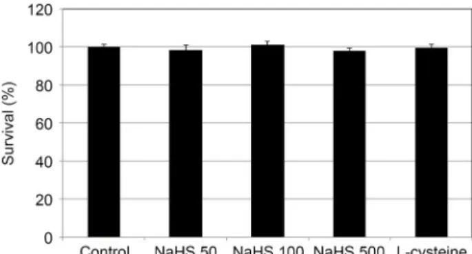
Figure 1. The Effect of sodium hydrogen sulfide (NaHS; 0, 50,
100, 500 μM) and L-cysteine (100 μM) on the survival of cul- tured human trabecular meshwork cells. Both NaHS and L-cys- teine did not affect the survival of trabecular meshwork cells compared to non-exposed controls (all p > 0.05).Figure 2. The Effect of sodium hydrogen sulfide (NaHS; 0, 50,
100, 500 μM) and L-cysteine (100 μM) on the production of ni- tric oxide in cultured human trabecular meshwork cells. Both NaHS and L-cysteine did not affect the production of nitric ox- ide compared to non-exposed controls (all p > 0.05).A B
Figure 3. The Effect of sodium hydrogen sulfide (NaHS; 0,
50, 100, 500 μM) and L-cysteine (100 μM) on the expression of endothelial nitric oxide synthase (eNOS) mRNA in cultured human trabecular meshwork cells. Both NaHS and L-cysteine did not affect the on the expression of eNOS mRNA compared to non-exposed controls (all p > 0.05).Carboxyfluorescein permeability assay
Transwell의 내측 chamber에 세포가 단일세포층으로 충 만하게 자란 것을 확인한 후 6시간 동안 각 약제에 노출시 킨 다음 투과도검사를 시행하였다.25-29 내측 chamber에 자 라고 있는 세포를 PBS로 3회 세척한 다음 50 mM carboxy- fluorescein (Sigma)을 노출시켰다. 노출 2시간 후 외측 chamber로 투과된 carboxyfluorescein의 농도를 532 nm에 서 분광형광계(Fluostar Optima, BMG Labtech)로 측정하여 백분율로 나타내었다.
통계적 처리
모든 실험은 3계대에서 5계대 사이의 세포를 이용하였 다. 세포의 생존과 NO의 생성은 평균 ± 표준오차로 나타내 었으며, 투과도와 eNOS mRNA의 변화는 백분율로 나타내 었다. Unpaired t-test를 사용하여 유의성을 비교하였으며 유의수준은 5%로 정하였다.
결 과
NaHS가 섬유주세포의 생존에 미치는 영향
배양된 섬유주세포에 0, 50, 100, 500 μM NaHS와 100 μM L-cysteine을 배양된 섬유주세포에 6시간 노출시켰을 때 모 든 약물이 섬유주 세포의 생존에 유의한 영향을 미치지 않 았다(모두 p>0.05) (Fig. 1). 따라서 실험에 사용된 농도에 서 측정된 아래 실험의 결과들은 세포의 생존 변화에 의한 것이 아님을 알 수 있다.
NaHS가 NO의 생성과 eNOS mRNA의 발현에 미치는 영향
배양된 섬유주세포에 0, 50, 100, 500 μM NaHS와 100 μM
L-cysteine을 6시간 노출시켰을 때 모든 약물이 섬유주 세 포에서 NO의 생성에 유의한 영향을 미치지 않았다(모두 p>0.05) (Fig. 2). 또한 약물에 노출되지 않은 대조군에 비 하여 섬유주 세포에서 eNOS mRNA의 발현에도 유의한 영 향을 미치지 않았다(모두 p>0.05) (Fig. 3).
NaHS가 섬유주단층세포층의 투과성에 미치는 영향 NaHS는 50 μM 농도에서 114.5%로 섬유주단층세포층의 투과도를 증가시켰으나 통계적으로 유의하지 않았다(p=0.08) (Fig. 4). 그러나 100 μM과 500 μM에서 농도에 비례하여 섬유주단층세포층의 투과도를 각각 유의하게 증가시켰다 (p=0.006, p=0.001). H2S 합성의 기질인 100 μM L-cysteine
Figure 4. The Effect of sodium hydrogen sulfide (NaHS: 0, 50,
100, 500 μM) and L-cysteine (100 μM) on the permeability of carboxyfluorescin through the trabecular meshwork cell monolayer. Both 100, 500 μM NaHS and 100 μM L-cysteine increased the permeabilty of carboxyfluorescein significantly (*p < 0.05). Carboxyfluorescein intensity of outer chamber normalized to the mean value obtained using non-exposed con- trol (permeability 100%).Figure 5. The Effect of sodium hydrogen sulfide (NaHS) at low
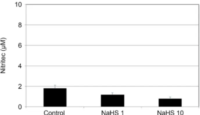
concentration (1, 10 μM) on the production of nitric oxide in cultured human trabecular meshwork cells. NaHS at low con- centration did not affect the production of nitric oxide (all p >0.05).
A B
Figure 6. Effect of sodium hydrogen sulfide (NaHS) at low
concentration (1, 10 μM) and Nω-Nitro-L-arginine methyl es- ter hydrochloride (L-NAME, 10 μM) on the expression of en- dothelial nitric oxide synthase (eNOS) mRNA in cultured hu- man trabecular meshwork cells. NaHS did not affect the on the expression of eNOS mRNA compared to non-exposed con- trols (all p > 0.05). In contrast, L-NAME decreased the ex- pression of eNOS mRNA significantly (p = 0.046).Figure 7. Effect of 10 μM sodium nitroprusside (SN) co-ex-
posed to sodium hydrogen sulfide (NaHS) at low concentration (1, 10 μM) on the permeability of carboxyfluorescein through the trabecular meshwork cell monolayer. Both 1, 10 μM NaHS co-exposed with SN did not affect the permeabilty of carboxy- fluorescein (p = 0.74, p = 0.511) compared with exposed to SN alone. Carboxyfluorescein intensity of outer chamber nor- malized to the mean value obtained using SN (permeability 100%).에 노출시킨 경우에는 126.4%로 섬유주단층세포층의 투과 도를 유의하게 증가시켰다(p=0.004). 이러한 결과로서 NaHS 를 외부에서 투여하거나 내인성 H2S의 생성을 증가시킨 경 우 모두 섬유주단층세포층의 투과도를 증가시킨다는 것을 알 수 있었다.
저농도의 NaHS와 NO의 상승 작용
1, 10 μM의 저농도에서 NaHS에 노출시켰을 때 NO의 생성량은 각각 약물에 노출되지 않은 대조군에 비해 차이를 보이지 않았으며(p=0.865, p=0.738) (Fig. 5), eNOS mRNA 의 발현 정도도 차이를 보이지 않았다(p=0.533, p=0.965) (Fig. 6). eNOS 합성저해제인 L-NAME에 노출시킨 경우에 는 eNOS mRNA의 발현이 91.4%로 감소하였다(p=0.046).
저농도에서 NaHS와 NO의 상승 작용이 일어나는지 알아 보기 위하여 10 μM SN과 1, 10 μM의 NaHS에 동시에 노 출시켜 섬유주단층세포층의 투과도를 측정한 결과 10 μM SN에 단독으로 노출시킨 경우에 비해 NaHS에 동시에 노 출시킨 경우 1, 10 μM NaHS 모두에서 유의한 투과도의 차 이를 나타내지 않았다(p=0.74, p=0.511) (Fig. 7).
고 찰
H2S의 기체송신기로서의 기능이 밝혀지면서30,31 인체 내 에서 다양한 기능을 수행하는 신호전달분자로서 평활근 이 완이나 염증 반응, 신경전달과 세포의 생존 등의 생리적 반 응에 중심적 역할을 하는 것으로 알려졌다.32 안구 내 여러 조직에서도 H2S 합성에 관여하는 효소들이 발견되며33 내 인성 경로로 H2S를 생산한다.10
본 연구에서 섬유주단층세포층의 투과도에 미치는 영향 을 조사한 결과 100 μM 이상의 고농도에서 NaHS는 투과 도를 유의하게 증가시켰는데, 이러한 결과는 이전의 동물 실험에서 H2S가 안압을 낮추는 효과가 있으며, H2S가 전안 부 관류 실험에서 방수 유출을 증가시킨다는 보고와16,17 일 치한다. 또한 H2S의 기질로 작용하는 L-cysteine을 투여하 였을 때 섬유주단층세포층의 투과도가 증가하였으므로 섬 유주세포에서 내인성으로 H2S가 합성됨을 알 수 있다.
섬유주세포는 형태학적 연구와 전기생리학적 연구에서 혈관내피세포 및 평활근 세포와 유사한 성질을 가지고 있
는데34,35 eNOS에서 생산하는 NO는 섬유주를 이완시켜 방
수 유출을 촉진하는 것으로 알려져 있고,36-38 H2S 역시 평 활근을 이완시키는 작용을 나타내므로 유사한 작용을 나타 낼 수 있을 것이다. 혈관내피세포를 이용한 이전 연구에서 H2S가 NO의 생성을 증가시키며18 NO 공여자가 H2S의 생 성을 촉진한다는 연구 결과가 보고되었지만,39,40 이와 반대 로 H2S가 eNOS의 활성을 감소시켜 NO의 합성을 억제한다 는 상반된 보고들도 있다.41,42 이에 따라 본 연구에서 섬유 주세포를 대상으로 H2S가 NO의 생성에 미치는 영향을 조 사하였는데, 고농도에서 NaHS는 투과도를 유의하게 증가 시켰음에도 불구하고 NO의 생성이나 NO 합성 효소의 발 현 정도에서 유의한 차이를 나타내지 않아 섬유주세포에서 는 H2S가 NO의 생성에 미치는 영향은 미약함을 알 수 있 었다.
H2S와 NO와의 상호작용에 관한 다양한 보고가 있는데 NO와 함께 상승적으로 작용하여 평활근 근육의 이완을 촉 진시킨다는 보고가 있었으나,19 이와 달리 H2S가 NO와 반 응하여 새로운 화합물을 생성하며, 이러한 새로운 화합물 은 H2S 또는 NO가 지닌 고유의 성질과는 다른 새로운 생 리적 작용을 나타낸다는 상반된 보고도 있다.21 예를 들어 심혈관계에서 NO 공여자와 NaHS를 혼합하여 투여할 경우 NO와 NaHS 단독으로 사용할 경우와는 전혀 다른 효과가 나타난다고 하며, 이러한 효과는 Nitroxyl같은 새로운 화합 물에 의해 그 작용이 나타나는 것으로 여겨진다.43 본 연구 에서 섬유주 투과도를 NO 공여자를 단독으로 처치한 경우 와 NaHS와 NO 공여자를 함께 투여한 경우와 비교하여 유
의한 차이를 나타내지 않았으므로 섬유주에서는 H2S와 NO 가 상승적으로 작용하지는 않는다고 생각된다. 그러나 위 에 언급한 바와 같이 새로운 화합물에 의해 다른 작용을 나 타낼 가능성이 있을 수 있으므로 이에 관해 향후 보다 자세 한 연구가 필요할 것이다.
심혈관계에서 H2S가 혈관긴장도에 미치는 영향은 농도 에 따라 다르게 나타나는데 100 μM 이상의 고농도 NaHS 는 혈관이완제로 작용하지만 그 이하의 저농도에서는 오히 려 eNOS에 의한 NO의 합성을 저하시켜 혈관수축제로 작 용한다고 한다.21,44 저농도의 NaHS가 NO의 생성을 억제한 다면 투과성을 저하시킬 수도 있으므로 본 실험에서 이를 규명하기 위해 저농도의 NaHS를 처치한 후 조사한 결과, 고농도로 처치한 경우와 마찬가지로 eNOS의 발현이나 NO 의 생성에 유의한 영향을 미치지 않았으므로 섬유주에서 농도에 따라 H2S가 NO에 생성에 미치는 영향은 적다고 볼 수 있다.
이전의 연구들과 본 연구의 결과를 함께 고려해 보면 H2S가 섬유주를 통한 방수 유출에 중요한 역할을 하므로 안압을 적절하게 유지하는 데 있어 방수 내에서 적정 농도 로 H2S가 유지되어야 할 것이다. 따라서 H2S를 합성하는 효소의 결함이 있는 경우 여러 안질환을 유발할 수 있는데 개방각녹내장이나 거짓비늘녹내장에서 방수 내 H2S 합성 기질의 농도가 높게 나타나며45,46 이는 H2S 합성이 제대로 되지 않아 나타나는 결과로서 기질 농도의 상승은 안압상 승뿐만 아니라 시신경 손상을 유발하여 개방각녹내장의 위 험인자가 될 수 있다.
결론적으로 H2S는 고농도에서 섬유주세포층의 투과도를 감소시켰으며, 이러한 효과는 NO의 생성과는 관련이 없으 며, H2S와 NO와의 상승 작용도 나타나지 않아 섬유주에서 H2S의 방수 유출 작용은 NO와 연관성이 적다고 볼 수 있 겠다. 최근 NaHS보다 작용시간이 길어진 GYY 4137 같은 새로운 H2S 공여자와 H2S를 분비하는 프로스타글란딘 결 합제제인 ACS67의 안압하강 효과가 보고되고 있는데47,48 H2S는 안압하강 효과와 더불어 신경보호작용을 함께 나타 낼 수 있으므로 녹내장 영역에서의 효과가 기대되고 있으 며, 이에 관해 향후 보다 자세한 임상적인 연구가 필요할 것이다.
REFERENCES
1) Łowicka E, Bełtowski J. Hydrogen sulfide (H2S) - the third gas of interest for pharmacologists. Pharmacol Rep 2007;59:4-24.
2) Chen CQ, Xin H, Zhu YZ. Hydrogen sulfide: third gaseous trans- mitter, but with great pharmacological potential. Acta Pharmacol Sin 2007;28:1709-16.
3) Li L, Rose P, Moore PK. Hydrogen sulfide and cell signaling. Annu Rev Pharmacol Toxicol 2011;51:169-87.
4) Kabil O, Banerjee R. Enzymology of H2S biogenesis, decay and signaling. Antioxid Redox Signal 2014;20:770-82.
5) Kimura H. Production and physiological effects of hydrogen sulfide. Antioxid Redox Signal 2014;20:783-93.
5) Kimura H. The physiological role of hydrogen sulfide and beyond.
Nitric Oxide 2014;41:4-10.
6) Elsey DJ, Fowkes RC, Baxter GF. Regulation of cardiovascular cell function by hydrogen sulfide (H2S). Cell Biochem Funct 2010;28:95-106.
7) Kimura Y, Kimura H. Hydrogen sulfide protects neurons from oxi- dative stress. FASEB J 2004;18:1165-7.
8) Wallace JL. Hydrogen sulfide-releasing anti-inflammatory drugs.
Trends Pharmacol Sci 2007;28:501-5.
9) Zhang Y, Tang ZH, Ren Z, et al. Hydrogen sulfide, the next potent preventive and therapeutic agent in aging and age-associated diseases. Mol Cell Biol 2013;33:1104-13.
10) Kulkarni, M, Njie-Mbye YF, Okpobiri I, et al. Endogenous pro- duction of hydrogen sulfide in isolated bovine eye. Neurochem Res 2011;36:1540-5.
11) Goldblum D, Mittag T. Prospects for relevant glaucoma models with retinal ganglion cell damage in the rodent eye. Vision Res 2002;42:471-8.
12) Lu M, Hu LF, Hu G, Bian JS. Hydrogen sulfide protects astrocytes against H2O2- induced neural injury via enhancing glutamate uptake.
Free Radic Biol Med 2008;45:1705-13.
13) Sakamoto K, Suzuki Y, Kurauchi Y, et al. Hydrogen sulfide attenu- ates NMDA-induced neuronal injury via its anti-oxidative activity in the rat retina. Exp Eye Res 2014;120:90-6.
14) Liu H, Anders F, Thanos S, et al. Hydrogen sulfide protects retinal ganglion cells against glaucomatous injury in vitro and in vivo.
Invest Ophthalmol Vis Sci 2017;58:5129-41.
15) Huang S, Huang P, Lin Z, et al. Hydrogen sulfide supplement at- tenuates the apoptosis of retinal ganglion cells in experimental glaucoma. Exp Eye Res 2018;168:33-48.
16) Salvi A, Bankhele P, Jamil J, et al. Effect of hydrogen sulfide do- nors on intraocular pressure in rabbits. J Ocul Pharmacol Ther 2016;32:371-5.
17) Robinson J, Okoro E, Ezuedu C, et al. Effects of hydrogen sul- fide-releasing compounds on aqueous humor outflow facility in porcine ocular anterior segments, Ex Vivo. J Ocul Pharmacol Ther 2017;33:91-7.
18) Kida M, Sugiyama T, Yoshimoto T, Ogawa Y. Hydrogen sulfide in- creases nitric oxide production with calcium-dependent activation of endothelial nitric oxide synthase in endothelial cells. Eur J Pharm Sci 2013;48:211-5.
19) Hosoki R, Matsuki N, Kimura H. The possible role of hydrogen sulfide as an endogenous smooth muscle relaxant in synergy with nitric oxide. Biochem Biophys Res Commun 1997;237:527-31.
20) Bruce King S. Potential biological chemistry of hydrogen sulfide (H2S) with the nitrogen oxides. Free Radic Biol Med 2013;55:1-7.
21) Nagpure BV, Bian JS. Interaction of hydrogen sulfide with nitric oxide in the cardiovascular system. Oxid Med Cell Longev 2016;2016:6904327.
22) Mosmann T. Rapid colorimetric assay for cellular growth and sur- vival: application to proliferation and cytotoxicity assays. J Immunol Methods 1983;65:55-63.
23) Fremoser FM, Jakob CA, Aebi M, Tuor U. The MTT [3-(4,5-dime- thylthiazol-2-yl)-2,5-diphenyltetrazolium bromide] assay is a fast and reliable method for colorimetric determination of fungal cell densities. Appl Environ Microbio 1999;65:3727-9.
24) Green LC, Wagner DA, Glogowski J, et al. Analysis of nitrites and nitrates in biological fluids. Analytical Biochem 1982;126:131-8.
25) Grimes PA, Stone RA, Laties AM, Li W. Carboxyfluorescein. A probe of the blood-ocular barriers with lower membrane perme- ability than fluorescein. Arch Ophthalmol 1982;100:635-9.
26) Araie M. Carboxyfluorescein. A dye for evaluating the corneal en- dothelial barrier function in vivo. Exp Eye Res 1986;42:141-50.
27) Grimes PA. Carboxyfluorescein transfer across the blood-retinal barrier evaluated by quantitative fluorescence microscopy: com- parison with fluorescein. Exp Eye Res 1988;46:769-83.
28) Nakagawa S, Usui T, Yokoo S, et al. Toxicity evaluation of anti- glaucoma drugs using stratified human cultivated corneal epi- thelial sheets. Invest Ophthalmol Vis Sci 2012;53:5154-60.
29) Lei Y, Stamer WD, Wu J, Sun X. Oxidative stress impact on barrier function of porcine angular aqueous plexus cell monolayers. Invest Ophthalmol Vis Sci 2013;54:4827-35.
30) Abe K, Kimura H. The possible role of hydrogen sulfide as an en- dogenous neuromodulator. J Neurosci 1996;16:1066-71.
31) Gadalla MM, Snyder SH. Hydrogen sulfide as a gasotransmitter. J Neurochem 2010;113:14-26.
32) Kimura H. Hydrogen sulfide: from brain to gut. Antioxid Redox Signal 2010;12:1111-23.
33) Persa C, Osmotherly K, Chao-Wei Chen K, et al. The distribution of cystathionine beta-synthase (CBS) in the eye: implication of the presence of a trans-sulfuration pathway for oxidative stress defense. Exp Eye Res 2006;83:817-23.
34) Wiederholt M, Dörschner N, Groth J. Effect of diuretics, channel modulators, and signal interceptors on contractility of the tra- becular meshwork. Ophthalmologica 1997;211:153-61.
35) Wiederholt M, Stumpff F. The trabecular meshwork and aqueous humor reabsorption. In: Civan MM, ed. Current topics in membranes.
The eye's aqueous Humor: from secretion to glaucoma, 1st ed. San Diego: Academic Press, 1998; 163-202.
36) Wiederholt M, Sturm A, Lepple-Wienhues A. Relaxation of tra- becular meshwork and ciliary muscle by release of nitric oxide.
Invest Ophthalmol Vis Sci 1994;35:2515-20.
37) Behar-Cohen FF, Goureau O, D’Hermies F, Courtois Y. Decreased intraocular pressure induced by nitric oxide donors is correlated to nitrite production in the rabbit eye. Invest Ophthalmol Vis Sci 1996;37:1711-5.
38) Dismuke WM, Mbadugha CC, Ellis DZ. NO-induced regulation of human trabecular meshwork cell volume and aqueous humor out- flow facility involve the BKCa ion channel. Am J Physiol Cell Physiol 2008;294:C1378-86.
39) Zhao W, Zhang J, Lu Y, Wang R. The vasorelaxant effect of H2S as a novel endogenous gaseous KATP channel opener. EMBO 2001;
20:6008-16.
40) Zhao W, Ndisang JF, Wang R. Modulation of endogenous pro- duction of H2S in rat tissues. Can J Physiol Pharmacol 2003;81:848-53.
41) Kubo S, Doe I, Kurokawa Y, et al. Direct inhibition of endothelial nitric oxide synthase by hydrogen sulfide: contribution to dual modulation of vascular tension. Toxicology 2007;232:138-46.
42) Geng B, Cui Y, Zhao J, et al. Hydrogen sulfide downregulates the aortic L-arginine/nitric oxide pathway in rats. Am J Physiol Regul
= 국문초록 =
황화수소가 섬유주단층세포층 투과도 증가에 미치는 영향과 일산화질소의 상호작용에 관한 연구
목적: 황화수소(hydrogen sulfide, H2S)가 섬유주단층세포층의 투과성에 미치는 영향과 일산화질소와의 상호작용에 대해 알아보고자 하였다.
대상과 방법: 섬유주세포를 sodium hydrogen sulfide (NaHS) 0, 50, 100, 500 μM에 6시간 노출시킨 다음 transwell과 carboxyfluorescein을 이용하여 섬유주 단층세포층의 투과도를 측정하였다. Griess assay로 일산화질소(nitrogen oxide, NO)의 생성을 측정하였고, reverse transcription-polymerase chain reaction로 eNOS mRNA의 발현을 측정하였다. 저농도의 H2S에서 NO와 상승 작용이 일어나는지 알아 보기 위하여 0, 1, 10 μM의 NaHS와 10 μM sodium nitropruside (SN)를 함께 투여하여 실험을 시행하였다.
결과: NaHS는 100 μM 이상에서 농도에 비례하여 섬유주단층세포층의 투과도를 증가시켰으며(p<0.05), NO의 생성과 eNOS mRNA의 발현에는 유의한 영향을 미치지 않았다(p>0.05). 0, 1, 10 μM의 NaHS와 10 μM SN을 함께 투여한 경우 섬유주단층세포층의 투과도와 NO의 생성, eNOS mRNA의 발현에는 각각 유의한 영향을 미치지 않았다(p>0.05).
결론: 고농도에서 H2S는 섬유주단층세포층의 투과도를 증가시켰으며 이러한 증가는 NO와는 무관하였다. 저농도에서 H2S는 NO와 상승 작용을 나타내지 않았다. 따라서 고농도의 H2S는 섬유주를 통한 방수 유출을 증가시킬 가능성이 있으며, 이러한 방수 유출의 증가는 NO와는 관련이 적을 것으로 생각된다.
<대한안과학회지 2020;61(3):267-273>
손명서 / Myung Seo Son
대구가톨릭대학교 의과대학 안과학교실 Department of Ophthalmology, Daegu Catholic University College of Medicine Integr Comp Physiol 2007;293:R1608-18.
43) Yong QC, Hu LF, Wang S, et al. Hydrogen sulfide interacts with nitric oxide in the heart: possible involvement of nitroxyl. Cardiovasc Res 2010;88:482-91.
44) Wu D, Hu Q, Zhu D. An update on hydrogen sulfide and nitric ox- ide interactions in the cardiovascular system. Oxid Med Cell Longev 2018;2018:4579140.
45) Roedl JB, Bleich S, Reulbach U, et al. Homocystein levels in aque- ous humor and plasma of patients with primary open-angle
glaucoma. J Neural Transm (Vienna) 2007;114:445-50.
46) Roedl JB, Bleich S, Reulbach U, et al. Homocysteine in tear fluid of patients with pseudoexfoliation glaucoma. J Glaucoma 2007;16:
234-9.
47) Patil A, Singh S, Opere C, Dash A. Sustained-release delivery sys- tem of a slow hydrogen sulfide donor, GYY 4137, for potential ap- plication in glaucoma. AAPS PharmSciTech 2017;18:2291-302.
48) Perrino E, Uliva C, Lanzi C, et al. New prostaglandin derivative for glaucoma treatment. Bioorg Med Chem Lett 2009;19:1639-42.