• 검색 결과가 없습니다.

Anti-Inflammatory Effect of Violapyrones B and C from a Marine-derived Streptomyces sp.

N/A
N/A
Protected

Academic year: 2022

Share "Anti-Inflammatory Effect of Violapyrones B and C from a Marine-derived Streptomyces sp."

Copied!
4
0
0

로드 중.... (전체 텍스트 보기)

전체 글

(1)

Natural Product Sciences 21(4) : 251-254 (2015)

http://dx.doi.org/10.20307/nps.2015.21.4.251

251

Anti-Inflammatory Effect of Violapyrones B and C from a Marine-derived Streptomyces sp.

Hwa-Sun Lee

1

, Bong-Jeun An

2

, Hyeon Jeong Kim

2,3

, Yong Hun Cho

2

, Dong In Kim

2

, Jae Yoon Jang

2

, Jae Hoon Kwak

2

, Hyi-Seung Lee

1

, Yeon-Ju Lee

1

, Jong Seok Lee

1

, and Hee Jae Shin

1,4,

*

1

Marine Natural Products Chemistry Laboratory, Korea Institute of Ocean Science and Technology, 787 Haeanro, Ansan 426-744, Korea

2

Department of Cosmeceutical Science, Dague Haany University, 285-10 Eobongji-gil, Gyeoungsan 712-220, Korea

3

Institute of technology, Herbnoori.Co.,Ltd. Daegu, 702-852, Korea

4

Department of Marine Biotechnology, University of Science and Technology, 217 Gajungro, Daejeon 305-350, Korea

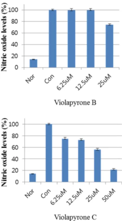
Abstract − Recently, we reported violapyrones B, C, H and I, unusual 3, 4, 6-trisubstituted α-pyrone derivatives, from the culture broth of the marine Streptomyces sp. 112CH148. In previous studies, violapyrones have been shown to have antibacterial and antitumor activities. However, the anti-inflammatory effect of violapyrones has not been reported yet. As part of our ongoing study for the discovery of bioactive metabolites from marine microorganisms, we found that violapyrones also have anti-inflammatory activity. In this study, we investigated the effect of violapyrones on LPS-induced inflammatory responses in vitro. Violapyrones B and C did not affect the viability of RAW 264.7 cells at concentrations up to 25 μM. However, violapyrones B and C inhibited the production of NO compared to the LPS-induced control. In addition, violapyrones B and C down-regulated the expression of iNOS protein in LPS-stimulated RAW 264.7 cells. To the best of our knowledge, this is the first report on the anti-inflammatory activity of violapyrones B and C.

Keywords − Violapyrones, α-Pyrones, Streptomyces sp., Anti-inflammatory

Introduction

Inflammation is part of non-specific immune response that caused by pathogens, physical injury or chemical irritants.

1

This complex biological response is a protective process to remove the injurious stimuli from the body tissues.

2

However, chronic inflammation is associated with the development of various diseases such as Alzhei- mer, atherosclerosis, rheumatoid arthritis and cancer.

3-6

During the inflammatory process, proinflammatory cytokines, nitric oxide (NO) and prostaglandin E

2

(PGE

2

), are produced by inducible nitric synthase (iNOS) and cyclooxygenase-2 (COX-2).

7

In addition, iNOS is activated by interferon- γ and lipopolysaccharide (LPS).

2

The high level of NO affects formation of peroxynitrite and cell toxicity.

8

In our recent study, we isolated violapyrones B, C, H and I from the culture broth of marine Streptomyces sp. 112CH148.

9,10

Violapyrones A-G have been also reported to show anti-

bacterial activities.

11

In this study, we report the anti- inflammatory effect of violapyrones B and C (Fig. 1) on the NO production and expression of iNOS and COX-2 proteins in LPS-induced RAW 264.7 cells.

Experimental

General experimental procedures − The general experi- mental procedures are described in detail in our previous paper.

10

Reagents and antibodies − Reagents for anti-inflam- matory activity such as dulbeco’s modified eagle medium (DMEM), fetal bovine serum (FBS), streptomycin and penicillin were supplied by Invitrogen (Carlsbad, California, USA); sodium dodesyl sulfate (SDS), acrylamide and bis- acrylamide were purchased from Bio-Rad Laboratories (Hercules, California, USA); NP-40, protease inhibitor, RIPA buffer and Griess reagent were obtained from Sigma-Aldrich (Saint Louis, Missouri, USA). Anti-iNOS (BD bioscience, San jose, California, USA), anti-COX-2 (Cayman Chemical, Ann Arbor, Michigan, USA), anti- β- actin, anti-rabbit Ig-G horseradish peroxidase (HRP)-

*Author for correspondence

Hee Jae Shin, Marine Natural Products Chemistry Laboratory, Korea Institute of Ocean Science and Technology, 787 Haeanro, Ansan 426-744, Korea

Tel: +82-31-400-6172; E-mail: [email protected]

(2)

252 Natural Product Sciences

conjugated antibody (Santa Cruz, California, USA) and ELISA kit (R&D systems Inc., Minneapolis, Minnesota, USA) were purchased as indicated. All solvents used in our study were guaranteed reagent grade.

Microorganism − The producing strain 112CH148 was isolated from a crown-of-thorns starfish, Acanthaster planci, collected from Chuuk, Federated States of Micronesia in 2011. The strain was identified as Streptomyces sp. on the basis of 16S rRNA sequence analysis. The sequence was deposited in the GenBank under the accession number KJ419328. This strain is currently preserved in the Microbial Culture Collection, KIOST, with the name of Streptomyces sp. 112CH148 under the curatorship of Hee Jae Shin.

Preparation of compounds − Violapyrones B and C were isolated from the EtOAc extract of culture broth and purified by chromatographic methods. In previous our paper, we reported the detailed experimental procedure including isolation of the strain 112CH148, seed and mass cultivation, extraction of the fermentation broth, purification of violapyrones B and C and their structural elucidation.

10

Violapyrone B − Yellowish amorphous solid; UV (MeOH) λ

max

(log ε) 286.5 (1.34) nm; IR (MeOH) ν

max

3347 (br), 2943, 1674 cm

−1

;

1

H NMR (CD

3

OD, 500 MHz) δ 5.97 (1H, s, H-5), 2.45 (2H, t, J = 7.5 Hz, H-7), 1.85 (3H, s, Me-3), 1.62 (2H, m, H-8), 1.54 (1H, m, H- 11), 1.35 (2H, m, H-9), 1.23 (2H, m, H-10), 0.88 (6H, d, J = 7.0 Hz, H-12);

13

C NMR (CD

3

OD, 125 MHz) δ 169.4 (C-2), 168.8 (C-4), 164.9 (C-6), 101.5 (C-5), 98.9 (C-3), 39.9 (C-10), 34.4 (C-7), 29.2 (C-11), 28.3 (C-8), 27.9 (C- 9), 23.1 (C-12), 8.4 (Me-3); HR-ESI-MS m/z 225.1485 [M+H]

+

; (calcd for C

13

H

21

O

3

, 225.1491).

Violapyrone C − Yellowish amorphous solid; : +50 ( c 0.1, MeOH); UV (MeOH) λ

max

(log ε) 288.0 (0.91) nm; IR (MeOH) ν

max

3343 (br), 2925, 1674 cm

−1

;

1

H NMR (CD

3

OD, 500 MHz) δ 5.98 (1H, s, H-5), 2.47 (2H, t, J = 7.5 Hz, H-7), 1.85 (3H, s, Me-3), 1.62 (2H, m, H-8), 1.36 (2H, overlapped, H-9), 1.34 (1H, overlapped, Ha-12), 1.13 (1H, overlapped, Hb-12), 1.33 (1H, over- lapped, Ha-10), 1.17 (1H, overlapped, Hb-10), 1.32 (1H,

overlapped, H-11), 0.88 (3H, t, J = 6.5 Hz, H-13), 0.87 (3H, d, J = 6.0 Hz, Me-11);

13

C NMR (CD

3

OD, 125 MHz) δ 169.5 (C-4), 169.2 (C-2), 164.9 (C-6), 101.8 (C- 5), 98.8 (C-3), 37.5 (C-12), 35.7 (C-11), 34.4 (C-7), 30.7 (C-10), 28.4 (C-8), 27.6 (C-9), 19.7 (Me-11), 11.9 (C-13), 8.4 (Me-3); HR-ESI-MS m/z 261.1461 [M+Na]

+

; (calcd for C

14

H

22

O

3

Na, 261.1467).

Cell culture − The murine macrophage RAW 264.7 cells were obtained from Korean Cell Line Bank (KCLB, Seoul, Korea). The cells were maintained in Dulbecco’s modified Eagle’s medium (DMEM) supplemented with 10% fetal bovine serum (FBS) and antibiotics (100 U/mL of penicillin, 100 μg/mL of streptomycin) at 37

o

C in humidified atmosphere with 5% CO

2

.

MTT assay for cell viability − The MTT assay was carried out to determine cell viability.

12

The RAW 264.7 cells (5 × 10

4

cells/mL) were seeded in 96-well plates.

Then, the cells were pretreated with various concentrations (6.25, 12.5, 25 and 50 μM) of compounds. After being incubated for 24 hours, each well was added to 20 μL of MTT solution and incubated for 4 hours at 37

o

C under a humidified atmosphere of 5% CO

2

. And then the medium was discarded and added DMSO in each well. After 30 min incubation, the plates were measured absorbance at 540 nm by a microplate reader. The relative cell viability (%) was measured as a percentage relative to the untreated control cells.

Measurement of nitric oxide (NO) production − NO production was assessed by measuring nitrite concentra- tions in medium using Griess reagents. The RAW 264.7 cells were seeded at a density of 1 × 10

5

cells/mL in 6- well plates. After pre-incubation of the cells for 24 hours at 37

o

C in 5% CO

2

, and then each well was added with lipopolysaccharide (LPS, 1 μg/mL) for 1 hour. The cells were treated with several concentrations (6.25, 12.5 25 and 50 μM) of samples and incubated for 24 hours. After treatment, the supernatant was mixed with Griess reagent and incubated at room temperature for 10 min. The absorbance at 540 nm was measured using a microplate reader. The NO production (%), relative percentage, was α

[ ]

D27

Fig. 1. The structures of violapyrones B and C.

(3)

Vol. 21, No. 4, 2015 253

determined by comparing the experimental group and only LPS-treated control group.

Evaluation of iNOS and COX-2 expressions − The protein expression rate of iNOS and COX-2 was deter- mined by western blot analysis.

13

The RAW 264.7 cells were pre-incubated at the density of 5 × 10

5

cells/mL in 100 mm cell culture dishes for 24 hours. And the cells were treated with 1 μg/mL of LPS in the prepared the various concentration of samples for 24 hours. The stimulated cells were washed with PBS and lysed in 100 μg/mL of lysis buffer, followed by centrifugation at 12,000 rpm, 4

o

C for 20 min. The protein concentrations of the supernatant were determined by Bradford assay.

Protein was separated on 10% sodium dodecylsulphate- polyacrylamide gel (SDS-PAGE) and transferred onto a nitrocellulose membrane. The membranes were blocked with 5% skim milk for 1 hour, and then incubated with a primary antibody (1:1000) at 4

o

C, overnight. Subsequently, the membranes were washed three times with TBST buffer, and then incubated with secondary antibody (1:1000) at room temperature for 2 hours. Again, it was washed three times with TBST buffer, and immuno- reactive proteins were detected using ECL reagent. The results of western blot analysis were quantified by measuring the relative intensity compared to the control using a LAS 4000 chemiluminescence detection system (Fuji, Tokyo, Japan). The results for iNOS and COX-2 were normalized to the band density of internal control

( β-actin), and the relative proteins expression was calculated according to the values of only LPS-treated group as 100%.

Statistical analysis − Data represent the mean ± S.D.

with three separate experiments. One-way ANOVA was used for comparisons of multiple group means followed by t-test (significant as compared to control. p < 0.05).

Results and Discussion

Violapyrones, bearing unusal 3, 4, 6-trisubstituted α- pyrone derivatives, were isolated from both marine and terrestrial Streptomyces sp.

10,11

Consequently, violapyrones seem to be biosynthesized through the same biological processes in the family of Streptomyces without being affected by the salinity. It is also plausible that 3, 4, 6- trisubstituted α-pyrone skeleton was biosynthesized by type III polyketide synthase.

14,15

To confirm the possibility that violapyrones B and C might have an adverse effect of anti-inflammatory activity, the cytotoxicity on RAW 264.7 cells was evaluated by MTT assay. As shown in Fig. 2, violapyrones B and C did not affect the viability of RAW 264.7 cells at concentrations up to 25 μM. However, violapyrones B and C inhibited the production of NO compared to the LPS-induced control (Fig. 3). Especially, violapyrone C potently inhibited NO production at 50 μM without significantly affecting the viability of RAW 264.7 cells. In addition, violapyrones B and C down-regulated Fig. 2. Effect of violapyrones B and C on the cell viablility of

RAW 264.7 cells.

Fig. 3. Effect of violapyrones B and C on the production of nitric

oxide (NO) in LPS-induced macrophage RAW 264.7 cells.

(4)

254 Natural Product Sciences

the expression of iNOS protein in LPS-stimulated RAW 264.7 cells (Fig. 4), but slightly inhibited the expression of COX-2 protein. Since NO and PGE

2

are produced by iNOS and COX-2, respectively, this result suggested that violapyrones B and C have a suppressive activity of NO production through inhibition of iNOS. In conclusion, this study revealed for the first time that violapyrones B and C possess anti-inflammatory property. However, further studies are needed to know the detailed mechanism of anti-inflammatory activity of violapyrones. Violapyrones can be new bioprobes for the development of new agents for the various inflammatory diseases.

Acknowledgments

This study was supported in part by the Ministry of Oceans and Fisheries, Korea (Grants PM58670 and PM58482).

References

(1) Ferrero-Miliani, L.; Nielsen, O. H.; Andersen, P. S.; Girardin, S. E.

Clin. Exp. Immunol. 2007, 147, 227-235.

(2) Moncada, S.; Palmer, R. M.; Higgs, E. A. Pharmacol. Rev. 1991, 43,

109-142.

(3) Azad, N.; Rojanasakul, Y.; Vallyathan, V. J. Toxicol. Environ. Health B Crit. Rev. 2008, 11, 1-15.

(4) Libby, P. Nature 2002, 420, 868-874.

(5) Tracey, K. J. Nature 2002, 420, 853-859.

(6) Weninger, S. C.; Yankner, B. A. Nat. Med. 2001, 7, 527-528.

(7) Posadas, I.; Terencio, M. C.; Guillén, I.; Ferrándiz, M. L.; Coloma, J.; Payá, M.; Alcaraz, M. Naunyn Schmiedebergs Arch. Pharmacol. 2000, 361, 98-106.

(8) Huang, G. J.; Pan, C. H.; Liu, F. C.; Wu, T. S.; Wu, C. H. Food Chem. Toxicol. 2012, 50, 1485-1493.

(9) Lee, J. S.; Shin, J.; Shin, H. J.; Lee, H. S.; Lee, Y. J.; Lee, H. S.;

Won, H. Eur. J. Org. Chem. 2014, 21, 4472-4476.

(10) Shin, H. J.; Lee, H. S.; Lee, J. S.; Shin, J.; Lee, M. A.; Lee, H. S.;

Lee, Y. J.; Yun, J.; Kang, J. S. Mar. Drugs 2014, 12, 3283-3291.

(11) Zhang, J.; Jiang, Y.; Cao, Y.; Liu, J.; Zheng, D.; Chen, X.; Han, L.;

Jiang, C.; Huang, X. J. Nat. Prod. 2013, 76, 2126-2130.

(12) Carmichael, J.; DeGraff, W. G.; Gazdar, A. F.; Minna, J. D.;

Mitchell, J. B. Cancer Res. 1987, 47, 936-942.

(13) Kim, H. J.; Kim, D. H.; Lee, J. Y.; Hwang, J. S.; Lee, J. H.; Lee, S.

G.; Jeong, H. G.; An, B. J. J. Life Sci. 2013, 23, 38-43.

(14) Shen, B. Curr. Opin. Chem. Biol. 2003, 7, 285-295.

(15) Sugiyama, Y.; Oya, A.; Kudo, T.; Hirota, A. J. Antibiot. 2010, 63, 365-369.

Received July 30, 2015

Revised September 22, 2015

Accepted September 30, 2015

Fig. 4. Effect of violapyrones B and C on the expression levels of iNOS and COX-2 proteins in LPS-stimulated macrophage RAW 264.7 cells.

수치

Fig. 3. Effect of violapyrones B and C on the production of nitric oxide (NO) in LPS-induced macrophage RAW 264.7 cells

참조

관련 문서

Effect of Oenanthe javanica ethanol extract (OJE) on LPS-induced p-IκBα, IκBα, iNOS, and COX-2 proteins expression in RAW 264.7 cells.. After pretreatment with the

Effect of Zostera marina ethanolic extract (ZMEE) on LPS-induced iNOS, COX-2, and NF-κB p65 expression in LPS-induced RAW246.7 cells.. The levels of iNOS, COX-2 in the

Modulation of LPS induced iNOS and COX-2 protein expression in RAW 264.7 cells by medicinal herb extracts.. Con,

Effect of Ulva pertusa Kjellman ethanol extract (UPKEE) on LPS-induced iNOS, COX-2, and NF-κB p65 expression in LPS- induced RAW 246.7 cells.. The levels of iNOS, COX-2 in

The anti-inflammatory effects of the ethanol extract of Undaria pinnatifida root (UPREE) were investigated using the lipopolysaccharide (LPS)-induced inflammatory response

Key words : Anti-inflammatory, cyclooxygenase-2 (COX-2), inducible NO synthase (iNOS), CopA3, cytokine, Copris tripartitus..

thunbergii methanol extract showed the anti-inflammatory effect on lipopolysaccharide- stimulated RAW 264.7 cells through inhibits iNOS and COX-2 expression.. Therefore, these

Effect of Oenanthe javanica ethanol extract (OJE) on LPS-induced p-IκBα, IκBα, iNOS, and COX-2 proteins expression in RAW 264.7 cells.. After pretreatment with the