P Pulsed and Color Doppler SonographicFindings of Penile Mondor’s Disease
3
0
0
전체 글
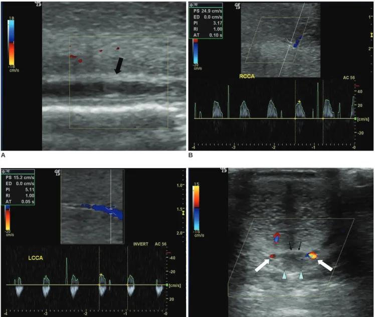
(2) Han et al.. was 24.9 cm/sec with a resistance index (RI) of 1.0 (Fig. 1B); the corresponding left-side values were 16 cm/sec and 1.0, respectively (Fig. 1C). Thirty minutes after an injection of an intracavernosal vasoactive agent, he showed a full erection with unbending rigidity. Doppler US indicated that an enlarged superficial dorsal vein was compressing the deep dorsal vein, and no flow signals were evident in either the superficial dorsal vein or the deep dorsal vein. Bilateral dorsal arterial flow signals were noted (Fig. 1D). The PSV of the cavernosal artery and its RI were unchanged by injection of the intracavernosal vasoactive. A. agent. He was treated with nonsteroidal anti-inflammatory drugs for six weeks and his symptom were much improved.. DISCUSSION Mondor’s disease of the penis is an uncommon disease that usually involves the superficial dorsal veins. In 1939, Henri Mondor first described a sclerosing thrombophlebitis of the subcutaneous veins of the anterior chest wall (1),. B. C D Fig. 1. Penile Mondor’s disease in 38-year-old man. A. Grey scale US revealed internal echogenicity in penile superficial dorsal vein (arrow). Color Doppler US failed to detect venous flow signals in this area. B, C. Pulsed wave Doppler US without intracavernosal vasoactive agent administration showed weak flow signal with low peak systolic velocity and high-resistance pattern. Right (B) and left side (C) peak systolic velocities of carvernosal artery were 24.9 and 16 cm/sec, respectively, and resistance indexes in both sides were 1.0. D. Thirty minutes after intracavernosal vasoactive agent injection, Doppler US showed flow signal in bilateral dorsal arteries (white arrows), but no flow in either superficial dorsal (arrowheads) or deep dorsal veins (black arrows).. 180. Korean J Radiol 9(2), April 2008.
(3) Pulsed and Color Doppler Ultrasonographic Findings of Penile Mondor’s Disease. and in 1955, Braun-Falco described phlebitis of the dorsal veins of the penis within the context of generalized phlebitis (2). Isolated penile Monor’s disease was first described in 1958 by Helm and Hodge (3). Mondor’s disease is a benign and usually a self-limited process. Patients complain of a cord-like induration, which is often painful, in the dorsal aspect of the penis, and this pain can be constant or episodic. The etiology of this condition is usually unknown, but various causative factors have been reported, e.g., penile trauma, excessive sexual activity, prolonged sexual abstinence, infection, pelvic tumors and the constrictive elements used during certain sexual practices; of these, the trauma caused by sexual intercourse appears to be the main etiologic factor (4, 5). For most patients, their symptoms completely resolve after 6 to 8 weeks of conservative management with antiinflammatory drugs and antibiotics. However, surgery is indicated when such symptoms persist after conservative management, although the long-term results after conservative or surgical treatment of superficial penile vein thrombosis have not been reported (6). In the present case, Doppler sonography showed a high flow resistance pattern, which resembled that of venous thrombosis after pancreatic or kidney transplantation. The Doppler sonographic findings of venous thrombosis in transplant graft recipients have revealed an absence of venous flow with a high resistance arterial waveform (7 9). Low flow priapism is a pathology that can produce a low-flow, high resistance flow pattern in the corpus cavernosal artery, and this presumably develops due to a disturbed venous outflow. On Doppler US, low flow priapism usually presents as the absence of cavernosal artery blood flow or as a very high-resistance cavernosal artery flow pattern. We consider that superficial dorsal vein thrombosis may result from a venous outflow disturbance and so it shows low-flow, priapism-like Doppler US findings. The primary venous drainage of the corpora cavernosa is into the deep dorsal vein (only the most distal portion of. Korean J Radiol 9(2), April 2008. the corpora cavernosa, skin and glans drain into the superficial dorsal vein); thus, it appears that superficial dorsal vein thrombosis does not significantly affect the cavernosal venous outflow. However, the enlargement of the superficial dorsal vein and the adjacent soft tissue edema that was caused by thrombophlebitis may cause compression and venous outflow disturbance of the deep dorsal vein. Nevertheless, this is insufficient to explain the high RI value observed in the present case. We suggests that further analysis of Doppler US findings in a larger number of cases needs to be done. In conclusion, the Doppler US findings of thrombus without blood flow in the superficial dorsal vein and the low-flow, high resistance in the cavernosal artery may be suggestive of penile Mondor’s disease.. References 1. Mondor H. Tronculite sous-cutanee subaigue de la paroi thoracigue antero-laterale. Mem Acad Chir 1939;65:1271-1278 2. Braun-Falco O. Clinical manifestations, histology and pathogenesis of the cordlike superficial phlebitis forms. Dermatol Wochenschr 1955;132:705-715 3. Helm JD Jr, Hodge IG. Thrombophlebitis of a dorsal vein of the penis: report of a case treated by phenylbutazone (Butazolidin). J Urol 1958;79:306-307 4. Rodriguez Faba O, Parra Muntaner L, Gomez Cisneros SC, Martin Benito JL, Escaf Barmadah S. Thrombosis of the dorsal penis vein (of Mondor’s phlebitis). Presentation of a new case. Actas Urol Esp 2006;30:80-82 5. Koh JS, Suh HJ, Choe HS, Jung JH, Kim YS, Kim JA, et al. Superficial thrombophlebitis of the dorsal vein of the penis (Penile Mondor’s disease). Korean J Urol 2004;45:399-401 6. Al-Mwalad M, Loertzer H, Wicht A, Fornara P. Subcutaneous penile vein thrombosis (Penile Mondor’s Disease): Pathogenesis, diagnosis, and therapy. Urology 2006;67:586-588 7. Foshager MC, Hedlund LJ, Troppmann C, Benedetti E, Gruessner RW. Venous thrombosis of pancreatic transplants: diagnosis by duplex sonography. AJR Am J Roentgenol 1997;169:1269-1273 8. Reuther G, Wanjura D, Bauer H. Acute renal vein thrombosis in renal allografts: detection with duplex Doppler US. Radiology 1989;170:557-558 9. Fitzgerald SW, Erickson SJ, Foley DW, Lipchik EO, Lawson TL. Color Doppler sonography in the evaluation of erectile dysfunction. Radiographics 1992;12:3-17. 181.
(4)
수치
관련 문서
1) If the fluid is real (viscous fluid) and if no energy is being added, then the energy line may never be horizontal or slope upward in the direction of flow.. 2) Vertical
웹 표준을 지원하는 플랫폼에서 큰 수정없이 실행 가능함 패키징을 통해 다양한 기기를 위한 앱을 작성할 수 있음 네이티브 앱과
_____ culture appears to be attractive (도시의) to the
이하선의 실질 속에서 하악경의 후내측에서 나와 하악지의 내측면을 따라 앞으로 간다. (귀밑샘 부위에서 갈라져 나와
Role of insulin resistance in human disease. 2) Expert panel on detection, evaluation and treatment of high blood cholesterol in adults: executive summary of
다양한 번역 작품과 번역에 관한 책을 읽는 것은 단순히 다른 시대와 언어, 문화의 교류를 넘어 지구촌이 서로 이해하고 하나가
The index is calculated with the latest 5-year auction data of 400 selected Classic, Modern, and Contemporary Chinese painting artists from major auction houses..
1 John Owen, Justification by Faith Alone, in The Works of John Owen, ed. John Bolt, trans. Scott Clark, "Do This and Live: Christ's Active Obedience as the
