• 검색 결과가 없습니다.

A Gallotannin from Cercidiphyllum japonicum Leaves

N/A
N/A
Protected

Academic year: 2021

Share "A Gallotannin from Cercidiphyllum japonicum Leaves"

Copied!
8
0
0

로드 중.... (전체 텍스트 보기)

전체 글

(1)

1. INTRODUCTION

Katsura tree (Cercidiphyllum japonicum Sieb.

Et Zucc), is the only species belonging to Cercidiphyllum genus, which is well represented in the fossil record, with the occurrence during the late Cretaceous and Tertiary of North America and Europe. However, it is now con- fined to East Asian countries (Manchester et al., 2009). The tree is a long-lived, deciduous,

wind-pollinated tree with dimorphic leaves and up to 30 to 45 m tall with a symmetrical can- opy and new growth is reddish turning a light pale green. Fall color is a spectacular yellow, with some red. Thus, it is valued as an orna- mental or a shade tree for landscape (Zhang et al., 2009). It is also a commercially and eco- logically valuable one and likely to become one of the medicinal tree species. The clustered pod-like fruits contain numerous small seeds

1 Date Received March 13, 2015, Date Accepted April 20, 2015

2 Samcheok City Agricultural Technology and Extension Center, Samcheok 245-802, Republic of Korea

3 Department of Forest Biomaterials Engineering, College of Forest and Environment Sciences, Kangwon National University, Chuncheon 200-701, Republic of Korea

Corresponding author: Young-Soo Bae (e-mail: [email protected])

A Gallotannin from Cercidiphyllum japonicum Leaves

1

Tae-Seong Lee

2

⋅Young-Soo Bae

3,†

ABSTRACT

Katsura tree (Cercidiphyllum japonicum Sieb. Et Zucc) leaves were collected, air-dried and extracted with 70% aqueous acetone, then concentrated and sequentially fractionated using n-hexane, methylene chloride (CH

2

Cl

2

), ethylacetate (EtOAc), and H

2

O. The EtOAc fraction was chromatographed on a Sephadex LH-20 column with various aqueous MeOH eluting solvents and finally treated with acetone-H

2

O (7:3, v/v) to isolate a gallotannin. According to the NMR analysis, including HSQC and HMBC, and with the comparison of authentic literature data, the isolate was elucidated as 6-m-digalloyl-1,2,3,4-tetra-O-galloyl β-D-(+)-glu- cose, one of hydrolyzable tannins and one of gallotannins. The compound was only gallotannin which was firstly isolated from the extracts of Katsura tree leaves, and has not been reported before in domestic tree sources.

Keywords :

Cercidiphyllum japonicum leaves, hydrolysable tannin, gallotannin, ethylacetate fraction, column

chromatography

(2)

which adapted for wind dispersal. The natural populations of the tree inhabit distribute sites (600 to 2000 m) of temperate deciduous forests scattered across East China and Japan (Isagi et al., 2005). Because of its extremely low ability of regeneration in natural population, the num- ber of its populations is very little. Therefore, the tree is now treated as “endangered” in China and recognized globally as lower risk un- der the International Union for the Conservation of Nature criteria.

Plants constitute a rich source of bioactive chemicals (Kador et al., 1985a; 1985b; Williamson et al., 1992). Many plants are largely free from adverse effects and have excellent pharmaco- logical actions and they could possibly lead to the development of new classes of safer functional agents and a hydrolyzable tannin is one of those sources.

A hydrolyzable tannin or pyrogallol-type tannin is a type of tannin that yields gallic or ellagic acids by hydrolysis (Bae, 1989; Haslam, 1989).

At the center of a hydrolyzable tannin mole- cule, there is a carbohydrate (usually D-glucose but also cyclitols like quinic or shikimic acids).

The hydroxyl groups of the carbohydrate are partially or totally esterified with phenolic groups such as gallic acid in gallotannins or el- lagic acid in ellagitannins.

Hydrolyzable tannins are mixtures of poly- galloyl glucoses and/or poly-galloyl quinic acid derivatives containing in between 3 up to 12 gallic acid residues per molecule (Haslam, 1989).

Gallotannins are polymers formed when gallic acid, a polyphenol monomer, esterifies and binds with the hydroxyl group of a polyol carbohydrate such as glucose (Cammann et al., 1989; Niehaus and Gross, 1997; Niemetz and Gross, 1998; Niemetz and Gross, 2001).

The ellagitannins are a class of hydrolyzable tannins, a type of polyphenol formed primarily from the oxidative linkage of galloyl groups in 1,2,3,4,6-pentagalloyl glucose (Sepulveda et al., 2011; Kwon and Bae, 2009; Steinmetz, 2010).

Ellagitannins contain various numbers of hexahydroxydiphenoyl (HHDP) units, as well as galloyl units and/or sanguisorboyl units bounded to sugar moiety (Yoshida et al., 2009).

Recently there have been many studies to evaluate biological activities of various natural resources, including plants and tree species, and to develop pharmaceutical or functional food or cosmetic products.

However, there are little studies on katsura tree extracts for functional uses in domestic or abroad (Towatari et al., 2002; Tada and Sakurai, 1991; Takasugi and Katui, 1986).

This work was carried out to investigate the chemical constituents of extracts of katsura tree leaves for future use, and to elucidate the struc- ture of a gallotannin from the leaves extracts.

2. MATERIALS and METHODS 2.1. Plant material

Fresh Cercidiphyllum japonicum leaves were

collected at Hwacheon, Gangwon-do in August

(3)

2013, air dried for two weeks and then ground to fine particles to be extracted.

2.2. Sample preparation

The ground leaves (3 kg) were immersed in 70% aqueous acetone at room temperature for 3 days. After three times extraction and filtration, the filtrates were combined together and evapo- rated on a rotary evaporator under the reduced pressure at 40 ℃. The aqueous crude residue was successively fractionated on a separatory funnel and freeze dried to give n-hexane (2.6 g), CH

2

Cl

2

(8.8 g), EtOAc (35.2 g), and H

2

O (45.2 g) soluble fractions.

2.3. Column chromatography

A portion of EtOAc fraction (10 g) was chro- matographed on a Sephadex LH-20 column, successively eluting with MeOH-H

2

O (1:9 → 3:7

→ 1:1 → 7:3 → 9:1, v/v) to divide 16 fractions.

However, the divided fractions did not contain any hydrolysable tannin compounds, and the column was finally washed with acetone-H
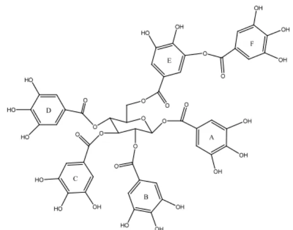
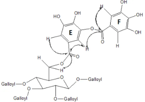
2

O (7:3, v/v) to isolate the gallotannin (Fig. 1), 6-m-digalloyl-1,2,3,4-tetra-O-galloyl β-D-(+)- glucose which is called digalloyl-1,2,3,4-tetra- O-galloyl β-D-(+)-glucose or hexa-O-galloyl β-D-(+)-glucose.

Yellowish amorphous powder, R

f

: 0.20 (TBAW) and 0.01 (6% HOAc).

MALDI-TOF-MS : Found m/z 1093 [M+H]

+

, 1115 [M+Na]

+

.

1

H (700 MHz) and

13

C (125 MHz) NMR : See Table 1.

2.4. Structure analysis

1

H and

13

C NMR spectra, including 2D-NMR such as HSQC (Heteronuclear Single Quantum Coherence) and HMBC (Heteronuclear Multiple Bond Correlation), were recorded on a Bruker (USA) Avance DPX 700 MHz spectrometers using TMS (Tetramethylsilane) as an internal standard and chemical shift was given in δ (ppm).

MALDI-TOF-MS were performed with a Micromass Autospec M363 spectrometer.

Thin layer chromatography (TLC) was done on DC-Plastikfolien Cellulose F (Merck) plates and developed with TBAW (t-BuOH-HOAc-H

2

O (3:1:1, v/v/v)) and 6% aqueous HOAc. The spot was detected by illuminating ultraviolet light (UV, 254 and 365 nm) and by spraying vanillin (Vanillin-EtOH-H

2

SO

4

(15:250:2.5, w/v/v)), then heating.

O O

O OO

O

O O

O O

O O

O HO OH

OH OH

OH

OH OH

OH

OH HO OH

HO OH HO HO

HO

HO A

B C

D

E

F

Fig. 1. Structure of the isolated compound.

(4)

Compound 1 Chemical Shift (δ) assignment

H-NMR 13C-NMR

Glucose

6.23 d 93.87 1

5.58 t 72.25 2

5.91 t 74.09 3

5.58 t 70.02 4

4.52 t 74.38 5

4.43 dd 63.60 6

Galloyl (A)

119.78 1

7.04 110.66 2

146.59 3

140.80 4

146.59 5

7.04 110.66 6

166.22 7

Galloyl (B)

120.25 1

6.97 110.50 2

146.47 3

140.39 4

146.47 5

6.97 110.50 6

167.01 7

Galloyl (C)

120.40 1

6.89 110.41 2

146.32 3

140.28 4

146.32 5

6.89 110.41 6

167.34 7

Galloyl (D)

120.28 1

6.94 110.44 2

146.42 3

140.34 4

146.42 5

6.94 110.44 6

167.07 7

Galloyl (E)

121.13 1

7.29 117.60 2

147.55 3

140.39 4

144.62 5

7.46 115.09 6

167.23 7

Galloyl (F)

120.54 1

7.23 110.91 2

146.64 3

140.54 4

146.64 5

7.23 110.91 6

166.70 7

Table 1.

1

H and

13

C-NMR chemical shifts of the isolated compound (CD

3

OD)

(5)

3. RESULTS and DISCUSSION

The compound was isolated from the EtOAc fraction of the extracts of katsura tree (Cerdidiphyllum japonicum Sieb, Et Zucc) leaves by column chromatography using Sephadex LH-20, and the structured were char- acterized by MALDI-TOF-MS analysis and NMR analysis including comparison with the other literature data.

MALDI-TOF-MS spectrum of the compound showed a sodium complex [M+Na]

+

at m/z 1115, corresponding to the molecular weight of 1115.

In the

1

H NMR spectrum, D-glucose of the compound showed a double doublet signal at δ4.43 for two H-6 protons and H-5 gave a trip- let signal at δ4.52. H-2 and H-4 indicated a triplet signal at δ5.58 and H-3 also showed a triplet signal at δ5.91. H-1 gave a doublet signal at δ6.23 with 8.37 Hz of the coupling constant suggesting β-anomeric glucose. These proton signals were similar to the previous lit- erature data (Si and Bae, 2007).

The five galloyl symmetrical protons in- dicated five singlet signals at δ7.04, δ6.97, δ6.89, δ6.94 and δ7.23, respectively, for H-2 and H-6 of the galloyl A, B, C, D, and F.

However, the galloyl E showed two signals at δ7.29 and δ7.46 for H-2 and H-6, respectively, due to the esterification between C-5 of the galloyl E and C-7 of the galloyl F. These proton chemical shifts of the galloyl groups were also similar to the literature values (Si and Bae, 2007).

In the

13

C NMR spectrum, C-6, C-5 and C-4 of D-glucose gave three signals at 63.60, 74.38, and 70.02 ppm, respectively. C-1 of D-glucose indicated at 93.87 ppm, and C-2 and C-3 also showed signals at 72.25 and 74.09 ppm, respectively. These carbon signals were similar to those of the authentic sample (Nishizawa and Yamagishi, 1982; Nishizawa et al., 1980; Si and Bae, 2007; Emam, 2010). The carbonyl gave six signals at between 166.20 to 167.34 ppm for the six carbonyl of the galloyl A, B, C, D, E, and F.

C-1 carbons of the galloyl groups were res- onated at between 119.78 to 120.54 ppm, and the symmetrical C-2 and C-6 pairs indicated at between 110.41 to 110.91 ppm. Another hy- droxyl containing symmetrical C-3 and C-5 pairs of the galloyl groups showed five signals at between 146.32 to 146.64 ppm for the gallo- yl A, B, C, D and F besides E.

In the galloyl E, C-1 and C-4 gave signals at 121.13 and 140.39 ppm, respectively. C-2 and C-6 showed signals at 117.60 and 115.09 ppm, respectively. Also C-3 and C-5 indicated signals at 147.55 and 144.62 ppm, respectively (Nishizawa and Yamagishi, 1982; Nishizawa et al., 1980; Si and Bae, 2007; Emam, 2010).

In the HSQC spectrum, δ4.43 (H-6) of D-glucose was correlated with 63.60 ppm (C-6).

Also δ4.52 (H-5) and 74.38 ppm (C-5), 5.58

(H-4) and 70.02 ppm (C-4), 5.91 (H-3) and

74.09 ppm (C-3), δ5.58 (H-2) and 72.25 ppm

(C-2), and 6.23 (H-1) and 93.87 ppm (C-1)

were correlated each other. In the galloyl rings,

δ6.89 was resonated with 110.41 ppm (C-2) of

(6)

the galloyl C. δ6.94, δ6.97, δ7.04 and δ7.23 had correlations with 110.44 ppm (C-2 of D), 110.50 ppm (C-2 of B), 110.66 ppm (C-2 of A), and 110.91 ppm (C-2 of F), respectively. In the galloyl E, δ7.29 and δ7.46 were correlated with 117.60 ppm (C-2) and 115.09 ppm (C-6), respectively.

In the HMBC spectrum on D-glucose, δ6.23 (H-1) had a correlation with 166.22 ppm (C-7 of A). δ5.58 (H-2 and H-4) also was resonated with 167.01 ppm (C-7 of B) and 167.07 ppm (C-7 of D). δ5.91 (H-3) was correlated with 167.34 ppm (C-7 of C) and δ4.43 (H-6) had a relationship with 167.23 ppm (C-7 of E).

In the HMBC correlation between the galloyl E and F (Fig. 2), δ7.23 (H-2 and H-6 of F) was resonated with 166.70 ppm (C-7 of F), and δ7.29 (H-2 of E) and δ7.46 (H-6 of E) were correlated with 167.23 ppm (C-7 of E). Also 4.43 (H-6 of glucose) had a correlation with 167.23 ppm (C-7 of E).

From the previous data, by comparison with those of authentic samples, and by the HSQC

and HMBC spectra, the compound was charac- terized as 6-m-digalloyl-1,2,3,4-tetra-O-galloyl β-D-(+)-glucose.

This type of gallotannin is firstly isolated from katsura tree leaves and has never been re- ported in domestic plants. Also it was the only hydrolyzable tannin from leaf extracts of the tree and can be used as an index marker to dis- tinct the tree.

4. CONCLUSION

Katsura tree (Cercidiphyllum japonicum Sieb.

Et Zucc) leaves were collected, air-dried and extracted with 70% aqueous acetone. The ex- tracts were concentrated and then sequentially fractionated with n-hexane, CH

2

Cl

2

, EtOAc, and H

2

O to be freeze dried.

A portion of EtOAc fraction was chromato- graphed on a Sephadex LH-20 column with various aqueous MeOH solvents and ace- tone-H

2

O (7:3, v/v) to isolate the gallotannin.

The structure was elucidated as 6-m-digalloyl- 1,2,3,4-tetra-O-galloyl β-D-(+)-glucose by NMR analysis and by the comparison with the other literature data.

To our best knowledge, this type of gallo- tannin is firstly isolated from katsura tree leaves and has not been reported in domestic tree species.

Also this compound was the only hydro- lyzable tannin from leaf extracts of the tree and can be used as an index mark to distinct the tree.

Fig. 2. Selected HMBC correlations observed be-

tween galloyl E and F.

(7)

ACKNOWLEDGEMENTS

This study was supported by the Basic Research Program for Forest Science of Korean Forest Service (No S211314L010130) and also partially supported by Kangwon National University.

REFERENCES

Bae, Y.S. 1989. Douglas-Fir Inner Bark Procyanidins : Sulfonation, Isolation and Characterization. Ph.

D. Dissertation. pp. 4. Oregon State University.

Cammann, J., Denzel, K., Schilling, G., Gross, G.G. 1989. Biosynthesis of gallotannins : β- Glucogallin-dependent formation of 1,2,3,4,6- pentagalloylglucose by enzymatic galloylation of 1,2,3,6-tetragalloylglucose. Archives of Biochemistry and Biophysics 273: 58 ∼63.

Edwin, H. 1989. Plant polyphenols: Vegetable tan- nins revisited. Chemistry and Pharmacology of Natural Products. 90 ∼146, University of Cambridge Oriental Publications.

Emam, S.S. 2010. Comparative study of the alka- loidal and tannin contents of some Reseda species. J. applied Sci. Research 6(7): 888 ∼896.

Isagi, Y., Kudo, M., Osumi, K., Sato, K., Sakio, H.

2005. Polymorphic microsatellite and markers for a relictual angiosperm Cercidiphyllum japoni- cum Sieb. et Zucc. and their utility for Cercidiphyllum magnificum. Molecular Ecology Notes 5: 596 ∼598.

Kador, P.F., Robison, W.G., Kinoshita, J.H. 1985a.

The pharmacology of aldose reductase inhibitors.

Annual Review of Pharmacology and Toxicology 25: 691 ∼714.

Kador, P.J., Konishita, J.H., Sharpless, N.E. 1985b.

Aldose reductase inhibitors: a potential new class of agents for the pharmacological control of certain diabetic complications. Journal of

Medicinal Chemistry 28: 841 ∼849.

Kwon, D.J., Bae, Y.S. 2009. Ellagitannins from bark of Juglans mandshurica. Journal of the Korean Wood Science and Technology 37(5): 480 ∼485.

Manchester, S.R., Chen, Z.D., Lu, A.M., Uemura, K.

2009. Eastern asian endemic seed plant genera and their paleogeographic history throughout the northern hemisphere. Journal of Systematics and Evolution 47: 1 ∼42.

Niehaus, J.U., Gross, G.G. 1997. A gallotannin de- grading esterase from leaves of Pedunculate oak.

Phytochemistry 45(8): 1555 ∼1560.

Niemetz, R., Gross, G.G. 1998. Gallotannin bio- synthesis: purification of β-glucogallin: 1,2,3,4,6 -pentagalloyl- β-D-glucose galloyltransferase from Sumac leaves. In honour of professor G.

H. Neil towers 75

th

birthday. Phytochemistry 49(2): 327 ∼332.

Niemetz, R., Gross, G.G. 2001. Gallotannin biosynthesis: β-glucogallin: hexagalloyl 3-O- galloyltransferase from Rhus typhina leaves.

Phytochemistry 58: 657 ∼661.

Nishizawa, M., Yamagishi, T. 1982. Tannins and related compounds. Part 5. Isolation and char- acterization of polygalloylglucoses from Chinese gallotannins. Journal of the Chemical Society, Perkin Transaction I, 2963 ∼2968.

Nishizawa, M., Yamagishi, T., Nonaka, G., Nishioka, I. 1980. Structure of gallotannins in Paeoniae Radix. Chemical and Pharmaceutical Bulletin 28(9): 2850 ∼2852.

Sepulveda, L., Ascacio, A., Raul, R.H., Antonio, A.C., Cristobal, N.A. 2011. Ellagic acid : Biological properties and Biotechnological devel- opment for production processes. African Journal of Biotechnology 10(20): 4518 ∼4523.

Si, C.L., Bae, Y.S. 2007. Antioxidant effect of

Juglans mandshurica bark gallotannins. Journal

of Forest Science 23(1): 1 ∼4.

(8)

Steinmetz, W.E. 2010 NMR assignment and charac- terization of proton exchange of the ellagitannin granatin B. Magnetic Resonance in Chemistry 48: 565 ∼570.

Tada, M., Sakurai, K. 1991. Antimicrobial compound from Cercidiphyllum japonicum. Phytochemistry 30(4): 1119 ∼1120.

Takasugi, M., Katui, N. 1986. A biphenyl phytoalexin from Cercidiphyllum japonicum.

Phytochemistry 25(12): 2751 ∼2752.

Towatari, K., Yoshida, K., Mori, N., Shimizu, K., Kondo, R., Sakai, K. 2002. Polyphenols from the heartwood of Cercidiphyllum japonicum and their effects on proliferation of mouse hair epithelial cells. Planta Medica 68: 995 ∼998.

Williamson, J., Kilo, C., Tilton, R.G. 1992.

Mechanism of glucose and diabetes-induced vascular dysfunction. In N. Ruderman, J.

Brownlee and J. Williamson (eds.), hyperglycemia, diabetes, and vascular disease. American physiology society. New York, pp. 107 ∼132.

Yoshida, T., Hatano, T., Ito, H., Okuda, T., Quideau, S. 2009. Structural diversity and antimicrobial activities of ellagitannins. Chemistry and Biology of Ellagitannins. 55 ∼93, World Scientific Publishing, Singapore.

Zhang, X.Y., Yuan, X.Y., Ma, J., Yuan, L.J. 2009.

Research on tissue culture and regeneration of

Ceridiphyllum japonicum. Northern horticulture

(9): 77 ∼79 (in Chinese).

수치

Fig. 1. Structure of the isolated compound.
Table 1.  1 H and  13 C-NMR chemical shifts of the isolated compound (CD 3 OD)
Fig. 2. Selected HMBC correlations observed be- be-tween galloyl E and F.

참조

관련 문서

4.14 Comparison of calculated hardness brinell by the model of Venugopalan et al.[8] and measured values from the present study and a literature[5].. 4.15 Comparison

• Only the single statement (simple or compound) is part of the loop.. • The statements runs from while to ; or }

The solidification process of metal alloy was expressed by the change of solid fraction, and the solid fraction was controlled by varying size and

A laboratory experiment was performed to investigate the removal of nitrogen and phosphorus from plating factory effluent using the soil column.. Soil,

웹 표준을 지원하는 플랫폼에서 큰 수정없이 실행 가능함 패키징을 통해 다양한 기기를 위한 앱을 작성할 수 있음 네이티브 앱과

_____ culture appears to be attractive (도시의) to the

• Since time is required in building up or depleting a charge distribution, the stored charge must inevitably lag behind the current in a time-dependent problem.. This is

1 John Owen, Justification by Faith Alone, in The Works of John Owen, ed. John Bolt, trans. Scott Clark, "Do This and Live: Christ's Active Obedience as the