Copyright ⓒ 2010, The Microbiological Society of Korea
해면 Spirastrella abata와 Cinachyrella sp.의 공생 세균의 계통학적 다양성
조현희1·심은정2·박진숙1*
1한남대학교 생명공학과, 2한남대학교 생명과학과
Phylogenetic Diversity of Bacteria Associated with the Marine Sponges, Spirastrella abata and Cinachyrella sp.
Hyun-Hee Cho1, Eun-Jung Shim2, and Jin-Sook Park1*
1Department of Biotechnology, Hannam University, Daejeon 305-811, Republic of Korea
2Department of Life Science, Hannam University, Daejeon 305-811, Republic of Korea (Received May 31, 2010/Accepted June 15, 2010)
The bacterial community structure of two marine sponges, Spirastrella abata and Cinachyrella sp.
collected from Jeju Island, in April 2009, was analyzed by 16S rDNA-denaturing gradient gel electrophoresis (DGGE). DGGE banding patterns indicated 8 and 7 bands for Spirastrella abata and Cinachyrella sp., respectively. Comparative sequence analysis of variable DGGE bands revealed from 92% to 100% similarity to the known published sequences. The bacterial groups associated with Spirastrella abata were Alphaproteobacteria and Deltaproteobacteria. The bacterial community of Cinachyrella sp. consisted of Alphaproteobacteria, Gammaproteobacteria, and Actinobacteria. Alphaproteo- bacteria was common and predominant in both the sponge species. Deltaproteobacteria was found only in Spirastrella abata while Actinobacteria and Gammaproteobacteria were found only in Cinachyrella sp.
The results revealed that though the common bacterial group was found in both the sponges, the bacterial community profiles differed between the two sponge species obtained from the same geographical location
Keywords: 16S rDNA, bacterial diversity, DGGE, marine sponge, sponge-associated bacteria
해면은 해양에서 열대, 온대, 극지에 이르기까지 범지구적인 넓은 분포를 나타내는 여과 섭식동물로써 해면 생체량의 40%
이상에 달하는 세균을 함유하며, 해면에 서식하는 공생미생물 은 해면의 표면, 혹은 해면 세포내, 혹은 mesohyl에 서식하며 일시적이거나 영구적인 공생관계를 유지하는 것으로 알려져 있다(15). 세균은 여과 섭식자인 해면동물의 먹이원이기도 하 지만, 해면의 면역 반응에 중요한 역할을 하기도 한다. 해면동 물의 소화과정과 면역반응에 저항하며 성공적으로 공생하는 세균은 이차대사산물의 생합성에 관련되어 있어 생리활성 물 질의 원생산자로 지목되고 있다(2, 23). 해면으로부터 많은 생 리활성 물질들이 분리되면서(17, 18), 최근 새로운 의약품 개발 을 위한 해양 생물자원으로써 해면에 많은 관심이 집중되고 있 다. 한편 해면의 종 채취에 의한 자연 훼손을 최소화하면서 해 면에서 생리활성물질을 생산해내기 위한 기초연구로 해면의
* For correspondence. E-mail: [email protected]; Tel: +82-42-629-8771;
Fax: +82-42-629-8769
인공양식(aquaculture)과 해면의 양식에 따른 미생물 군집 구조 의 변화에 관한 연구(5, 12, 13), 그리고 공생세균의 다양성과 생리활성 물질에 관한 연구(10, 24)도 활발히 수행되고 있다.
일반적으로 자연계의 미생물 중 실제 배양 가능한 종류는 전체 자연계 미생물 군집의 1% 이하인 것으로 알려져 있다 (15). 자연계로부터 배양을 거치지 않고 직접 DNA를 분리하 고, 이로부터 현재 미생물 분류의 토대가 되고 있는 16S ribosomal RNA 유전자 (16S rDNA)를 확보하여 시료 내 종 다양성을 연구하여 미생물 다양성을 파악하려는 방법들이 많 이 사용되고 있다. 최근 16S rDNA 염기서열 분석에 기초한 미생물 종류에 대한 조사 및 새로운 배양방법의 개발에 의해 엄청난 범위의 미생물 다양성이 밝혀지고 있다(1). 특히, 해면 의 공생미생물 중 세균이 가장 많은 분포를 나타내므로 세균의 16S rDNA를 이용한 군집 구조의 파악은 해면의 전체 미생물 군집을 평가하는데 매우 중요하다(15).
해면 공생세균에 관한 다양성 및 군집구조에 관한 연구는 FISH (fluorescence in situ hybrydization), ARDRA (amplified
rDNA restriction analysis), T-RFLP (terminal-restriction fragment length polymorphism), PCR-DGGE (polymerase chain reaction- denaturing gradient gel electrophoresis) 등을 포함하는 분자적 방법에 의해(4, 5) 활발히 이루어지고 있으며, 해면 미생물 군 집과 다양성 연구에 PCR-DGGE와 PCR-RFLP 방법은 비교적 간편하며 효과적인 연구 방법으로 알려져 있다. 분자적 방법을 사용한 해면 미생물 군집의 다양성에 관한 연구는 해면 공생 세균의 산업적 응용과 해면 공생 세균의 특성 및 생태적 기능 의 연구에 있어 매우 중요하다(13, 16). 본 연구에서는 우리나 라의 제주도 운진항에서 2009년 4월에 채집한 해양 해면 Spirastrella abata와 Cinachyrella sp.의 공생 세균의 주요 군 집구조를 비배양법(culture-independent methods)에 기초한 16S rDNA- denaturing gradient gel electrophoresis (DGGE) 방법 을 이용하여 비교·분석하였다. Spirastrella abata와 Cinachyrella sp.는 모두 Demospongia에 속하는 해면으로 S. abata의 경우 우리나라의 제주 해역에 널리 분포하는 해면인 반면, Cinachyrella sp.는 지금까지 우리나라에서는 거의 보고된 바가 없는 미기록 종으로 알려져 있다. 본 연구에서는 분포 특성이 다른 두 종의 해면이 동일 지역에 서식하는 경우에 있어 공생세균 군집 구조 의 차이를 파악하고자 하였다.
재료 및 방법
해면의 채집
해양 해면 S. abata와 Cinachyrella sp.에 공생하는 세균의 다양성 조사를 위하여 2009년 4월, 제주도의 운진항에서 스쿠 버 다이빙을 이용하여 25 m깊이의 바다에서 각각의 해면을 2 개체씩 채집하였다. 채집한 해면은 멸균된 인공해수(ASW)로 3 회 세척 후 -20°C에서 동결하여 운반하였다.
Total genomic DNA 추출
채집된 해면은 -70°C에서 동결 후 동결건조기에서 -50°C, 0.033 M bar기압으로 14시간 건조하였다. 동결 건조된 해면은 멸균된 막자 사발에 넣고 액체질소를 부은 후 분쇄하였으며 CTAB 방법을 이용하여 total genomic DNA를 추출하였다 (25). 1.5 ml tube의 100 μl선까지 분쇄된 시료를 넣고 CTAB buffer 800 μl을 첨가하여 섞은 후 동결(-70°C)과 융해(60°C) 를 각각 10분씩 5회 반복 한 후, 50 μl proteinase K (10 mg/ml, Biosesang, Korea)와 50 μl lysozyme (50 mg/ml, Sigma, USA)을 첨가하여 37°C에서 2시간 처리하였다. 이어 20%
SDS를 0.1 ml를 첨가한 후 65°C에서 1시간처리하고 0.6배의 Phenol:Chroloform:Isoamylalcohol (25:24:1, Sigma, USA) 넣고 12,000 rpm에서 5분간 원심 분리하여 상등액을 수거하고 이 과정을 2회 반복하였다. 수거된 상등액에 0.54배의 isopropanol 을 첨가하여 냉동고에서 15분간 방치 후 12,000 rpm에서 10분 간 원심 분리한 후 상등액은 버리고 침전물을 얻었다. 여기에 70% 에탄올 500 μl를 넣어 같은 조건으로 원심 분리하여 상등 액을 제거한 후 공기 중에서 건조하여 50 μl의 DW에 녹여 DGGE를 위한 PCR 반응의 주형으로 사용하였다.
16S rDNA의 DGGE-PCR 증폭
GC clamp가 추가된 341f와 518r primer쌍을 이용하여 16S rRNA gene의 V3 영역을 증폭하였다. PCR 반응은 GeneAmp PCR system 2700 thermal cycler (Applied Biosystems, Version 2.0, USA)을 이용하였다. PCR 반응 혼합물의 조성은 5 μl의 10× reaction buffer, 1 μl의 dNTPs, 5 unit/μl Taq polymerase (Solgent, Korea), 각각 10 pmol, 그리고 30 ng의 시료 DNA 를 1 μl첨가하여 최종부피가 50 μl가 되도록 하였다. PCR조건 은 94°C에서 3분간 초기 변성시킨 후, 94°C에서 40초간 변성, 65°C (1 cycle당 0.5°C touch down)에서 40초간 냉각, 72°C 에서 1분간 신장, 이 과정을 30 cycle 반복 수행한 후 최종적 으로 72°C에서 10분간 신장시켰다. 증폭된 DNA의 확인을 위 해서 PCR 반응액 2 μl를 취하여 2% agarose gel (Bio-Rad, USA)을 이용하여 Mupid-ex (ADVANCE, Japan)로 100 V에 서 25분간 1× TAE buffer (40 mM Tris, 20 mM acetic acid, 50 mM EDTA, pH 8.0)에서 전기 영동하였다. 전기 영동 후 Ethidium Bromide (50 ng/ml)로 염색하여 Gel Logic 200 (Kodak, USA)을 이용, 자외선 조사로 확인하였다. 증폭된 DNA의 크기를 확인하기 위한 marker로는 100 bp ladder (Intron, Korea)를 사용하였다.
DGGE 전기영동
DGGE는 Bio-Rad Dcode system (Bio-Rad, USA)을 이용 하여 수행하였다(15). 증폭된 PCR 산물은 bis acrylamide (Bio- Rad, USA)를 포함한 10% polyacrylamide를 이용하여 35%에 서 65%의 농도 구배 조성(7 M urea, 40% formamide)으로 전 기영동을 수행하였다. Polyacrylamide gel은 크기 20×13 (W×H cm), 두께 1 mm로 작성하여 PCR 시료는 2× loading dye와 혼합하여 40 μl의 시료를 loading 하였다. 1× TAE buffer 를 사용하여 60°C, 30 V에서 30분간 안정화한 후 60 V로 전압을 올려 16시간 전기 영동을 수행하였다. 전기영동 후 DGGE gel 은 ethidium bromide로 1시간 염색 후 Gel Logic 200 (Kodak, USA)을 이용, 자외선 조사로 확인하였다.
염기서열 분석 및 계통학적 분석
DGGE로부터 확인된 각각의 band는 적출하여 50 μl의 DW 로 DNA를 추출한 후 이것을 PCR 반응의 주형으로 사용하였 다. GC가 제거된 341f와 518r primer 쌍을 이용하여 DGG- PCR과 동일한 조건으로 PCR을 수행하였다. PCR증폭 산물은 PCR-purification kit (Solgent, Korea)를 이용하여 정제 후 염 기서열 분석(Macrogen, Korea)을 의뢰하였다. 염기서열은 DNA- star program을 이용하여 편집하였으며 National Center for Biotechnology Information (NCBI)의 Basic Local Alignment Search Tool (BLAST)를 이용하여 염기서열의 상동성을 검색 하였다. 계통수는 Molecular Evolutionary Genetics Analysis (MEGA) software version 4.0의 neighbor-joining 방법을 이용 하였고, 1,000회 반복 bootstrap 분석에 의해 계통수의 안정성 을 조사하였다.
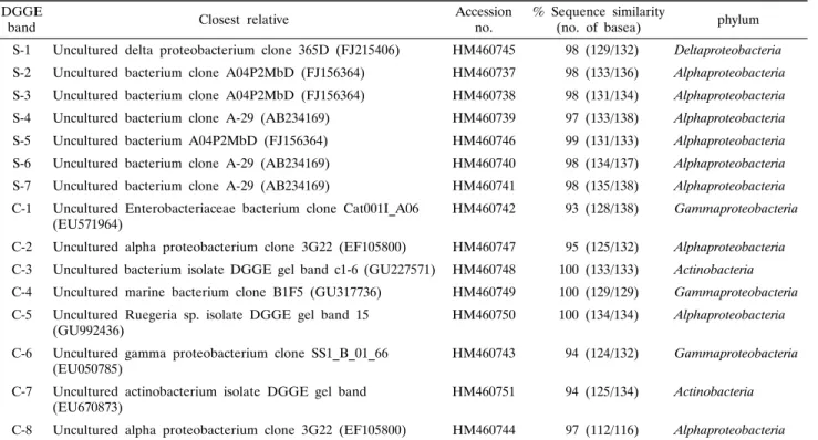
Table 1. Phylogenetic affiliation of re-amplified denaturing gradient gel electrophoresis (DGGE) bands derived from the sponges DGGE
band Closest relative Accession
no. % Sequence similarity
(no. of basea) phylum S-1 Uncultured delta proteobacterium clone 365D (FJ215406) HM460745 98 (129/132) Deltaproteobacteria S-2 Uncultured bacterium clone A04P2MbD (FJ156364) HM460737 98 (133/136) Alphaproteobacteria S-3 Uncultured bacterium clone A04P2MbD (FJ156364) HM460738 98 (131/134) Alphaproteobacteria S-4 Uncultured bacterium clone A-29 (AB234169) HM460739 97 (133/138) Alphaproteobacteria S-5 Uncultured bacterium A04P2MbD (FJ156364) HM460746 99 (131/133) Alphaproteobacteria S-6 Uncultured bacterium clone A-29 (AB234169) HM460740 98 (134/137) Alphaproteobacteria S-7 Uncultured bacterium clone A-29 (AB234169) HM460741 98 (135/138) Alphaproteobacteria C-1 Uncultured Enterobacteriaceae bacterium clone Cat001I_A06
(EU571964) HM460742 93 (128/138) Gammaproteobacteria
C-2 Uncultured alpha proteobacterium clone 3G22 (EF105800) HM460747 95 (125/132) Alphaproteobacteria C-3 Uncultured bacterium isolate DGGE gel band c1-6 (GU227571) HM460748 100 (133/133) Actinobacteria C-4 Uncultured marine bacterium clone B1F5 (GU317736) HM460749 100 (129/129) Gammaproteobacteria C-5 Uncultured Ruegeria sp. isolate DGGE gel band 15
(GU992436) HM460750 100 (134/134) Alphaproteobacteria
C-6 Uncultured gamma proteobacterium clone SS1_B_01_66
(EU050785) HM460743 94 (124/132) Gammaproteobacteria
C-7 Uncultured actinobacterium isolate DGGE gel band
(EU670873) HM460751 94 (125/134) Actinobacteria
C-8 Uncultured alpha proteobacterium clone 3G22 (EF105800) HM460744 97 (112/116) Alphaproteobacteria
a The numbers in parentheses are the part of the total bases used to calculate the levels of sequences similarity. S, S. abata ; C, Cinachyrella sp.
S C
Fig. 1. DGGE banding patterns of amplified 16S rDNA obtained from S. abata (S) and Cinachyrella sp. (C).
결과 및 고찰
DGGE band 양상
제주도의 동일 지역에 서식하는 두 종의 해양 해면 S. abata 와 Cinachyrella sp.의 해면 공생세균의 계통학적 다양성을 알 아보기 위하여 비 배양 지문법인 16S rDNA의 PCR-DGGE를 수행하였다. 해면 시료로부터 추출한 gDNA를 주형으로 16S rRNA gene의 V3 영역을 증폭한 결과, 194 bp의 예상된 크기 의 PCR 산물을 얻었다. 동일 지역에서 같은 시기에 채집한 해 면 S. abata와 Cinachyrella sp.의 공생세균의 DGGE band 패 턴 분석 결과, 해면 S. abata에서 7개, Cinachyrella sp.에서 8 개의 band가 나타나 총 15개의 band를 확인할 수 있었으며 S.
abata와 Cinachyrella sp.의 주요 DGGE band들은 각각의 해 면에 특징적인 것으로 나타났다(Fig. 1). 각각의 해면 종의 다 른 개체에서 주요 DGGE band 패턴은 동일하였으며, 이는 동 일지역에서 채취한 동일 해면 종의 다른 개체인 경우, 동일한 밴드패턴을 나타내는 것으로 보고한 연구(15) 결과와 유사한 것이었다. 두 해면 종의 주요 DGGE band들이 나타내는 세균 군집 다양성을 파악하기 위하여 16S rDNA의 부분 염기서열을 분석하였다.
염기서열 분석 및 계통학적 분석
세균 군집의 다양성을 분석하기 위하여 DGGE band로부터 DNA를 추출하여 341f와 518r을 이용하여 재 증폭하여 염기서 열을 분석하였다. 15개의 DGGE band에 대하여 염기 서열을 결정한 결과, Table 1에 보는 바와 같이 S. abata에서 7개,
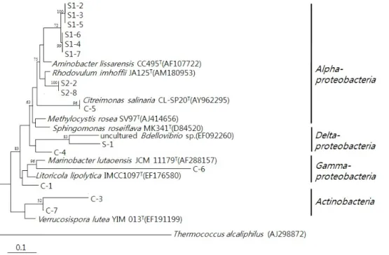
Cinachyrella sp.에서 8개, 총 15개의 주요 band들에 대하여 부분 염기서열을 확인할 수 있었다. 이 서열들을 Blast search 를 통하여 기존의 보고된 서열들과 상동성(similarity)을 검색 하였다. 각각의 DGGE band에서 유래한 염기서열을 이용하여 계통수를 작성한 결과는 Fig. 3과 같다.
두 해면의 DGGE band로부터 결정된 서열들은 기존의 서열 들과 93%에서 100%의 상동성을 나타내었다. DGGE band의 패턴은 다양하였으며, band로부터 밝혀진 모든 서열들은 배양 되지 않은 세균 클론(uncultured bacterial clone)들과 높은 상
S C
Fig. 2. Comparison of predominant bacterial community based on 16S rDNA-V3 in S. abata (S) and Cinachyrella sp. (C).
Actinobacteria Deltaproteobacteria Gammaproteobacteria Alphaproteobacteria 9
8 7 6 5 4 3 2 1 0
Fig. 3. Phylogenetic tree from analysis of 16S rDNA sequences of DGGE bands from S. abata (S) and Cinachyrella sp. (C). Numbers above branches indicate bootstrap values of neighbor-joining analysis (>50%) from 1,000 replicates. The scale bar represents 0.1 substitution per nucleotide position.
동성을 나타내었다. 일반적으로 자연계의 미생물 중 실제 배양 가능한 종은 전체 자연계 미생물 군집의 1% 이하인 것으로 알 려져 있는 바(15)와 같이 본 연구에서 조사된 해면에 존재하는 세균 군집 역시 배양되지 않는 세균 군집이 높은 비율로 존재 함을 알 수 있었다.
S. abata의 주요 공생세균 그룹은 해수 유래의 uncultured Alphaproteobacteria로 밝혀졌으며, 기존의 서열과 93%에서 100%의 상동성을 나타내었다. DGGE band S-1은 uncultured Deltaproteobacteria (FJ215406)와 98%의 상동성을 나타내었 으며, S-2, S-3, S-4, S-5, S-6 그리고 S-7은 uncultured Alphapro- teobacteria (FJ156364, AB234169)와 97%에서 98%의 상동성 을 나타내었다. Cinachyrella sp.의 주요 공생세균 그룹은 uncultured Gammaproteobacteria로 나타났으며 기존의 서열과
93%에서 100%의 상동성을 보였다. DGGE band C-1, C-4와 C-6는 uncultured Gammaproteobacteria (EU571964, GU317736, EU050785)와 93% 이상의 상동성을 나타내었으며, C-2, C-5와 C-8은 uncultured Alphaproteobacteria (EF105800, GU992436) 와 95%에서 100%의 상동성을 나타내었고, C-3과 C-7은 uncultured Actinobacteria (EU670873, GU227571)과 94%와 100%의 상동성을 각각 나타내었다(Table 1).
16S rRNA gene염기서열로부터 계통학적 다양성을 분석한 결과, 두 종의 해면에서 나타난 전체 세균 군집 구조는 S.
abata에서 Alphaproteobacteria와 Deltaproteobacteria가, Cinachyrella sp.에서는 Alphaproteobacteria, Gammaproteo- bacteria 그리고 Actinobacteria를 포함하는 것으로 나타났다.
두 종의 해면에서 공통되는 공생세균 그룹은 Alphaproteo- bacteria였으며 이 세균 그룹은 두 종의 해면 모두에서 우점하 는 것으로 나타났다. 두 해면 종의 공생세균 군집 구조에서 우 점하는 그룹으로 나타난 Proteobacteria 문(phylum)의 세균은 해양환경에서 일반적으로 서식하며, 해양 해면에서도 높은 비 율로 분포하는 것으로 알려져 있다(9).
두 해면은 공통 세균 그룹으로 Alphaproteobacteria를 포함 하고 있으나 Deltaproteobacteria는 오직 S. abata에서, 그리고 Actinobacteria와 Gammaproteobacteria는 Cinachyrella sp.에 서만 관찰되어 두 종의 해면은 서로 다른 공생세균 군집구조를 갖는 것으로 나타났다(Fig. 2). 즉, 해면 종에 따른 미생물 군집 의 차이가 분명하여 두 해면 종의 공통 세균 군집의 존재에도 불구하고 군집 구조는 숙주 특이적(host-specific)인 것으로 파 악된다. 최근 보고된 홍해로부터 채집한 해면 Hyrtios erectus
의 경우 Amphimedon sp.는 동일한 지역에 서식하는 해면 종 이지만 두 종에서 나타나는 세균 군집은 서로 다르며 이차대사 산물에 관련되어 보고된 바 있는 Hyrtios erectus는 Amphimedon sp.에 비해 세균 군집의 다양성이 더 높은 것으로 보고된 바 있다(16). 중국 남지나해에 서식하는 Stelletta tenui, Halichon- dria sp., Dysidea avara, Craniella australiensis의 해면 공생 세균 군집에 관한 연구에서도 Selletta tenui와 Halichondria sp.의 경우 Proteobacteria 문(phylum) 만이 나타난 반면, Dysidea avara와 Craniella australiensis에서는 Proteobacteria 문(phylum)을 포함하여 Flavobacteria, Bacillales, Actinobacteria 가 나타나 해면 종에 따라 세균 군집 구조가 다르다는 연구 결 과가 제시되었다(9). 제주도 운진항에서 2007년 9월에 채집되 어 보고된 S. abata의 공생세균의 군집구조와 2009년 4월에 채집하여 본 연구에서 이용한 S. abata의 경우(15), 주요 우점 세균 군집이 동일한 것으로 확인되었다. 이는 동일한 해면 종 의 개체를 서로 다른 지역에서 복수로 채집하여 공생세균 다양 성을 연구한 결과, 해면 종에 따른 특이적 세균 군집을 나타낸 다는 연구 결과들과도 유사하다(7, 21, 22). 한편 동일 해면일 지라도 온대와 열대의 지리적 분포에 따라 세균군집이 다르거 나(20), 혹은 지리적 분포가 다르고 계통적 유연관계가 먼 해 면에서 유사한 세균군집을 갖는다는(6) 보고도 이루어지고 있 다. 해면 공생미생물의 다양성에 관한 연구는 주로 미생물의 군집구조와 해면 종류에 따른 숙주 특이성 및 공생관계의 안정 성에 관한 측면에서 연구가 수행되었으나(1, 5, 7, 19), 숙주 특 이성에 관한 근거는 아직 명확히 밝혀져 있지 않다. 한편 지역 적 분포를 달리 하는 다양한 해면 종에 관한 미생물 다양성이 밝혀지면서 해면 숙주 특이적인 미생물 외에 해면에 공통적인 미생물 그룹이 존재한다는 보고도 점차 증가하고 있다(6, 14).
해면 공생세균의 다양성에 관한 연구는 주로 열대와 지중해 에 분포하는 해면을 중심으로 이루어져 왔으나(3, 21, 22), 최 근, 중국 남지나해에 분포하는 해면에 관한 연구도 활발히 수 행되고 있다(8, 9, 26). 또한 남극에 서식하는 해면에 관한 보 고(11)도 이루어지고 있으나 우리나라 해역을 포함하여 보다 광범위한 지역의 다양한 해면에 관하여 연구가 수행되어야 할 것으로 사료된다. 특히 우리나라의 해양에 서식하는 해면에 관 한 공생 세균의 군집구조에 관한 보고는 매우 적기 때문에 이 에 관하여 많은 연구가 필요한 실정이다.
본 연구에서는 제주도에 서식하는 두 종의 해면을 같은 시 기에 동일 지역에서 채집하여 DGGE fingerprint를 이용하여 공생세균 군집의 다양성을 살펴본 결과, S. abata와 Cinachyrella sp.의 공생세균 군집은 Alphaproteobacteria를 공통의 우점 세 균 군집으로 포함하나 해면 종에 따라 서로 다른 공생세균 군 집을 나타내어 숙주 특이적 분포를 보이는 것으로 나타났다.
이는 해면 종에 따라 해면에 서식하는 공생세균의 군집 구조가 서로 다르다는 보고(7, 8)와 일치하는 결과로써 해면 종에 따 른 해양 환경에서의 생태적 기능에 따른 결과로 생각된다. 나 아가 본 연구에 사용한 두 종의 해면에 대하여 공생세균 군집 구조를 보다 면밀하게 분석하기 위하여는 다양한 지역으로부 터 동일한 해면 종을 채취하여 분석할 필요가 있을 것으로 사
료된다.
16S rRNA PCR-DGGE 방법은 비교적 신속하게 해면 공생 세균의 군집구조를 분석하기 위한 효과적인 방법이나 DNA 추 출방법에 따라 우점 그룹이 달라진다는 연구결과도 제시되어 있어(4) 연구 방법에 따른 차이를 고려하면서 해양의 환경적 요인과 지리적 요인 등과 연계하여 해면 공생세균 다양성에 관 한 더 많은 연구가 더 수행되어야 할 것으로 사료된다.
적요
2009년 4월 제주도의 운진항에서 채집한 해양 해면 Spirastrella abata와 Cinachyrella sp.의 공생 세균의 주요 군집구조를 16S rDNA-denaturing gradient gel electrophoresis (DGGE) 방법 을 이용하여 비교 분석하였다. S. abata와 Cinachyrella sp.는 각 각 7개와 8개의 DGGE 밴드를 나타내었으며 이들을 적출하여 염기서열을 분석한 결과, NCBI에 등록된 서열들과 92-100%
의 유사도를 나타내었다. S. abata의 주요 공생세균은 Alphaproteo- bacteria와 Deltaproteobacteria에 속하였으며, Cinachyrella sp.
의 경우 Alphaproteobacteria, Gammaproteobacteria와 Actino- bacteria에 속하는 세균으로 구성되었다. 두 종의 해면에서 공 통되는 공생세균 그룹은 Alphaproteobacteria였으며 이 세균 그룹은 두 종의 해면 모두에서 우점하였다. Deltaproteobacteria 는 S. abata에서, Actinobacteria와 Gammaproteobacteria는 Cinachyrella sp.에서만 관찰되었다. 동일지역에서 채집한 서로 다른 두 종의 해면은 각각 다른 공생세균 군집구조를 나타내 었다.
감사의 말
이 논문은 2009년 정부(국토해양부)의 재원으로 한국해양수 산기술진흥원의 지원을 받아 수행된 연구(R20800809H010000110) 이며, 이에 감사 드립니다.
참고문헌
1. Anderson, S.A., P.T. Northcote, and M.J. Page. 2010. Spatial and temporal variability of the bacterial community in different chemotypes of the New Zealand marine sponge Mycale hentscheli.
FEMS Microb. Ecol. 72, 328-342.
2. Cho, H.H. and J.S. Park. 2009. Comparative analysis of the community of culturable bacteria associated with sponges, Spirastrella abata and Spirastrella panis by 16S rDNA-RFLP.
Kor. J. Microbiol. 45, 155-162.
3. Friedrich, A.B., J. Hacker, I. Fischer, P. Proksch, and U.
Hentschel. 2001. Temporal variations of the microbial community associated with the Mediterranean sponge Aplysina aerophoba.
FEMS Microbiol. Ecol. 38, 105-113.
4. Hardoim, C.C., R. Costa, F.V. Araujo, E. Hajdu, R. Peixoto, U.
Lins, A.S. Rosado, and J.D. van Elsas. 2009. Diversity of bacteria in the marine sponge Aplysina fulva in Brazilian coastal waters.
Appl. Environ. Microbiol. 75, 3331-3343.
5. Isaacs, L.T., J. Kan, L. Nguyen, P. Videau, M.A. Anderson, T.L.
Wright, and R.T. Hill. 2009. Comparison of the bacterial communi-
ties of wild and captive sponge Clathria prolifera from the Chesapeake Bay. Mar. Biotechnol. (NY) 11, 758-770.
6. Lafi, F.F., M.J. Garson, and J.A. Fuerst. 2005. Culturable bacterial symbionts isolated from two distinct sponge species (Pseudoceratina clavata and Rhabdastrella globostellata) from the Great Barrier Reef display similar phylogenetic diversity.
Microb. Ecol. 50, 213-220.
7. Lee, O.O., Y.H. Wong, and P.Y. Qian. 2009. Inter- and intraspecific variations of bacterial communities associated with marine sponges from San Juan Island, Washington. Appl. Environ.
Microbiol. 75, 3513-3521.
8. Li, Z., L. He, and X. Miao. 2007. Cultivable bacterial community from South China Sea sponge as revealed by DGGE finger- printing and 16S rDNA phylogenetic analysis. Curr. Microbiol.
55, 465-472.
9. Li, Z.Y., L.M. He, J. Wu, and Q. Jiang. 2006. Bacterial community diversity associated with four marine sponges from the South China Sea based on 16S rDNA-DGGE fingerprinting.
J. Exp. Mar. Biol. Ecol. 329, 75-85.
10. Li, Z.Y. and Y. Liu. 2006. Marine sponge Craniella austrialiensis- associated bacterial diversity revelation based on 16S rDNA library and biologically active actinomycetes screening, phylo- genetic analysis. Lett. Appl. Microbiol. 43, 410-416.
11. Mangano, S., L. Michaud, C. Caruso, M. Brilli, V. Bruni, R.
Fani, and A.L. Giudice. 2009. Antagonistic interactions between psychrotrophic cultivable bacteria isolated from Antarctic sponges:
A preliminary analysis. Res. Microbiol. 160, 27-37.
12. Mohamed, N., J.J. Enticknap, J.E. Lohr, S.M. McIntosh, and R.T.
Hill 2008. Changes in bacterial communities of the marine sponge Mycale laxissima on transfer into aquaculture. Appl. Environ.
Microbiol. 74, 1209-1222.
13. Mohamed, N.M., V. Rao, M.T. Hamann, M. Kelly, and R.T. Hill.
2008. Monitoring bacterial diversity of the marine sponge Ircinia strobilina upon transfer into aquaculture. Appl. Environ. Microbiol.
74, 4133-4143.
14. Muscholl-Silberhorn, A., V. Thiel, and J.F. Imhoff. 2008. Abundance and bioactivity of cultured sponge-associated bacteria from the Mediterranean Sea. Microb. Ecol. 55, 94-106.
15. Park, J.S., J.J. Sim, and K.D. An. 2009. Community structure of bacteria associated with two marine sponges from Juju Island based on 16S rDNA-DGGE profile. Kor. J. Microbiol. 45, 170-176.
16. Radwan, M., A. Hanora, J. Zan, N.M. Mohamed, D.M. Abo-
Elmatty, S.H. Abou-El-Ela, and R.T. Hill. 2010. Bacterial community analyses of two Red Sea sponges. Mar. Biotechnol. 12, 350-360.
17. Ramm, W., W. Schatton, I. Wagner-Dobler, V. Wray, M. Nimtz, H. Tokuda, F. Enjyo, H. Nishino, W. Beil, R. Heckmann, V.
Lurtz, and S. Lang. 2004. Diglucosyl-glycerolipids from the marine sponge-associated Bacillus pumilus strain AAS3: Their production, enzymatic modification and properties. Appl. Microbiol.
Biotechnol. 64, 497-504.
18. Selvin, J. 2009. Exploring the antagonistic producer Streptomyces MSI051: Implications of polyketide synthase gene type II and a ubiquitous defense enzyme phospholipase A2 in the host sponge Dendrilla nigra. Curr. Microbiol. 58, 459-463.
19. Sipkema, D. and H.W. Blanch. 2009. Spatial distribution of bacteria associated with the marine sponge Tethya californiana.
Mar. Biol. 157, 627-638.
20. Taylor, M.W., P.J. Schupp, R. de Nys, S. Kjelleberg, and P.D.
Steinberg. 2005. Biogeography of bacteria associated with the marine sponge Cymbastela concentrica. Environ. Microbiol. 7, 419-433.
21. Thiel, V., S. Leininger, R. Schmaljohann, F. Brummer, and J.F.
Imhoff. 2007. Sponge-specific bacterial associations of the Mediterranean sponge Chondrilla nucula (demospongiae, tetracti- nomorpha). Microb. Ecol. 54, 101-111.
22. Thiel, V., S.C. Neulinger, T. Staufenberger, R. Schmaljohann, and J.F. Imhoff. 2007. Spatial distribution of sponge-associated bacteria in the Mediterranean sponge Tethya aurantium. FEMS Microbiol. Ecol. 59, 47-63.
23. Thoms, C., M. Horn, W. Wagner, U. Hentschel, and P. Proksch.
2003. Monitoring microbial diversity and natural products profiles of the sponge Aplysina cavernicola following trasplantation. Mar.
Biol. 142, 685-692.
24. Weisz, J.B., U. Hentschel, N. Lindquist, and C.S. Martens. 2007.
Linking abundance and diversity of sponge-associated microbial communities to metabolic differences in host sponges. Mar.
Biotechnol. 152, 475-483.
25. Wichels, A., S. Wurtz, H. Dopke, C. Schutt, and G. Gerdts. 2006.
Bacterial diversity in the Breadcrumb sponge Halichondria panicea (pallas). FEMS Microbiol. Ecol. 56, 102-118.
26. Zhang, H., W. Zhang, Y. Jin, M. Jin, and X. Yu. 2008. A comparative study on the phylogenetic diversity of culturable actinobacteria isolated from five marine sponge species. Antonie van Leeuwenhoek 93, 241-248.