Artemisia scoparia Inhibits Adipogenesis in 3T3-L1 Pre-adipocytes by Downregulating the MAPK Pathway
Jung Hwan Oh1, Fatih Karadeniz1,2, Youngwan Seo3,4 and Chang-Suk Kong1,2*
1Department of Food and Nutrition, College of Medical and Life Sciences, Silla University, Busan 46958, Korea
2Marine Biotechnology Center for Pharmaceuticals and Foods, College of Medical and Life Sciences, Silla University, Busan 46958, Korea
3Division of Marine Bioscience, College of Ocean Science and Technology, Korea Maritime and Ocean University, Busan 49112, Korea
4Department of Convergence Study on the Ocean Science and Technology, Ocean Science and Technology School, Korea Maritime and Ocean University, Busan 49112, Korea
Received April 30, 2018 /Revised June 29, 2018 /Accepted July 4, 2018
Obesity is epidemic worldwide and has reportedly been linked to the progression of several metabolic and cardiovascular diseases. The natural products are decreasing the side effects of medicines used for obesity and also have health benefits dut to their numerous bioactive compounds. In this context, Artemisia scoparia is a widespread plant that has been suggested as possessing various types of bioactivity. In this study, the crude extract from A. scoparia (ASE) was tested for its ability to suppress adipogenesis in mouse 3T3-L1 pre-adipocytes. The molecular pathway by which ASE affects differ- entiation of 3T3-L1 cells was also investigated. The introduction of ASE to differentiating 3T3-L1 pre-adipocytes resulted in suppressed adipogenesis, as confirmed by decreased intracellular lipid accumulation. The differentiating cells treated with 10 and 100 μg/ml of ASE showed 21.9 and 29.0%
less lipid accumulation, respectively, than untreated adipocytes. In addition, the results indicated that ASE treatment lowered the expression of the adipogenesis-related factors PPARγ, C/EBPα, and SREBP-1c. Furthermore, treating with ASE notably decreased levels of phosphorylated p38, ERK, and JNK in 3T3-L1 adipocytes. These results indicate that ASE exhibits significant anti-adipogenesis activ- ity by downregulating the MAPK and PPARγ pathways during the differentiation of 3T3-L1 pre- adipocytes. Therefore, A. scoparia may be a potential source of natural products against obesity.
Key words : Adipogenesis, Artemisia scoparia, MAPK, PPARγ, 3T3-L1
*Corresponding author
*Tel : +82-51-999-5429, Fax : +82-51-999-5457
*E-mail : [email protected]
This is an Open-Access article distributed under the terms of the Creative Commons Attribution Non-Commercial License (http://creativecommons.org/licenses/by-nc/3.0) which permits unrestricted non-commercial use, distribution, and reproduction in any medium, provided the original work is properly cited.
Journal of Life Science 2018 Vol. 28. No. 9. 999~1006 DOI : https://doi.org/10.5352/JLS.2018.28.9.999
Introduction
Obesity is characterized as irregular fat metabolism of the body expressed as persistent fat accumulation. Obesity is the main risk factor for most of major diseases such as Type II diabetes [17], heart diseases [15], hypertension [31] and cancer [1]. In metabolic phenotype of obesity, the adipose tissue function is abnormal and affected by several genetic and environmental factors [18, 23]. Triacylglycerols are high- ly efficient sources of energy in the body, and mammals have developed intricate mechanisms to store triacylglycer- ols as fats in adipocytes to minimize the loss of energy [30].
Adipose tissue is formed by specialized cell types called adi-
pocytes, which are capable of storing excessive energy as fat and secreting adipose tissue-specific hormones that affect almost all metabolic pathways of the body [5]. During the onset of obesity, the number of adipocytes rises redundantly as the adipose tissue expanses. The role of adipocytes, there- fore, is gaining increasing interest towards the efforts to pre- vent and treat obesity along other metabolic diseases linked with deteriorated adipocyte function [8]. Adipocyte cells are developed from pre-adipocytes through adipogenesis in- volving the conversion of mesenchymal stem cells (MSCs) to the pre-adipocytes, and differentiation of pre-adipocytes into mature adipocytes.
Differentiation of the pre-adipocytes is strictly regulated by transcription factors and enzymes, mainly peroxisome proliferator-activated receptor γ (PPARγ) and mitogen acti- vated protein kinase (MAPK) pathways [6, 24]. PPARγ is a member of the nuclear-receptor superfamily and has been considered as the master regulator in adipogenesis along CCAAT-enhancer-binding protein α (C/EBPα). Their se- quential activation induces the expression of important pro-
teins and enzymes in order to attain and maintain adipocyte characteristics. Several on the market obesity drugs, acting as PPARγ ligands, target and inhibit this pathway of PPARγ through preventing the upregulation [4]. Recently, consid- erable attention has been directed to development of active ingredients from natural sources with minor side effects and high biocompatibility for preventing and alleviating obesity.
Artemisia scoparia is a widespread plant that belongs to a very large flowering plant family of Asteraceae and grow- ing natively across Eurasia. Leaves and flowers of A. scoparia are referred in traditional medicine sources with activities such as diuretic, antiphlogistic and for treatment of hepatitis [25]. In addition, several studies reported antioxidant, in- secticidal, phytotoxic and anti-inflammatory properties of A.
scoparia as well as chemical constituents derived from it such as essential oils, flavonoids and coumarins [3, 7, 22, 28]. In the present study, therefore, A. scoparia was analyzed to eval- uate its effect on the differentiation of 3T3-L1 adipocytes and possible mechanism of action during adipogenesis.
Materials and Methods
Reagents
Fetal bovine serum (FBS) and Dulbecco’s modified Eagle’s medium (DMEM) were purchased from Gibco BRL (Grand Island, NY). Antibodies for Western blotting were procured from Cell Signaling Technology (Danvers, MA, USA).
Primers for reverse transcription polymerase chain reaction (RT-PCR) were obtained from Bio-RAD (Hercules, CA, USA). Remaining reagents were purchased from Sigma- Aldrich (St. Tropez, MI, USA) unless otherwise specified.
Crude extract
The sample (3 kg) of A. scoparia was air dried and cut into small pieces prior to maceration. Ground sample was extracted twice with methylene chloride (CH2Cl2) for 24 hr at room temperature. The extraction solution was dried in vacuo. The remains were then subjected to extraction again, twice with methanol (MeOH), using the same procedure as above. Lastly, both extracts were combined and used for fur- ther experiments.
Cell culture and adipocyte differentiation
Murine 3T3-L1 pre-adipocytes (ATCC® CL-173TM) were cultured in DMEM supplemented with 10% FBS at 37℃ in a humidified atmosphere of 5% CO2. Differentiation of the
pre-adipocytes was induced with a differentiation mixture containing methylisobutylxanthine (0.5 mM), dexameth- asone (0.25 μM) and insulin (5 μg/ml) in culture medium after 2 days following the confluence as described earlier [11]. This was replaced with DMEM containing 10% FBS supplemented with insulin only (5 μg/ml) after 2 days of incubation. Initial culture medium was introduced after 2 days incubation and replaced with fresh one every 2 days until the maturation of adipocytes. Successful adipocyte dif- ferentiation was confirmed by intracellular lipid droplets ob- served under a light microscope (Nikon Instruments, Tokyo, Japan). Sample was administered to the cell culture medium starting with introduction of differentiation medium and in- cluded in all medium changes. Cytotoxicity level of sample in 3T3-L1 cells was evaluated by MTT assay as previously described [11].
Oil-Red O staining
Accumulation of triglycerides as lipid droplets in 3T3-L1 adipocytes were confirmed by Oil-Red O staining of the tri- glycerides with common staining procedures reported in our previous study [11]. Following the confirmation of adipo- genesis in cultured cells (6-well plate), culture medium was removed, and cells were washed with PBS. Cells were then fixed on wells with 3.7% fresh formaldehyde in PBS at room temperature for 1 hr. Oil-Red O (0.5% w/v) solution (60%
isopropanol and 40% water) was filtered and 1 ml of solution was transferred to the wells. After staining incubation of 1 hr, the Oil-Red O solution was aspired from the plates and the plates were washed with distilled water prior to ob- servation. Images of lipid droplets in 3T3-L1 adipocytes were collected by a Nikon Instruments microscope (Tokyo, Japan). Finally, Oil-Red O stain retained in the cells was elut- ed with 100% isopropanol and quantified by optical absorb- ance value at 500 nm using a microplate reader (Tecan Austria GmbH, Austria).
RT-PCR
Analysis of mRNA levels was carried out by RT-PCR fol- lowing the method from previous reports [14]. Briefly, Total RNA was isolated from 3T3-L1 adipocytes using Trizol re- agent (Invitrogen Co., CA, USA) following the manu- facturer’s instructions. For synthesis of cDNA from total RNA isolates, RNA (1 μg) was added to RNase-free water and oligo (dT), denaturated at 70°C for 5 min and cooled immediately. RNA was reverse transcribed in a master mix
Concentration (μg/ml)
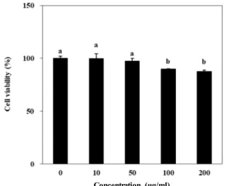
Fig. 1. Effect of A. scoparia crude extract on the viability of 3T3- L1 cells. Cytotoxicity of sample in 3T3-L1 cells was eval- uated by MTT assay. Cell viability following sample treatment was evaluated by ability to form MTT-for- mazan crystals and measured by the absorbance values at 540 nm. Viability of the cells was quantified as a per- centage of untreated control. Values are means ± SD (n=3). a-bMeans with the different letters are significantly different (p<0.05) by Duncan’s multiple range test.
containing 1X RT buffer, 1 mM dNTPs, 500 ng oligo (dT), 140 U M-MLV reserve transcriptase and 40 U RNase in- hibitor at 42°C for 60 min and at 72°C for 5 min using an automatic T100 Thermo Cycler (Bio-Rad, UK). The target gene in synthesized cDNA was amplified using specific fol- lowing sense and antisense primers as following: forward 5'-TTT-TCA-AGG-GTG-CCA-GTT-TC-3' and reverse 5'-AAT- CCT-TGG-CCC-TCT-GAG-AT-3' for PPARγ; forward 5‘- TGT-TGG-CAT-CCT-GCT-ATC-TG-3’ and reverse 5‘-AGG- GAA-AGC-TTT-GGG-GTC-TA-3’ for SREBP-1c; forward 5‘- TTA-CAA-CAG-GCC-AGG-TTT-CC-3’ and reverse 5‘-GGC- TGG-CGA-CAT-ACA-GTA-CA-3’ for C/EBPα; forward 5'- CCA-CAG-CTG-AGA-GGG-AAA-TC-3' and reverse 5'-AA G-GAA-GGC-TGG-AAA-AGA-GC-3' for β-actin. The ampli- fication cycles were set as 95℃ for 45 sec, 60℃ for 1 min and 72℃ for 45 sec. After the completion of 30 cycles, the amplified products were separated on 1.5% agarose gel with electrophoresis for 30 min at 100 V. Gels were then stained with 1 mg/mL ethidium bromide and the bands were vi- sualized by UV light using Davinch-Chemi imagerTM (CAS- 400SM, Seoul, Korea).
Western blotting
Immunoblotting was performed according to standard procedures [11]. Briefly, cells were lysed in RIPA lysis buffer (Sigma–Aldrich Corp., St. Louis, USA) at 4℃ for 30 min.
Cell lysates (25 μg) were separated by 12% SDS-poly- acrylamide gel electrophoresis followed by transfer onto a polyvinylidene fluoride membrane (Amersham Pharmacia Biotech., England, UK). The membrane was then blocked with 5% skim milk and hybridized with primary antibodies (diluted 1:1,000) for 1 hr. After incubation with horse- radish-peroxidase-conjugated secondary antibody at room temperature for 1 hr, protein bands were visualized using a chemiluminescence ECL assay kit (Amersham Pharmacia Biosciences, England, UK) according to the manufacturer's instructions. Membrane images were captured using a Davinch-Chemi imagerTM (CAS-400SM, Seoul, Korea).
Statistical Analysis
The data were presented as mean ± SD. Differences be- tween the means of the individual groups were analyzed using the analysis of variance (ANOVA) procedure of Statistical Analysis System, SAS v9.1 (SAS Institute, Cary, NC, USA) with Duncan’s multiple range tests. The signifi- cance of differences was defined at the p<0.05 level.
Results and Discussion
Crude extract of A. scoparia (ASE) was used for evaluation of the anti-adipogenesis effect. Prior to in vitro experiments using mouse pre-adipocyte 3T3-L1 cells, any cytotoxic prop- erties of ASE were analyzed with MTT formazan assay. The cells were treated with or without ASE in various concen- trations (10, 50, 100, 200 μg/ml). For tested concentrations, ASE did not exhibit any significant cytotoxicity as expected (Fig. 1). Results from MTT assay revealed that ASE is safe to be used in vitro experiments up to 100 μg/ml, which was decided as the upper limit of treatment concentration.
3T3-L1 pre-adipocytes were differentiated with differ- entiation medium treatment in the absence or presence of ASE. Differentiation medium was changed with feeding me- dium containing insulin only at day 2 and medium was re- placed with fresh one every 2 days. Under these conditions, it was assumed that adipocytes were fully matured by day 6. In order to confirm that the cells were fully differentiated and ready for further experiments, intracellular lipid accu- mulation, an indicator for successful adipogenesis was con- trolled under a light microscope. At day 6, 70 to 90% of the ASE-untreated control cells were showed adipocyte char- acteristics by accumulating lipid droplets and accepted as successfully differentiated into mature adipocytes. There-
A. scoparia (μg/ml)
A. scoparia (μg/ml)
Fig. 2. Effect of A. scoparia crude extract on the intracellular lipid accumulation of the differentiated 3T3-L1 cells. Cells were induced to adipogenesis with a differentiation mix- ture with different concentrations (10 and 100 μg/ml).
Following the successful differentiation of the cells, intra- cellular lipid droplets were stained by Oil-Red O staining and images were taken. Lipid accumulation was meas- ured by the absorbance values of the eluted stain from cells at 500 nm. Accumulated lipid droplets were quanti- fied as a percentage of the fully differentiated untreated control cells. Values are means ± SD (n=3). a-eMeans with the different letters are significantly different (p<0.05) by Duncan’s multiple range test.
fore, it was accepted to be sufficient to carry out further experiments. Progression of obesity occurs with redundant accumulation of lipid by adipose tissue. During the for- mation of new adipocytes through adipogenesis, ability to accumulate lipid droplets is the main factor that marks the successful adipogenic differentiation [10]. Accordingly, one of the main approaches to prevent and treat obesity is in- hibition of fat storage in adipocytes [19]. In this respect, lipid accumulation of differentiated cells under ASE treatment was assessed by staining the lipid droplets with Oil Red O staining assay. As seen in Fig. 2, the amount of lipid droplets in ASE treated cells was decreased gradually in a dose-de- pendent manner. Staining of lipid droplets was quantified by elution of the Oil Red O stain that was bind to the lipid droplets. As expected from cell images, amount of stain bound by intracellular lipid was relatively low in ASE-treat- ed cells compared to untreated control. Ability of ASE to decrease the storage of triglycerides in differentiating pre-
adipocytes was regarded as a marker for intervening the ei- ther adipogenesis or lipid accumulation pathways of 3T3-L1 cells. Additionally, inhibiting the possible fat storage of adi- pocytes suggested that ASE might contain substances that were able to act against obesity-related characteristics of the adipose tissue.
The adipocyte differentiation is comprehensively studied and reported to be activated and regulated by a complicated signaling cascades of transcription factors and enzymes.
PPARs, especially PPARγ, are ligand-activated transcription factors and reported to play pivotal roles in energy metabo- lism and, in this context, in differentiation of adipocytes [6].
Along PPARs, C/EBPs are also key proteins in the pathway that activate PPARγ resulting in the progression and matura- tion of differentiating pre-adipocytes. Following the in- troduction of adipogenic stimuli, activated C/EBPα induces the further activation and expression of PPARγ, which se- quentially progress differentiation of adipocytes and regu- late insulin sensitivity in mature adipocytes. During final steps of adipogenesis another transcription factor, SREBP-1c, is closely linked with fatty acid metabolism where the stor- age of fat in differentiated adipocytes occur [26]. The effect of ASE on the PPARγ pathway regulation of adipogenesis, was analyzed by reverse transcription polymerase chain re- action and immunoblotting of PPARγ, C/EBPα and SREBP- 1c for gene and protein expression, respectively.
At the beginning of the differentiation 3T3-L1 cells were treated with different concentrations of ASE (10, 50 and 100 μg/mL) during the differentiation period and its effect was observed after the maturation into adipocytes. The mRNA expression levels of PPARγ, C/EBPα and SREBP-1c were all increased in the mature adipocytes compared to non-differ- entiated pre-adipocytes (Fig. 3A). Treatment with ASE dose-dependently suppressed the mRNA levels. At the con- centration of 100 μg/ml, ASE lowered the mRNA expre- ssion levels of PPARγ, C/EBPα and SREBP-1c to 71.9, 71.9 and 54.4% of the untreated control cells, respectively. These results suggested that ASE was able to hinder the activation of PPARγ pathway which resulted in the suppressed adipo- genesis in 3T3-L1 cells. In order to strengthen this sugges- tion, protein levels of transcription factors were evaluated by immunoblotting. Similarly, presence of ASE ameliorated the overexpressed levels of adipogenesis markers. Following ASE treatment of 100 μg/ml, PPARγ, C/EBPα and SREBP- 1c protein levels were lowered by 68.7, 55.5 and 68.2% com- pared to untreated control cells (Fig. 3B). Combines results
A B
A. scoparia (μg/ml) A. scoparia (μg/ml)
A. scoparia (μg/ml) A. scoparia (μg/ml)
Fig. 3. Effect of A. scoparia crude extract on the expression of key adipogenesis transcription factors; PPARγ, SREBP-1c and C/EBPα:
(A) Effect of the sample on the mRNA levels of the transcription factors was analyzed by RT-PCR. Regulation of the mRNA levels was quantified by the density of the bands and normalized against the housekeeping gene β-actin. Effect of the sample on mRNA levels was given as the percentage of the fully differentiated untreated control cells; (B) Effect of the sample on the protein levels of the transcription factors was analyzed by Western blotting. The protein levels was quantified by the density of the bands and normalized against the housekeeping protein β-actin. Effect of sample on protein levels was given as the percentage of the fully differentiated untreated control cells. Values are means ± SD (n=3). a-eMeans with the different letters are significantly different (p<0.05) by Duncan’s multiple range test.
were confirmed that ASE possessed ability to intervene with the PPARγ pathways and lowered the expression of asso- ciated crucial proteins.
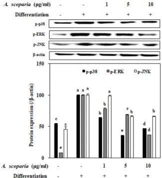
Various transcription factors like PPARγ and C/EBP fam- ily are involved in MAPK pathways during adipocyte differ- entiation [2]. In order to provide insights towards the action mechanism during inhibitory effect of AES, activation levels of other crucial MAPK cascade proteins, namely p38, ERK and JNK, were observed. Effect of AES treatment (1, 5 and 10 μg/mL) on the phosphorylated (p-) p38, ERK and JNK levels were evaluated by protein levels through Western blotting. In fully differentiated mature adipocytes these pro- tein levels were increased significantly while AES treatment suppressed the levels substantially in a dose-dependent manner (Fig. 4). Adipogenesis of 3T3-L1 cells resulted in ele- vated levels of activated p38, ERK and JNK compared to non-differentiated blank cells. Treatment with AES lowered the levels of phosphorylated MAPK proteins to 46.4, 36.3 and 65.6% of the untreated fully differentiated control cells for p38, ERK and JNK, respectively.
Natural products, especially the plants from traditional
medicine references are gaining much interest in alternative and complementary medicine studies due to their various benefits such as few side effects and high biocompatibility.
In this context, A. scoparia is a widely distributed plant that has been reported to possess several bioactivities including but not limited to antimicrobial, anti-inflammatory, anti- oxidant and antihepatotoxic [7, 22, 28]. Current results in- dicated that A. scoparia suggestively disrupts the differ- entiation of pre-adipocytes into adipocytes. For prevention and treatment of obesity, hindering the expansion ability of the adipose tissue by means of stopping formation of new adipocytes is a viable target. Several PPARγ ligands that act as adipogenesis suppressors are on the market for their ben- eficial effects against obesity [21]. Effect of A. scoparia on adi- pogenesis in 3T3-L1 adipocytes indicated a promising chem- ical composition that may yield substances that can be used natural products against obesity. Previous phytochemical in- vestigations of A. scoparia resulted in the isolation of numer- ous chemical compounds such as coumarins, essential oils and flavonoids where all of them were previously reported adipogenesis inhibitory substances from different sources [9,
A. scoparia (μg/ml)
A. scoparia (μg/ml)
Fig. 4. Effect of A. scoparia crude extract on the protein levels of the phosphorylated (p-) MAPK pathway proteins p38, ERK and JNK analyzed by Western blotting. The protein levels was quantified by the density of the bands and normalized against the housekeeping protein β-actin.
Effect of sample on protein levels was given as the per- centage of the fully differentiated untreated control cells.
Values are means ± SD (n=3). a-eMeans with the different letters are significantly different (p<0.05) by Duncan’s multiple range test.
13].
Effect of A. scoparia was suggested to be raised from its content of flavonoids and essential oils, as current results were also exhibited an inhibitory effect on the MAPK path- way during adipogenesis similar to that of flavonoids and essential oils, observed by suppressed levels of PPARγ, SREBP1c and C/EBPα along with activated levels of key MAPK proteins p38, ERK and JNK. A study by Cha et al.
[3] reported that A. scoparia and A. capillaris were rich in essential oils. Study showed that these species dominantly contain camphor (11.0%), 1,8-cineole (21.5%), and β-caryo- phyllene (6.8%) as the major essential oils Among them, β -caryophyllene [16] and camphor [29] were suggested to in- hibit adipogenesis via similar pathway suggested in this study. Another study by Xie et al. [32] investigated the chemical composition of A. scoparia and isolated 7-methox- ycoumarine, isosabandin, 6,7-dimethylesculetin, 7-methyl- esculetin, scopoletin, capillarisin, chlorogenic acid butyl es- ter, isoscopoletin-β-D-glucoside and β-sitosterol. Yang et al.
[33] and Shin et al. [27] reported adipogenesis inhibitory ef- fects of similar compounds from different sources in 3T3-L1 cells. Using A. scoparia, Nam et al. [20] isolated and charac- terized 3,5-dicaffeoyl-epi-quinic acid as an active compound against inflammation. Kim et al. [12] showed that same com- pound isolated from Ainsliaea acerifolia exerted a strong an- ti-obesity effect in 3T3-L1 cells. In this context, the anti-adi- pogenesis effect of A. scoparia extracts in current study may be credited to its chemical composition of bioactive essential oils and derivatives of coumarines and flavonoids. There- fore, further studies to elucidate the possible active con- stituents of A. scoparia are urged. Nevertheless, the results presented here showed that A. scoparia was a potential source of natural products that may be used to reduce obe- sity by preventing the forming of adipocytes and hence, re- ducing the fat accumulation.
Acknowledgment
This research was supported by Basic Science Research Program through the National Research Foundation of Korea (NRF) funded by the Ministry of Education (NRF- 2017R1A2B4009588).
References
1. Anderson, A. S. and Caswell, S. 2009. Obesity management - an opportunity for cancer prevention. Surgeon 7, 282-285.
2. Bost, F., Aouadi, M., Caron, L. and Binétruy, B. 2005. The role of MAPKs in adipocyte differentiation and obesity.
Biochimie 87, 51-56.
3. Cha, J. D., Jeong, M. R., Jeong, S. I., Moon, S. E., Kim, J.
Y., Kil, B. S. and Song, Y. H. 2005. Chemical composition and antimicrobial activity of the essential oils of Artemisia scoparia and A. capillaris. Planta Med. 71, 186-190.
4. Choi, J. H., Banks, A. S., Estall, J. L., Kajimura, S., Bostrom, P., Laznik, D., Ruas, J. L., Chalmers, M. J., Kamenecka, T.
M., Bluher, M., Griffin, P. R. and Spiegelman, B. M. 2010.
Obesity-linked phosphorylation of PPARγ by cdk5 is a di- rect target of the anti-diabetic PPARγ ligands. Nature 466, 451-456.
5. de Ferranti, S. and Mozaffarian, D. 2008. The perfect storm:
Obesity, adipocyte dysfunction, and metabolic consequences.
Clin. Chem. 54, 945-955.
6. Fajas, L., Fruchart, J. C. and Auwerx, J. 1998. Transcriptional control of adipogenesis. Curr. Opin. Cell Biol. 10, 165-173.
7. Habib, M. and Waheed, I. 2013. Evaluation of anti-noci- ceptive, anti-inflammatory and antipyretic activities of Arte- misia scoparia hydromethanolic extract. J. Ethnopharmacol.
145, 18-24.
8. Hajer, G. R., van Haeften, T. W. and Visseren, F. L. J. 2008.
Adipose tissue dysfunction in obesity, diabetes, and vas- cular diseases. Eur. Heart J. 29, 2959-2971.
9. Hsu, C. L. and Yen, G. C. 2007. Effects of flavonoids and phenolic acids on the inhibition of adipogenesis in 3T3-L1 adipocytes. J. Agric. Food Chem. 55, 8404-8410.
10. Kawada, T., Takahashi, N. and Fushiki, T. 2001. Biochemical and physiological characteristics of fat cell. J. Nutr. Sci.
Vitaminol. 47, 1-12.
11. Kim, J., Karadeniz, F., Ahn, B., Kwon, M. S., Mun, O., Bae, M. J., Seo, Y., Kim, M., Lee, S., Kim, Y. Y., Mi Soon, J. and Kong, C. 2016. Bioactive quinone derivatives from the ma- rine brown alga Sargassum thunbergii induce anti‐adipo- genic and pro‐osteoblastogenic activities. J. Sci. Food Agric.
96, 783-790.
12. Kim, T., Jo, C., Kim, H. S., Park, Y. M., Wu, Y. X., Cho, J. H. and Kim, T. H. 2016. Chemical constituents from Ainsliaea acerifolia as potential anti-obesity agents. Phytochem.
Lett. 16, 146-151.
13. Kim, Y. and Lee, J. 2015. Esculetin, a coumarin derivative, suppresses adipogenesis through modulation of the AMPK pathway in 3T3-L1 adipocytes. J. Func. Foods 12, 509-515.
14. Kim, J. A., Karadeniz, F., Ahn, B. N., Kwon, M. S., Mun, O. J., Kim, M., Lee. S. H., Yu, K. H., Kim, Y. Y. and Kong, C. S. 2014. Sargassum sp. attenuates oxidative stress and sup- presses lipid accumulation in vitro. J. Life Sci. 24, 274-283.
15. Lavie, C. J., Milani, R. V. and Ventura, H. O. 2009. Obesity and cardiovascular disease: Risk factor, paradox, and impact of weight loss. J. Am. College Cardiol. 53, 1925-1932.
16. Lee, M. H., Chen, Y. Y., Tsai, J. W., Wang, S. C., Watanabe, T. and Tsai, Y. C. 2011. Inhibitory effect of β-asarone, a com- ponent of Acorus calamusessential oil, on inhibition of adipo- genesis in 3T3-L1 cells. Food Chem. 126, 1-7.
17. Malnick, S. D. and Knobler, H. 2006. The medical complica- tions of obesity. QJM. 99, 565-579.
18. Marti, A., Martinez-Gonzalez, M. A. and Martinez, J. A.
2008. Interaction between genes and lifestyle factors on obesity. Proc. Nutr. Soc. 67, 1-8.
19. Mcpherron, A. and Lee, S. 2002. Suppression of body fat accumulation in myostatin-deficient mice. J. Clin. Investig.
109, 595-602.
20. Nam, S. Y., Han, N. R., Rah, S. Y., Seo, Y., Kim, H. M. and Jeong, H. J. 2017. Anti-inflammatory effects of Artemisia sco- paria and its active constituent, 3,5-dicaffeoyl-epi-quinic acid against activated mast cells. Immunopharm. Immunotoxicol.
40, 52-58.
21. Nawrocki, A. R. and Scherer, P. E. 2005. Keynote review:
the adipocyte as a drug discovery target. Drug Discov. Today 10, 1219-1230.
22. Negahban, M., Moharramipour, S. and Sefidkon, F. 2006.
Chemical composition and insecticidal activity of Artemisia scoparia essential oil against three coleopteran stored-prod- uct insects. J. Asia-Pac. Entomol. 9, 381-388.
23. Ordovas, J. M. and Shen, J. 2008. Gene–environment inter- actions and susceptibility to metabolic syndrome and other chronic diseases. J. Periodontol. 79, 1508-1513.
24. Otto, T. C. and Lane, M. D. 2005. Adipose development:
from stem cell to adipocyte. Crit. Rev. Biochem. Mol. Biol.
40, 229-242.
25. Park, J. H. 1999. Korean Folk Medicine pp. 68, Busan National University Publisher: Busan, Korea.
26. Pettinelli, P. and Videla, L. A. 2011. Up-regulation of PPAR- γ mRNA expression in the liver of obese patients: An addi- tional reinforcing lipogenic mechanism to SREBP-1c in- duction. J. Clin. Endocrinol. Metab. 96, 1424-1430.
27. Shin, E., Choi, M. K., Yoo, H. S., Lee, C. K., Hwang, B. Y.
and Lee, M. K. 2010. Inhibitory effects of coumarins from the stem barks of Fraxinus rhynchophylla on adipocyte differ- entiation in 3T3-L1 cells. Biol. Pharm. Bull. 33, 1610-1614.
28. Singh, H. P., Mittal, S., Kaur, S., Batish, D. R. and Kohli, R. K. 2009. Chemical composition and antioxidant activity of essential oil from residues of Artemisia scoparia. Food Chem. 114, 642-645.
29. Song, Y., Lee, S. J., Jang, S. H., Kim, T. H., Kim, H. D., Kim, S. W., Won, C. K. and Cho, J. H. 2017. Annual wormwood leaf inhibits the adipogenesis of 3T3-L1 and obesity in high-fat diet-induced obese rats. Nutrients 9, 554.
30. Sun, K., Kusminski, C. M. and Scherer, P. E. 2011. Adipose tissue remodeling and obesity. J. Clin. Investig. 121, 2094- 2101.
31. Wofford, M. R. and Hall, J. E. 2004. Pathophysiology and treatment of obesity hypertension. Curr. Pharm. Des. 10, 3621-3637.
32. Xie, T., Liang, J. Y., Liu, J., Wang, M., Wei, X. L. and Yang, C. H. 2004. Chemical study on Artemisia scoparia. J. China Pharm. Univ. 35, 401-403.
33. Yang, Y., Yang, X., Xu, B., Zeng, G., Tan, J., He, X., Hu, C. and Zhou, Y. 2014. Chemical constituents of Morus alba L. and their inhibitory effect on 3T3-L1 preadipocyte pro- liferation and differentiation. Fitoterapia 98, 222-227.
초록:비쑥 추출물이 3T3-L1 지방세포 분화 및 MAPK 신호 전달 경로에 미치는 영향
오정환1․파티 카라데니즈2․서영완3,4․공창숙1,2*
(1신라대학교 의생명과학대학 식품영양학과, 2신라대학교 해양식의약소재융합기술연구소, 3한국해양대학교 해양
과학기술대학 해양환경·생명과학부, 4한국해양대학교 해양과학기술전문대학 해양과학기술융합학과)
비쑥(Atermisia scoparia)은 국화과에 속하는 한해살이 풀로서 유라시아 지역 분포하며, 염생습지에 자생하는 염 생식물의 일종이다. 비쑥은 민간요법에서 이뇨제, 소염제, 간염치료제로 사용되어 왔으며, 비쑥에서 분리한 플라 보노이드, 쿠마린 화합물의 항산화, 항염증 등의 생리활성이 보고되어 있다. 본 연구에서는 비쑥 추출물이 3T3-L1 지방전구세포 모델에서 지방세포 내에서의 중성지방 생성 및 지방세포 분화조절 인자 발현에 미치는 영향을 검토 하였다. 지방전구세포 3T3-L1을 지방세포로 분화하여 Oil Red O 염색법으로 지방세포 분화 정도를 측정한 결과, 비쑥 추출물 처리군에서 농도 의존적으로 지방세포 형성이 억제되었다. 또한 지방세포 분화 관련 인자인 PPARγ, C/EBPα, SREBP-1c의 발현을 mRNA 및 단백질 수준에서 확인한 결과, 비쑥 추출물을 처리한 군에서 지방세포 분화 인자 발현이 감소하는 것으로 나타났다. 지방세포 분화 및 증식에 관여하는 것으로 알려져 있는 MAPK 신호 전달 경로를 확인한 결과 비쑥 추출물을 처리한 군에서 p38, ERK, JNK의 인산화가 억제되었다. 이를 통해 비쑥 추출물은 MAPK 신호전달 경로를 통한 지방세포 분화 인자 조절을 통해 지방 생성과 합성을 억제하는 것으로 사료된다. 따라서 본 연구 결과로부터 비쑥 추출물의 MAPK 신호전달 경로 억제를 통한 항비만 효과를 확인하였 으며, 나아가 건강 기능성 식품 소재로서의 개발 가능성이 기대된다.