Microtubule-damaging Chemotherapeutic Agent-mediated Mitotic Arrest and Apoptosis Induction in Tumor Cells
Do Youn Jun and Young Ho Kim*
Laboratory of Immunobiology, School of Life Science and Biotechnology, College of Natural Sciences, Kyungpook National University, Daegu 41566, Korea
Received March 18, 2016 /Revised March 25, 2016 /Accepted March 29, 2016
Apoptosis induction has been proposed as an efficient mechanism by which malignant tumor cells can be removed following chemotherapy. The intrinsic mitochondria-dependent apoptotic pathway is fre- quently implicated in chemotherapy-induced tumor cell apoptosis. Since DNA-damaging agent (DDA)- induced apoptosis is mainly regulated by the tumor suppressor protein p53, and since more than half of clinical cancers possess inactive p53 mutants, microtubule-damaging agents (MDAs), of which apop- totic effect is mainly exerted via p53-independent routes, can be promising choice for cancer chemo- therapy. Recently, we found that the apoptotic signaling pathway induced by MDAs (nocodazole, 17α- estradiol, or 2-methoxyestradiol) commonly proceeded through mitotic spindle defect-mediated prom- etaphase arrest, prolonged Cdk1 activation, and subsequent phosphorylation of Bcl-2, Mcl-1, and Bim in human acute leukemia Jurkat T cells. These microtubule damage-mediated alterations could render the cellular context susceptible to the onset of mitochondria-dependent apoptosis by triggering Bak activation, Δψm loss, and resultant caspase cascade activation. In contrast, when the MDA-induced Bak activation was inhibited by overexpression of anti-apoptotic Bcl-2 family proteins (Bcl-2 or Bcl-xL), the cells in prometaphase arrest failed to induce apoptosis, and instead underwent mitotic slippage and endoreduplication cycle, leading to formation of populations with 8N and 16N DNA content.
These data indicate that cellular apoptogenic mechanism is critical for preventing polyploid formation following MDA treatment. Since the formation of polyploid cells, which are genetically unstable, may cause acquisition of therapy resistance and disease relapse, there is a growing interest in developing new combination chemotherapies to prevent polyploidization in tumors after MDA treatment.
Key words : Apoptotic cell death, endoreduplication, microtubule-damaging agents, polyploid Formation, prometaphase arrest
*Corresponding author
*Tel : +82-53-950-5378, Fax : +82-53-955-5522
*E-mail : [email protected]
This is an Open-Access article distributed under the terms of the Creative Commons Attribution Non-Commercial License (http://creativecommons.org/licenses/by-nc/3.0) which permits unrestricted non-commercial use, distribution, and reproduction in any medium, provided the original work is properly cited.
Journal of Life Science 2016 Vol. 26. No. 3. 376~386 DOI : http://dx.doi.org/10.5352/JLS.2016.26.3.376
Introduction
Significant advances have recently been made in our un- derstanding on cancer biology, techniques for early diag- nosis of cancer, and treatment modalities such as surgery, radiation, and chemotherapy. Cancer, however, is still a leading cause of mortality and morbidity of worldwide, ac- counting for 8.2 million deaths or 14.6% of all human death in 2012 [79]. Since cancer arises from one single cell, the final goal of the treatment regimen for the cancer patient should be to remove all cancer cells throughout the whole body.
Treatment for cancer patients comprises, in general, one or
more modalities including radiation therapy, surgery, and chemotherapy. While chemotherapy is employed either alone or in combination with radiotherapy and/or surgery depending on the cancer status, the efficacy of chemotherapy in cancer patients is mainly exerted by the cytostatic and cytotoxic effects of chemotherapeutic drugs on tumor cells, which can systemically react with tumor cells.
Because one of the main characteristics of tumor cells is uncontrolled growth and proliferation resulting from defects in cell cycle regulation, a number of anticancer drugs have been developed to target cell cycle control mechanism [10, 23, 29, 44, 63]. Mammalian cells are known to duplicate their contents and then divide into two cells via completing the cell cycle, which is proceeded by four successive stages such as the periods of DNA synthesis (S phase) and mitosis (M phase) separated by gaps called G1 and G2 phases. Progre- ssion through each phase in dividing cells is controlled by the sequential activation and inactivation of a series of cy- clin-dependent kinases (Cdks) [33]. To ensure the fidelity of
- Review -
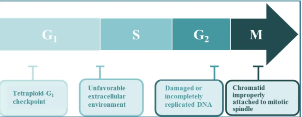
Fig. 1. Mammalian cell cycle progression and checkpoints.
cell division, the passage through a series of cell cycle check- points that act as molecular breaks to verify the accuracy of cell cycle progression is required [54]. Three major check- points such as the G1 checkpoint, the G2 checkpoint, and the mitotic spindle assembly checkpoint are involved in the cell cycle control (Fig. 1). The G1 checkpoint functions in G1
phase to confirm that the environment is favorable for com- mitting to S phase, the G2 checkpoint functions in G2 phase and prevents entry into mitosis until DNA damage is re- paired and DNA replication is completed, and the mitotic spindle assembly checkpoint functions during mitosis and ensures proper attachment of the replicated chromosomes to the mitotic spindles [7, 8, 19, 39]. Although the existence of tetraploid checkpoint remains controversial, several stud- ies have shown that mammalian cells have a p53-dependent tetraploid checkpoint that blocks cell cycle progression in the G1 phase in response to failure of cytokinesis during cell division. During G1 or G2 checkpoint activation, a combina- tion of several distinct molecular mechanisms operates to negatively regulate Cdk activity. These include increased cy- clin destruction, decreased cyclin gene expression, Cdk in- hibition by Cdk inhibitors (CKIs; p21CIP1/WAF1, p27KIP1, and p16INK4A) that bind and inactivate Cdk/cyclin complexes, and Cdc25 inactivation. Additionally, when the mitotic spin- dle assembly checkpoint becomes activated, Cdk1/cyclin B is kept in an active state by inhibiting the role of the ana- phase-promoting complex (APC) in order to prevent the on- set of anaphase. While checkpoint activation delays or halts cell cycle progression, the repair mechanisms operate to re- cover cellular integrity. However, if the damage is beyond repair, the cells generate a signal to undergo apoptosis [1, 32].
The efficacy of chemotherapy that targets cell cycle regu- lation depends on the status of cell cycle progression of tu- mor cells, because chemotherapeutic agents mainly impact
on actively proliferating cells that are in the S phase or M phase rather than cells in the resting (G0) phase. However, many chemotherapeutic drugs fail to distinguish the differ- ence between proliferating cells of normal tissues and tumor cells, and thus may cause normal cell damage which results in side-effects. With respect to the efficacy of chemotherapy, which induces cancer cell death, several biochemical mecha- nisms including apoptosis, necrosis, and autophagy are im- plicated [3, 47, 61]. Autophagy is known to be a catabolic degradation process that proceeds by sequestering un- necessary or dysfunctional cellular components via the for- mation of double-membrane vesicles (autophagosomes), and targeting them for degradation via the fusion of autophago- somes with lysosomes to generate single-membrane autoly- sosomes. A number of studies have reported that autophagic response is enhanced in tumor cells than in normal cells, which allows tumor cells to survive under inadequate con- ditions such as nutritional deprivation and chemotherapy [17]. In addition, since tumor cells can activate autophagy in response to cellular stress associated with chemo- therapeutic treatment, it has been suggested that inhibition of autophagy is an effective chemotherapeutic approach to accelerate tumor cell death [36]. The mechanism of chemo- therapeutic drug-induced cell death needs to be studied fur- ther in order to clarify whether the antitumor effect of the drug is confined to tumor cells rather than normal cells.
These efforts will improve the survival rates of cancer pa- tients receiving chemotherapy [53].
Chemotherapy-induced apoptosis in tumor cells results in their own destruction into apoptotic bodies that can be cleared by phagocytic cells without accompanying a local inflammatory response, whereas necrosis in tumor cells leads to release of their intracellular contents that possibly provoke a damaging inflammatory response. In this context, apoptotic cell death has been proposed as an efficient mecha-
nism by which malignant tumor cells can be removed fol- lowing chemotherapeutic treatments [30, 40]. In chemo- therapeutic drug-induced apoptosis of tumor cells, three dif- ferent death signaling pathways, such as the extrinsic death receptor-dependent pathway [74], the intrinsic mitochon- dria-dependent pathway [18] and the intrinsic endoplasmic reticulum (ER) stress-mediated pathway [55], are likely to be implicated. The intrinsic mitochondria-dependent path- way is most frequently associated with tumor cell apoptosis caused by chemotherapeutic drugs that are DNA-damaging and microtubule-damaging agents [49].
Since numerous studies have reported that the apoptosis induction of tumor cells following treatment with DNA- damaging agents (DDAs) is associated with the regulatory role of tumor suppressor protein p53, and since more than half of clinical cancers possess mutations in the p53 gene so that p53 cannot be functional as tumor suppressor pro- tein, it is reasonable to assume that microtubule-damaging agents (MDAs), of which cytotoxic effect can be exerted in a p53-independent manner, are better choice for chemo- therapeutic treatment of cancers with p53 mutations [26, 35, 62].
In this review, the molecular and cellular mechanisms un- derlying MDA-induced apoptosis of tumor cells are discussed. These understandings will provide new insights and opportunities for better treatment of cancer patients as well as for drug development.
MDA-induced cell cycle arrest is the prometaphase arrest
The segregation of the replicated chromosomes during M phase of the cell cycle is governed by a complex cytoskeletal structure of microtubules and associated proteins, which is known as the mitotic spindle [24, 76]. These microtubules are highly dynamic polymers, composed of α- and β-tubulin heterodimers, and they tend to switch between states of rap- id growth and rapid shrinkage in a situation of dynamic instability [20, 71]. Microtubule dynamics increases sig- nificantly during mitosis, compared with interphase, to ach- ieve the proper alignment of the duplicated chromosomes at the metaphase plate and accurate segregation of the chro- mosomes to the daughter cells. Based on the important role of microtubule dynamics in controlling mitosis, micro- tubules have been considered a critical target for the devel- opment of anticancer drugs [51, 52, 64]. Among the MDAs, the microtubule-polymerizing drugs such as paclitaxel and
docetaxel promote microtubule assembly and stabilize mi- crotubules, whereas the microtubule-depolymerizing drugs including vinblastine, colchicine and nocodazole inhibit mi- crotubule assembly and promote depolymerization. Tumor cells treated with these MDAs are known to commonly ex- hibit loss of microtubule function, leading to the disruption of mitotic spindle, mitotic arrest of the cell cycle, and mitotic catastrophe-mediated apoptotic cell death [38, 52, 64].
The M phase of the cell cycle can be subdivided into the prophase, the prometaphase, the metaphase, the anaphase, and the telophase, based on the microscopic characteristics of the microtubule organization, chromosomes, and nuclear envelop [26, 58]. The sophisticated regulation of Cdk1 activ- ity is known to be critical for the G2/M transition and M phase-progression of the cell cycle. When cells enter into the prophase from the G2 phase, the Cdk1/cyclin B complex, which is held in an inactive state by phosphorylation at Thr-14 and Tyr-15 mediated by the kinases Myt1 and Wee1, becomes activated as a result of phosphorylation at Thr-161 by the Cdk-activating kinase (Cak) as well as dephosphor- ylation at Thr-14 and Tyr-15 by the phosphatase Cdc25 [21, 75]. The activated Cdk1 phosphorylates a large number of substrates that are associated with nuclear envelope break- down, centrosome separation, spindle assembly, and chro- mosome condensation [34]. Subsequently, the activated Cdk1 should be inactivated through the degradation of cy- clin B by anaphase promoting complex (APC) to progress from the metaphase to the anaphase.
In relation to the chemotherapy-mediated G2/M arrest ac- companying apoptosis in tumor cells, two different molec- ular mechanisms, the G2 checkpoint pathway and the mitotic spindle assembly checkpoint pathway, have been implicated [8, 39, 43]. Following exposure to DDAs, the cells can be arrested at the G2 checkpoint by inactivating Cdk1/cyclin B through the action of Wee1 and Cdc25, and by p53-medi- ated transcriptional repression of cyclin B. When the cells being treated with MDAs fail to align all the chromosomes on the metaphase plate, the mitotic spindle assembly check- point becomes activated to prevent the onset of anaphase and Cdk1/cyclin B is kept in an active state by inhibiting the role of APC. As the mechanism underlying MDA-in- duced apoptotic cell death, it was reported that a prolonged activation of Cdk1, resulting from an enforced arrest at the mitotic spindle assembly checkpoint by MDAs, could act as an apoptotic mediator [46, 68, 78].
Although numerous studies reported that MDAs induce
Fig. 2. The apoptogenic activity of NOC, 17α-E2, or 2-MeO-E2 was attributable to mitotic spindle damage, prometaphase arrest, Cdk1 activation, phosphorylation of Bcl-2, Mcl-1, and Bim, and activation of Bak and mitochondria-dependent caspase cascade.
G2/M-arrest of the cell cycle and apoptotic cell death in tu- mor cells, the link between mitotic spindle assembly check- point activation-mediated mitotic arrest and apoptotic death signaling pathway was not fully understood. Furthermore, little information was known regarding the sub-stage of the M phase, at which cells were arrested due to the mitotic spindle assembly checkpoint activation following treatment with MDAs. Recently, we have investigated the effects of MDAs such as nocodazole (NOC), 17α-estradiol (17α-E2), and 2-methoxyestradiol (2-MeO-E2) on microtubule network, mitotic spindle assembly checkpoint activation, and sub- sequent induction of apoptosis by employing human Jurkat T cell clone stably transfected with an empty expression vec- tor (JT/Neo) or a Bcl-2 expression construct (JT/Bcl-2). The analysis of the immunofluorescence microscopy of NOC-, 17α-E2-, or 2-MeO-E2-induced mitotic arrest in Jurkat T cells showed that the chromosomes failed to congress at the meta- phase plate; however, aberrant bipolar array of microtubules and nuclear envelop breakdown were provoked [27, 28, 45].
These hallmarks of prometaphase arrest were more appa- rently observed in JT/Bcl-2 cells compared to JT/Neo cells, because the prometaphase arrest was induced without ac- companying apoptotic cell death in JT/Bcl-2 cells.
Induction of apoptosis following treatment with MDAs is mediated by Cdk1-dependent phosphorylation of
Bcl-2 family proteins (Bcl-2, Bim, and Mcl-1) and sub- sequent activation of mitochondrial apoptotic pathway In our studies, comparative analysis of NOC-induced apoptotic events between JT/Neo and JT/Bcl-2 cells showed that Bak activation, mitochondrial membrane potential (Δψ m) loss, mitochondrial cytochrome c release and resultant activation of caspase-9 and -3, which could be abrogated in the presence of Bcl-2 overexpression, were crucial for NOC-induced apoptosis [28]. Additionally, the results showed that NOC-induced prometaphase arrest, Cdk1 acti- vation, and phosphorylation of Bcl-2, Mcl-1, and Bim were upstream of the Bcl-2-sensitive Bak activation and mitochon- dria-dependent caspase cascade activation. Similar prom- etaphase arrest, Cdk1 activation, phosphorylation of Bcl-2 family proteins (Bcl-2, Mcl-1 and Bim), and mitochondria- dependent apoptotic events were observed in Jurkat T cells treated with 17α-E2 or 2-MeO-E2 [27, 45]. In Jurkat T cells treated with MDAs (NOC, 17α-E2, or 2-MeO-E2), Bcl-2 phos- phorylation at Thr-56 and Ser-70, Mcl-1 phosphorylation at Ser-159/Thr-163, and Bim phosphorylation were commonly detected along with prometaphase arrest and prolonged Cdk1 activation (Fig. 2). However, Bcl-xL phosphorylation at Ser-62 was detected only in Jurkat T cell clone over- expressing Bcl-xL (J/Bcl-xL). Under these conditions, 2- MeO-E2-induced Mcl-1 phosphorylation was accompanied by a significant reduction in the level of Mcl-1; however,
17α-E2-induced phosphorylation of Mcl-1 failed to cause a remarkable down-regulation of Mcl-1 level. These results suggest that the phosphorylation of Bcl-2, Mcl-1, and Bim rather than that of Bcl-xL might be the more critical apop- totic signals for the MDA-induced apoptosis.
In the literature, the phosphorylation of anti-apoptotic Bcl-2 proteins such as Bcl-2, Bcl-xL, and Mcl-1 by Cdk1 is required for coupling prolonged mitotic arrest to micro- tubule inhibitor-induced apoptosis [12, 13, 31, 68]. The phos- phorylation of Bcl-2 is known to cause its conformational changes that inactivate the anti-apoptotic function of Bcl-2, leading to the activation of the pro-apoptotic multidomain Bcl-2 family members Bax and/or Bak to provoke mitochon- drial cytochrome c release [12, 16, 60, 64]. The BH3-only pro-apoptotic proteins, such as Bim or tBid, can trigger the activation of Bax and Bak either directly or indirectly by an- tagonizing the anti-apoptotic Bcl-2 proteins [11, 15]. When we performed a co-immunoprecipitation assay to examine if prometaphase arrest-mediated phosphorylation of Bcl-2 and Bim can alter the protein-protein associations among the Bcl-2 family proteins to allow Bak activation, a reduction in the association of Bcl-2 with Bak or Bim following the prometaphase arrest-dependent phosphorylation of Bcl-2 and Bim was easily detected in JT/Bcl-2 cells overexpressing Bcl-2 [28]. Under these conditions, however, the association of Bcl-2 with Bax or p53 was relatively constant regardless of NOC treatment causing the prometaphase arrest. Since the mitochondrial Bax level increased by ~2.6-fold after NOC treatment, it raised the possibility that Bax could contribute indirectly to NOC-induced Bak activation by binding with Bcl-2. It is noteworthy that the association of Bcl-2 with Bak or Bim was barely detected in JT/Neo cells that underwent apoptosis after NOC treatment. Consequently, these results suggest that a reduction in the association of Bcl-2 with Bak or Bim following the prometaphase arrest-dependent phos- phorylation of Bcl-2 and Bim might be underlying factors responsible for the Bak activation by MDAs.
Because Jurkat T cells treated with NOC were arrested at prometaphase, prior to metaphase-anaphase transition, it was likely that NOC treatment could cause prolonged Cdk1 activation. Along with NOC-induced prometaphase arrest, the upregulation of the cyclin B1 levels, the decrease in the Tyr-15-phosphorylation, and the increase in the Thr-161- phosphorylation of Cdk1, all of which were previously shown to be required for the activation of Cdk1 kinase at the G2/M boundary [21, 75], were detected. An in vitro kin-
ase assay using histone H1 as the substrate revealed that the Cdk1 kinase activity in mitotic-arrested cells by NOC appeared to be 4.8-fold higher than that in exponentially growing cells. Western blot analysis also showed NOC-in- duced increase in the phosphorylation of cellular histone H1, which is known to be catalyzed by Cdk1 during G2/M phase [14]. NOC-induced phosphorylation of cellular histone H3 at Ser-10 further reflected the activation of Cdk1 during NOC-induced mitotic prometaphase arrest, because the Aurora B kinase activation responsible for the histone H3 phosphorylation at Ser-10 during G2/M phase is known to be dictated by Cdk1 [50]. Previously, taxol and several other MDAs were reported to induce Cdk1 kinase-mediated serine phosphorylation of Bcl-2, resulting in abrogation of its anti- apoptotic function, suggesting a pro-apoptotic role of Cdk1 kinase as the Bcl-2 kinase [37, 59]. However, there was much controversy over the protein kinases responsible for the Bcl-2 phosphorylation because a number of protein kinases, in ad- dition to Cdk1 kinase, including Raf-1, PKA, JNK, and p38 MAPK, were also reported to mediate the phosphorylation of Bcl-2 during the mitotic arrest caused by various MDAs [6, 16, 22, 59, 65, 80]. In our recent studies, we investigated the effects of a Cdk1 kinase inhibitor (roscovitine), a JNK inhibitor (SP600125), and a p38 MAPK inhibitor (SB203580) on MDA (NOC, 17α-E2, or 2-MeO-E2)-induced phosphor- ylation of Bcl-2 by western blot analysis using specific anti- bodies for phospho-Bcl-2 (Thr-56) and phospho-Bcl-2 (Ser-70) [28]. At the same time, to obtain evidence for Bim phosphorylation, the phosphorylation-induced retardation of the SDS-polyacrylamide gel electrophoretic mobility of Bim isoforms (BimEL and BimL) was measured in JT/Neo and JT/Bcl-2 cells following NOC treatment. As the results, roscovitine, but not SP600125 or SB203580, could prevent the MDA-induced phosphorylation of Bcl-2 and Bim, Δψm loss, and apoptotic cell death. These our data demonstrate that Cdk1 kinase is the protein kinase responsible for the MDA-induced phosphorylation of Bcl-2 (Thr-56 and Ser-70) and Bim, and the resultant Δψm loss and caspase cascade activation.
Prometaphase arrest is the casual event of MDA- induced apoptosis
To examine whether MDA-induced prometaphase arrest of the cell cycle is a prerequisite for the phosphorylation of Bcl-2 and Bim, Bak activation, and mitochondria-depend- ent caspase cascade activation in Jurkat T cells, we inves-
tigated if forced arrest of cell cycle progression at the G1/S boundary by hydroxyl urea (HU) can protect cells from NOC-induced apoptotic processes. HU inhibits the ribonu- cleotide reductase that converts ribonucleotides to deoxy- ribonucleotides required for DNA synthesis, thereby inhibit- ing entry into S phase in a variety of cells [2, 41]. While Jurkat T cells in the concomitant presence of NOC and HU exhibited G1/S arrest rather than prometaphase arrest, none of the apoptotic events including Cdk1 activation and the phosphorylation of Bcl-2 and Bim were induced, demon- strating the dependency of MDA-induced apoptotic events on prometaphase arrest of the cell cycle [28]. In addition, when Jurkat T cells concomitantly treated with 2-MeO-E2
and aphidicolin were arrested at the G1/S boundary by the action of aphidicolin, which blocks the cell cycle at the G1/S boundary by inhibiting DNA polymerase α [42, 67], 2-MeO- E2-induced apoptotic events were completely abrogated, confirming that the prometaphase arrest is the casual event of MDA-induced apoptosis [45].
Our results demonstrated that the MDA-induced apop- totic signaling pathway, which leads to apoptotic DNA frag- mentation in Jurkat T cells, was provoked by mitotic prom- etaphase arrest of the cell cycle and resultant Cdk1 kin- ase-mediated phosphorylation of Bcl-2 family members (Bcl -2, Bim, and Mcl-1). These effects rendered the cell suscep- tible to the onset of Bak activation, leading to mitochondrial cytochrome c release and subsequent activation of the cas- pase cascade via a reduction of the association of Bcl-2 with Bak or Bim [28]. Consequently, these results provide an in- sight into the molecular and cellular mechanism that under- lies the pro-apoptotic role of prolonged Cdk1 activation in the prometaphase-arrested tumor cells by MDAs including NOC, 17α-E2, and 2-MeO-E2.
Endoreduplication induction can be a fate of tumor cells possessing the failure in apoptotic mechanism when treated with MDAs
Previously, it was reported that following treatment with MDAs, tumor cells deficient of p53 [48], pRb [72], APC [69], Lats2 [5], Bax [9], p21Waf1/Cip1 [73], or p16Ink4 [72] were able to undergo mitotic slippage and endoreduplication, and thus generate polyploid such as tetraploid (4N), and octaploid (8N), and hexadecaploid (16N). Although these results ini- tially suggested that negative regulatory mechanism of cell cycle progression and apoptosis-inducing mechanism might be associated with preventing endoreduplication and re-
sultant polyploid generation in tumor cells treated with MDAs, the precise mechanisms responsible for the gen- eration of polyploid cells remain to be elucidated. Further- more, because the polyploid cells being generated after che- motherapy are apt to cause genetic instability that leads to anticancer drug resistance and to become a factor for cancer recurrence following chemotherapy [56, 57], further inves- tigation are urgently required for a better understanding of endoreduplication, which occurs and causes genetically un- stable polyploids in tumor cells following treatment with MDAs.
Several studies reported the existence of a cytokinesis checkpoint (tetraploid checkpoint) which functions to pre- vent polyploid generation in cells with chromosome segre- gation errors [4, 48, 66]. In this regard, it has been indicated that if the binucleate cells exit mitosis without completing the cytokinesis, the cytokinesis checkpoint is activated to ar- rest cells in the following G1 phase in a p53-dependent manner. By contrast, Wong and Stearns have reported that there might be no tetraploid checkpoint in mammalian cells, as evidenced by that tetraploid cells created from primary human diploid fibroblasts after treatment with 2 μM cy- tochalasin could traverse the G1/S transition point and com- plete S phase [70, 77]. Additionally, it has been suggested that all previous reports of a tetraploid checkpoint might be due to side effects of the drug treatments used to observe them [77].
In our recent studies, to obtain direct evidence for con- tribution of apoptosis-inducing system to prevention of pol- yploid formation in tumor cells treated with MDAs, we de- cided to take advantage of Bcl-2 overexpression, which pro- tects cells from the drug-induced apoptosis without affecting cell cycle progression. When Jurkat T cell clone stably trans- fected with a Bcl-2 expression vector (JT/Bcl-2) were treated with MDAs (NOC or 2-MeO-E2) for 6 days, 5~10% cells were <2N, 1~3% cells were 2N, 10~20% cells were 4N, 40~
50% cells were 8N and 10~20% cells were 16N, respectively (Fig. 3). At the same time, the control Jurkat T cell clone stably transfected with an empty expression vector (JT/Neo) showed that most cells (~90%) were <2N resulting from accumulation of apoptotic sub-G1 cells, and significantly lower level of cells were 2N (6.3%), 4N (1.0%), 8N (0.5%), and 16N (0.3%). These results demonstrate that cellular apoptogenic mechanism, which can be blocked by anti-apop- totic Bcl-2 or Bcl-xL, plays a key role in preventing polyploid formation following MDA treatment (Fig. 4).
Fig. 3. Time kinetics of polyploid formation and apoptosis induction in Jurkat T cell clone stably transfected with an empty expression vector (JT/NeO) and Jurkat T cell clone stably transfected with a Bcl-2-expression vector (JT/Bcl-2) following treatment with 2-methoxyestradiol (2-MeO-E2) at a concentration of 5 μM.
Fig. 4. Apoptotic cell death is the key regulator in preventing tetraploid formation in Jurkat T cells following MDA treatment.
Based on these results, it can be speculated that chemo- therapeutic treatment of tumor cells, which possess defect(s) in the apoptogenic pathway, should be performed using MDA plus an S phase inhibitor, in order to abrogate the occurrence of genetically unstable polyploid cells.
Conclusions
Our recent results show that the apoptotic cell death pro- voked in Jurkat T cells treated with MDAs such as NOC, 17α-E2, and 2-MeO-E2 was attributable to activation of the mitotic spindle assembly checkpoint, which causes prom- etaphase arrest of the cell cycle, prolonged activation of
Cdk1, and subsequent phosphorylation of Bcl-2, Mcl-1, and Bim. The Cdk1-dependent phosphorylation of Bcl-2 resulted in a reduction of the association of Bcl-2 with Bak or Bim.
These microtubule damage-mediated alterations in prom- etaphase-arrested cells could render the cellular context sus- ceptible to the onset of mitochondria-dependent apoptosis by triggering Bak activation, Δψm loss, and resultant caspase cascade activation. These MDA-induced apoptotic events in- cluding Cdk1 activation and phosphorylation of Bcl-2, Mcl-1 and Bim were completely abrogated, when Jurkat T cells were concomitantly treated with MDAs and the G1/S block- ing agent (hydroxyurea or aphidicolin) and thus arrested at the G1/S boundary of the cell cycle, confirming that the prometaphase arrest is the initial causal event of MDA-in- duced apoptosis. In contrast, when the MDA-induced apop- tosis was blocked in Jurkat T cells by overexpression of an- ti-apoptotic Bcl-2 family members such as Bcl-2 or Bcl-xL, the prometaphase-arrested cells with mitotic spindle defect appeared to eventually undergo mitotic slippage to enter the G1 phase. Under these conditions, however, the cells failed to induce tetraploid-G1 arrest and instead underwent endor- eduplication cycle. This allowed generation of populations with 8N and 16N DNA content following incubation with MDA for 2-6 days. These findings suggest that elimination of prometaphase-arrested cells by inducing apoptotic cell death is the key regulator rather than the 4N-G1 checkpoint causing tetraploid-G1 arrest in preventing polyploid for- mation in Jurkat T cells treated with MDAs. Since the for- mation of polyploid cells following chemotherapeutic MDA treatment of tumor cells is known to be connected with can- cer recurrence in cancer patients undergoing chemotherapy, and since the final goal of chemotherapy for the cancer pa- tient is to eliminate all cancer cells throughout the whole body, microtubule-targeting chemotherapy for tumors pos- sessing apoptotic defect(s) needs to be combined with new therapeutic approaches to prevent polyploidization rather than a single-agent MDA therapy.
Acknowledgement
This research was supported by Basic Science Research Program through the National Research Foundation of Korea (NRF) funded by the Ministry of Science, ICT &
Future Planning (2010-0006341).
References
1. Abraham, R. T. 2001. Cell cycle checkpoint signaling through the ATM and ATR kinases. Genes Dev. 15, 2177-2196.
2. Adams, R. L. and Lindsay, J. G. 1967. Hydroxyurea: reversal of inhibition and use as a cell synchronizing agent. J. Biol.
Chem. 242, 1314-1317.
3. Amaravadi, R. K. and Thompson, C. B. 2007. The roles of therapy-induced autophagy and necrosis in cancer treat- ment. Clin. Cancer Res. 13, 7271-7279.
4. Andreassen, P. R., Lohez, O. D., Lacroix, F. B. and Margolis, R. L. 2001. Tetraploid state induces p53-dependent arrest of nontransformed mammalian cells in G1. Mol. Biol. Cell 12, 1315-1328.
5. Aylon, Y., Michael, D., Shmueli, A., Yabuta, N., Nojima, H.
and Oren, M. 2006. A positive feedback loop between the p53 and Lats2 tumor suppressors prevents tetraploidization.
Genes Dev. 20, 2687-2700.
6. Blagosklommy, M. V., Schulte, T., Nguyen, P., Trepel, J. and Neckers, L. M. 1996. Taxol-induced apoptosis and phos- phorylation of Bcl-2 protein involves c-Raf-1 and represents a novel c-Raf-1 signal transduction pathway. Cancer Res. 56, 1851-1854.
7. Brito, D. A. and Rieder, C. L. 2006. Mitotic checkpoint slip- page in humans occurs via cyclin B destruction in the pres- ence of an active checkpoint. Curr. Biol. 16, 1194-1200.
8. Burke, D. J. and Stukenberg, P. T. 2008. Linking kinetochore- microtubule binding to the spindle checkpoint. Developmental Cell 14, 474-479.
9. Castedo, M., Coquelle, A., Vivet, S., Vitale, I., Kauffmann, A., Dessen, P., Pequignot, M. O., Casares, N., Valent, A., Mouhamad, S., Schmitt, E., Modjtahedi, N., Vainchenker, W., Zitvogel, L., Lazar, V., Garrido, C. and Kroemer, G.
2006. Apoptosis regulation in tetraploid cancer cells. EMBO J. 25, 2584-2595.
10. Cavenee, W. K. and White, R. L. 1995. The genetic basis of cancer. Sci. Am. 272, 72-79.
11. Chipuk, J. E. and Green, D. R. 2008. How do BCL-2 proteins induce mitochondrial outer membrane permeabilization?
Trends Cell Biol. 18, 157-164.
12. Chipuk, J. E., Moldoveanu, T., Llambi, F., Parsons, M. J. and Green, D. R. 2010. The BCL-2 family reunion. Mol. Cell 37, 299-310.
13. Chu, R., Terrano, D. T. and Chambers, T. C. 2010. Cdk1/
cyclin B plays a key role in mitotic arrest-induced apoptosis by phosphorylation of Mcl-1, promoting its degradation and freeing Bak from sequestration. Biochem. Pharmacol. 83, 199- 206.
14. Contreras, A., Hale, T. K., Stenoien, D. L., Rosen, J. M., Mancini, M. A. and Herrera, R. E. 2003. The dynamic mobi- lity of histone H1 is regulated by cyclin/CDK phosphor- ylation. Mol. Cell. Biol. 23, 8626-8636.
15. Czabotar, P. E., Colman, P. M. and Huang, D. C. 2009. Bax activation by Bim? Cell Death Differ. 16, 1187-1191.
16. De Chiara, G., Marcocci, M. E., Torcia, M., Lucibello, M., Rosini, P., Bonini, P., Higashimoto, Y., Damonte, G.,
Armirotti, A., Amodei, S., Palamara, A. T., Russo, T., Garaci, E. and Cozzolino, F. 2006. Bcl-2 phosphorylation by p38 MAPK: identification of target sites and biologic con- sequences. J. Biol. Chem. 281, 21353-21361.
17. Degenhardt, K., Mathew, R., Beaudoin, B., Bray, K., Ander- son, D., Chen, G., Mukherjee, C., Shi, Y., Gelinas, C., Fan, Y., Nelson, D. A. and Jin, S. 2006. White E. Autophagy pro- motes tumor cell survival and restricts necrosis, inflamma- tion, and tumorigenesis. Cancer Cell 10, 51-64.
18. Desagher, S. and Martinou, J. C. 2000. Mitochondria as the central control point of apoptosis. Trends Cell Biol. 10, 369- 377.
19. Donzelli, M. and Draetta, G. F. 2003. Regulating mammalian checkpoints through Cdc25 inactivation. EMBO Rep. 4, 671- 677.
20. Etienne-Manneville, S. 2010. From signaling pathways to microtubule dynamics: the key players. Curr. Opin. Cell Biol.
22, 104-111.
21. Fesquet, D., Labbe, J. C., Derancourt, J., Capony, J. P., Galas, S., Girard, F., Lorca, T., Shuttleworth, J., Doree, M. and Cavadore, J. C. 1993. The MO15 gene encodes the catalytic subunit of a protein kinase that activates cdc2 and other cyclin-dependent kinases (CDKs) through phosphorylation of Thr161 and its homologues. EMBO J. 12, 3111-3121.
22. Furukawa, Y., Iwase, S., Kikuchi, J., Terui, Y., Nakamura, M., Yamada, H., Kano, Y. and Matsuda, M. 2000. Phosphor- ylation of Bcl-2 protein by CDC2 kinase during G2/M phas- es and its role in cell cycle regulation. J. Biol. Chem. 275, 21661-21667.
23. Gabrielli, B., Brooks, K. and Pavey, S. 2012. Defective cell cycle checkpoints as targets for anti-cancer therapies. Front.
Pharmacol. 3, 9.
24. Gadde, S. and Heald, R. 2004. Mechanisms and molecules of the mitotic spindle. Curr. Biol. 14, R797-805.
25. Goshima, G. and Scholey, J. M. 2010. Control of mitotic spin- dle length. Annu. Rev. Cell Dev. Biol. 26, 21-57.
26. Hainaut, P. and Hollstein, M. 2000. p53 and human cancer:
the first ten thousand mutations. Adv. Cancer Res. 77, 81-137.
27. Han, C. R., Jun, D. Y., Kim, Y. H., Lee, J. Y. and Kim, Y.
H. 2013. Prometaphase arrest-dependent phosphorylation of Bcl-2 family proteins and activation of mitochondrial apop- totic pathway are associated with 17α-estradiol-induced apoptosis in human Jurkat T cells. Biochim. Biophys. Acta 1833, 2220-2232.
28. Han, C. R., Jun, D. Y., Lee, J. Y. and Kim, Y. H. 2014.
Prometaphase arrest-dependent phosphorylation of Bcl-2 and Bim reduces the association of Bcl-2 with Bak or Bim, provoking Bak activation and mitochondrial apoptosis in nocodazole-treated Jurkat T cells. Apoptosis 19, 224-240.
29. Hanahan, D. and Weinberg, R. A. 2000. The hallmarks of cancer. Cell 100, 57-70.
30. Hannun, Y. A. 1997. Apoptosis and dilemma of cancer chemotheraphy. Blood 89, 1845-1853.
31. Harley, M. E., Allan, L. A., Sanderson, H. S. and Clarke, P. R. 2010. Phosphorylation of Mcl-1 by CDK1-cyclin B1 ini- tiates its Cdc20-dependent destruction during mitotic arrest.
EMBO J. 29, 2407-2420.
32. Hartwell, L. H. and Weinert, T. A. 1989. Checkpoints: con- trols that ensure the order of cell cycle events. Science 246, 629–634.
33. Hochegger, H., Takeda, S. and Hunt, T. 2008. Cyclin-de- pendent kinases and cell-cycle transitions: does one fit all?
Nat. Rev. Mol. Cell Biol. 9, 910-916.
34. Holloway, S. L., Glotzer, M., King, R. W. and Murray, A.
W. 1993. Anaphase is initiated by proteolysis rather than by the inactivation of maturation-promoting factor. Cell 73, 1393-1402.
35. Hollstein, M., Rice, K., Greenblatt, M. S., Soussi, T., Fuchs, R., Sorlie, T., Hovig, E., Smith-Sorensen, B., Montesano, R.
and Harris, C. C. 1994. Database of p53 gene somatic muta- tions in human tumors and cell lines. Nucleic Acids Res. 22, 3551-3555.
36. Huang, J. and Klionsky, D. J. 2007. Autophagy and human disease. Cell Cycle 6, 1837-1849.
37. Ibrado, A. M., Kim, C. N. and Bhalla, K. 1998. Temporal relationship of CDK1 activation and mitotic arrest to cyto- solic accumulation of cytochrome c and caspase-3 activity during Taxol-induced apoptosis of human AML HL-60 cells.
Leukemia 12, 1930-1936.
38. Jordan, M. A. and Wilson, L. 2004. Microtubules as a target for anticancer drugs. Nat. Rev. Cancer 4, 253-265.
39. Kastan, M. B. and Bartek, J. 2004. Cell-cycle checkpoints and cancer. Nature 432, 316-323.
40. Kaufman, S. H. and Earnshaw, W. C. 2000. Induction of apoptosis by cancer chemotherapy. Exp. Cell Res. 256, 42-49.
41. Kim, Y. H., Proust, J. J., Buchholz, M. J., Chrest, F. J. and Nordin, A. A. 1992. Expression of the murine homologue of the cell cycle control protein p34cdc2 in T lymphocytes.
J. Immunol. 149, 17-23.
42. Krokan, H., Wist, E. and Krokan, R. H. 1981. Aphidicolin inhibits DNA synthesis by DNA polymerase alpha and iso- lated nuclei by a similar mechanism. Nucleic Acids Res. 9, 4709-4719.
43. Kuntz, K. and O'Connell, M. J. 2009. The G2 DNA damage checkpoint: could this ancient regulator be the Achilles heel of cancer? Cancer Biol. Ther. 8, 1433-1439.
44. Lapenna, S. and Giordano, A. 2009. Cell cycle kinases as therapeutic targets for cancer. Nat. Rev. Drug Discov. 8, 547-566.
45. Lee, S. T., Lee, J. Y., Han, C. R., Kim, Y. H., Jun, D. Y., Taub, D. and Kim, Y. H. 2015. Dependency of 2-methox- yestradiol-induced mitochondrial apoptosis on mitotic spin- dle network impairment and prometaphase arrest in human Jurkat T cells. Biochem. Pharmacol. 94, 257-269.
46. Li, Y. M. and Broome, J. 1999. Arsenic targets tubulins to induce apoptosis in myeloid leukemia cells, Cancer Res. 59, 776-780.
47. Mansilla, S., Llovera, L. and Portugal, J. 2012. Chemother- apeutic targeting of cell death pathways. Anticancer Agents Med. Chem. 12, 226-238.
48. Margolis, R. L. 2005. Tetraploidy and tumor development.
Cancer Cell 8, 353-354.
49. Mathias, S. and Holger, B. 2007. Mitotic drug targets and the development of novel anti-mitotic anticancer drugs.
Drug Resist. Update 10, 162-181.
50. Maton, G., Thibier, C., Castro, A., Lorca, T., Prigent, C. and Jessus, C. 2003. Cdc2-cyclin B triggers H3 kinase activation of Aurora-A in Xenopus oocytes. J. Biol. Chem. 278, 21439- 21449.
51. McGrogan, B. T., Gilmartin, B., Carney, D. N. and McCann, A. 2008. Taxanes, microtubules and chemoresistant breast cancer. Biochim. Biophys. Acta. 1785, 96-132.
52. Mollinedo, F. and Gajate, C. 2003. Microtubules, micro- tubule-interfering agents and apoptosis. Apoptosis 8, 413-450.
53. Morgan, G., Wardy, R. and Bartonz, M. 2004. The con- tribution of cytotoxic chemotherapy to 5-year survival in adult malignancies. Clin. Oncol. 16, 549-560.
54. Murray, A. 1994. Cell cycle checkpoints. Curr. Opin. Cell Biol.
6, 872-876.
55. Nakagawa, T., Zhu, H., Morishima, N., Li, E., Xu, J., Yankner, B. A. and Yuan, J. 2000. Caspase-12 mediates endoplas- mic-reticulum-specific apoptosis and cytotoxicity by amy- loid-beta. Nature 403, 98-103.
56. Nitta, M., Tsuiki, H., Arima, Y., Harada, K., Nishizaki, T., Sasaki, K., Mimori, T., Ushio, Y. and Saya, H. 2002. Hyper- ploidy induced by drugs that inhibit formation of micro- tubule promotes chromosome instability. Genes Cells 7, 151-162.
57. Ogden, A., Rida, P. C., Knudsen, B. S., Kucuk, O. and Aneja, R. 2015. Docetaxel-induced polyploidization may underlie chemoresistance and disease relapse. Cancer Lett. 367, 89-92.
58. Ohi, R. and Gould, K. L. 1999. Regulating the onset of mitosis. Curr. Opin. Cell Biol. 11, 267-273.
59. Pathan, N., Aime-Sempe, C., Kitada, S., Haldar, S. and Reed, J. C. 2001. Microtubule-targeting drugs induce Bcl-2 phos- phorylation and association with Pin1. Neoplasia 3, 70-79.
60. Rathinasamy, K. and Panda, D. 2006. Suppression of micro- tubule dynamics by benomyl decreases tension across ki- netochore pairs and induces apoptosis in cancer cells. FEBS J. 273, 4114-4128.
61. Ricci, M. S. and Zong, W. X. 2006. Chemotherapeutic ap- proaches for targeting cell death pathways. Oncologist 11, 342-357.
62. Robles, A., Linke, S. P. and Harris, C. C. 2002. The p53 net- work in lung carcinogenesis. Oncogene 21, 6898-6907.
63. Sherr, C. J. 1996. Cancer cell cycles. Science 274, 1672-1677.
64. Singh, P., Rathinasamy, K., Mohan, R. and Panda, D. 2008.
Microtubule assembly dynamic: An attractive target for an- ticancer drugs. IUBMB Life 60, 368-375.
65. Srivastava, R. K., Srivastava, A. R., Korsmeyer, S. J., Nesterova, M., Cho-Chung, Y. S. and Longo, D. L. 1998.
Involvement of microtubules in the regulation of Bcl2 phos- phorylation and apoptosis through cyclic AMP-dependent protein kinase. Mol. Cell Biol. 18, 3509-3517.
66. Steigemann, P., Wurzenberger, C., Schmitz, M. H., Held, M., Guizetti, J., Maar, S. and Gerlich, D. W. 2009. Aurora B- mediated abscission checkpoint protects against tetraploid- ization. Cell 136, 473-484.
67. Takahashi, Y., Ogra, Y. and Suzuki, K. T. 2004. Synchronized generation of reactive oxygen species with the cell cycle.
Life Sci. 75, 301-311.
68. Terrano, D. T., Upreti, M. and Chambers, T. C. 2010.
Cyclin-dependent kinase 1-mediated Bcl-xL/Bcl-2 phos- phorylation acts as a functional link coupling mitotic arrest and apoptosis. Mol. Cell Biol. 30, 640-656.
69. Tighe, A., Johnson, V. L. and Taylor, S. S. 2004. Truncating APC mutations have dominant effects on proliferation, spin- dle checkpoint control, survival and chromosome stability.
J. Cell Sci. 117, 6339-6353.
70. Uetake, Y. and Sluder, G. 2004. Cell cycle progression after cleavage failure: mammalian somatic cells do not possess a tetraploidy checkpoint. J Cell Biol 165, 609-615.
71. van der Vaart, B., Akhmanova, A. and Straube, A. 2009.
Regulation of microtubule dynamic instability. Biochem. Soc.
Trans. 37, 1007-10013.
72. Vogel, C., Kienitz, A., Hofmann, I., Muller, R. and Bastians, H. 2004. Crosstalk of the mitotic spindle assembly check- point with p53 to prevent polyploidy. Oncogene 23, 6845- 6853.
73. Waldman, T., Lengauer, C., Kinzler, K. W. and Vogelstein, B. 1996. Uncoupling of S phase and mitosis induced by anti- cancer agents in cells lacking p21. Nature 381, 713-716.
74. Wallach, D., Boldin, M., Varfolomeev, E., Beyaert, R., Van- denabeele, P. and Fiers, W. 1997. Cell death induction by receptors of the THF family: towards a molecular under- standing. FEBS Lett. 410, 96-106.
75. Watanabe, N., Broome, M. and Hunter, T. 1995. Regulation of the human WEE1Hu CDK tyrosine 15-kinase during the cell cycle. EMBO J. 14, 1878-1891.
76. Wittmann, T., Hyman, A. and Desai, A. 2001. The spindle:
a dynamic assembly of microtubules and motors. Nat. Cell Biol. 3, E28-34.
77. Wong, C. and Stearns, T. 2005. Mammalian cells lack check- points for tetraploidy, aberrant centrosome number, and cy- tokinesis failure. BMC Cell Biol. 6, 6.
78. Woods, C. M., Zhu, J., McQuenet, P. A., Bollag, D. and Lazarides, E. 1995. Taxol-induced mitotic block triggers rap- id onset of a p53-independent apoptotic pathway. Mol. Med.
1, 506-526.
79. World Health Organization. World cancer report, 2014. In:
WHO, eds. WHO Report. Geneva: WHO; 2014.
80. Yamamoto, K., Ichijo, H. and Korsmeyer, S. J. 1999. BCL-2 is phosphorylated and inactivated by an ASK1/Jun N-ter- minal protein kinase pathway normally activated at G2/M.
Mol. Cell Biol. 19, 8469-8478.
초록:미세소관-손상 항암제 처리에 의한 세포주기의 정지 및 에폽토시스 유도
전도연․김영호*
(경북대학교 자연과학대학 생명과학부 면역학연구실)
에폽토시스에 의한 세포자멸사는 암세포에 대한 항암제 효능의 핵심적 기전이다. 항암제의 대표적인 두 종류로 알려진 DNA-손상 약제(DNA-damaging agents, DDAs)와 미세소관-손상 약제(microtubule-damaging agents, MDAs)가 암세포에 야기하는 초기 항암신호전달 기전은 다르지만, 최종적으로는 대부분 미토콘드리아 의존-에폽 토시스를 통해 암세포를 사멸시킨다. 한편, DDAs에 의한 에폽토시스 유도에는 wild-type 종양억제 단백질 p53의 역할이 매우 중요하다. 그러나 인체 암의 약 50% 이상이 p53유전자의 돌연변이 때문에 종양억제 단백질로서의 p53 기능이 불활성화 되어 있다. 따라서 p53과 무관하게 에폽토시스를 유도할 수 있는 MDAs를 이용한 항암치료 는 돌연변이 p53을 지닌 암세포에 대해 유리한 화학요법으로 이해된다. 최근 본 연구진은 인체 급성 백혈병 세포 주인 Jurkat T 세포를 모델로 하여, MDAs (nocodazole, 17-α-estradiol, 혹은 2-methoxyestradiol)의 항암작용과 관련된 세포주기 정지 및 에폽토시스 유도 기전을 구명하였다. 그 결과, Jurkat T 세포를 MDAs로 처리할 경우, 유사분열방추사의 결함에 의한 세포주기(전중기, prometaphase) 정지, 장시간에 걸친 Cdk1의 활성화, 활성화된 Cdk1에 의한 에폽토시스 조절인자들(Bcl-2, Bcl-xL, Mcl-1 및 Bim)의 인산화, 이에 따른 Bak 활성화, 미토콘드리아 막 손상 및 카스파아제 연쇄 활성화에 의해 에폽토시스가 유도됨을 밝혔다. 또한 동일한 MDA 처리 조건하에서 Bcl-2 혹은 Bcl-xL의 과발현시켜 에폽토시스 진행을 차단할 경우, Jurkat T 세포는 약제처리 후에 전중기 정지된 4N 상태에 도달하지만, 이어서 유사분열 불이행(mitotic slippage) 및 내재복제(endoreduplication)가 진행되어 다 배수체들(polyploids; 8N, 16N)을 생성하게 됨을 확인하였다. 이러한 결과는 MDAs처리에 따른 다배수체들의 생 성을 차단하는 세포 내 기전으로서, 전중기 정지된 4N 세포의 에폽토시스에 의한 제거가 매우 중요함을 보여준다.
특히, 다배수체는 유전적으로 매우 불안정하여 암세포의 항암제 내성 획득 및 암 재발과 직접 연관되는 것으로 알려져 있으므로, 에폽토시스 기전에 결함이 있는 암세포를 대상으로 MDAs를 이용한 항암 화학요법을 시행할 경우에는 다배수체 세포의 생성을 차단하기 위한 새로운 수단이 반드시 병행되어야 할 것으로 사료된다.